
Alima orientalis Manning, 1978
|
publication ID |
https://doi.org/ 10.11646/zootaxa.3722.1.2 |
|
publication LSID |
lsid:zoobank.org:pub:0F28CE70-F4D5-489C-AB19-21284714A98D |
|
DOI |
https://doi.org/10.5281/zenodo.5662770 |
|
persistent identifier |
https://treatment.plazi.org/id/B34B7F7D-D41D-FFD8-FF02-FF5EAE9EFA6C |
|
treatment provided by |
Plazi |
|
scientific name |
Alima orientalis Manning, 1978 |
| status |
|
Alima orientalis Manning, 1978
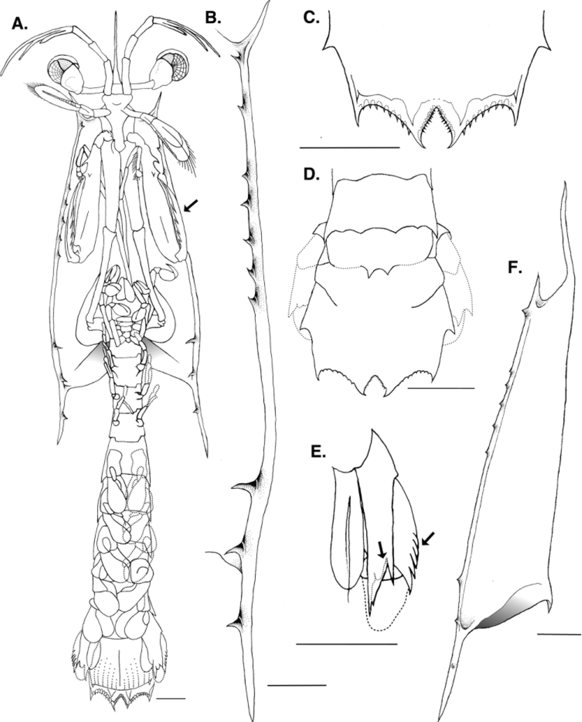
( Fig. 3 View FIGURE 3 )
Material examined. Alima orientalis last stage larva, captured northeast side of Lizard Island, Queensland, Australia; intertidal, collected at night by M.L. Porter and K.D. Feller; 3 June, 2012. All measurements and drawings reported in text are from a single reference specimen ( KF214292 View Materials , P.90969). An additional specimen ( KF205337 View Materials , P.90970) is included in Table 1 View TABLE 1 for mean character measurements.
Description. Last larval stage 20.2 mm in total length. Carapace slightly less than half of total length at 8.8 mm long, extending posteriorly along midline to thoracic somite 6; strong posterolateral spines, 2.1 mm in length, extending to abdominal somite 1; 3.2 mm long anterior spine extending past antennular peduncle to antennular article 3; anterolateral spines strong but smaller than posterolateral, extending to base of eyestalk; short, dorsally directed spine placed on hump just in front of posterior margin of carapace; lateral margins of carapace straight or convex, with 9 ventral spinules in groups of 1 or 2 large, 5 small, 3 large spinules along carapace margin; carapace width 3.3 mm.
Eyestalks elongated, not extending past anterolateral spines; evidence of both larval, adult retinae in single cornea. Adult retina with visible midband emerging medial to larval retina.
Antennules with 3-segmented stalk and 3 setiferous flagella, with lower 2 shortest. Antenna with elongated stalk, scale lying lateral to anterolateral angles of carapace; flagellum present.
Maxillipeds 1–5 well developed, first 2 elongated, last 3 short, all segments developed; epipod present on maxillipeds 1–4. Maxilliped 2 greatly enlarged, forming raptorial claw; with spine on basal portion; propodus 8 or 9 times as long as deep, distal portion of upper margin pectinate; dactylus unarmed, 5 teeth visible under cuticle.
Thoracic somites elongated, not armed posterolaterally. Pereopods 1–3 well developed. Abdominal somites 1– 5 with posterolateral angles each drawn into strong upcurved spine; each somite with pair of well-developed pleopods; pleopods with gill buds.
Abdominal somite 6 dorsally with submedian spines only; strong spine anterior to uropodal articulation; somite 6 not separated laterally from telson, demarcation visible mid-dorsally; outline of postlarval somite 6 clearly visible through cuticle.
Uropods well developed, with 5 or 6 spines on outer margin of proximal segment of exopod, all visible through cuticle with last 1 or 2 spines free. Uropodal protopod with inner spine longer than outer, without distinct lobe between spines but with rounded single lobe of postlarva clearly visible through cuticle.
Telson approximately as long as wide, without median carina or dorsal spine; submedian, intermediate, and lateral primary spines with apices fixed. Larval denticles: submedian 9–13 submedian, intermediate 7–9, none lateral. Postlarval denticles, movable apices of submedian teeth clearly visible through cuticle, about 2 submedian denticles present for every postlarval denticle; intermediate denticles 1:1.
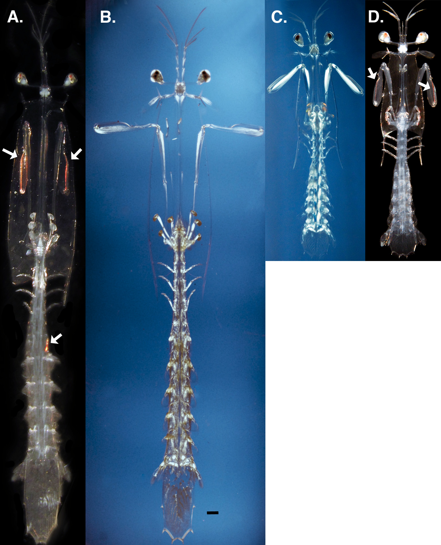
Coloration. With the exception of the retinal tissue, all other tissue types in specimens were originally observed as completely transparent. Epi-illumination produced small, red patches of iridescent color along edges of raptorial appendages and abdominal segments ( Fig. 5 View FIGURE 5 ).
Remarks. The last larva can be distinguished by the presence of postlarval features evident beneath the larval cuticle, which include the carapace, raptorial dactylus, telson armature, and uropod structure.
No known copyright restrictions apply. See Agosti, D., Egloff, W., 2009. Taxonomic information exchange and copyright: the Plazi approach. BMC Research Notes 2009, 2:53 for further explanation.
|
Kingdom |
|
|
Phylum |
|
|
Class |
|
|
Order |
|
|
Family |
|
|
Genus |