
Phyllomedusa tarsius (Cope, 1868)
|
publication ID |
https://doi.org/ 10.11646/zootaxa.5223.1.1 |
|
publication LSID |
lsid:zoobank.org:pub:2AF3B77E-408A-4104-A058-108101993EBC |
|
DOI |
https://doi.org/10.5281/zenodo.7518204 |
|
persistent identifier |
https://treatment.plazi.org/id/B31987BB-FFA9-FF82-E0D0-50FB8DC7FC31 |
|
treatment provided by |
Plazi |
|
scientific name |
Phyllomedusa tarsius |
| status |
|
Phyllomedusa tarsius View in CoL View at ENA
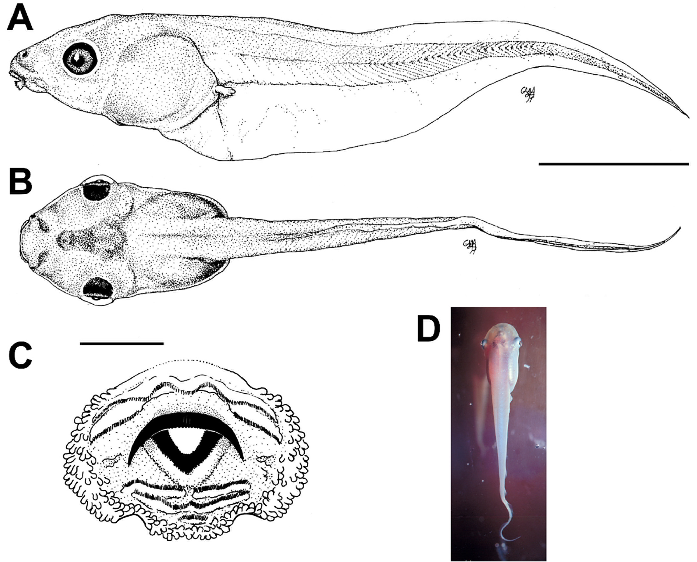
External morphology. Description based on three tadpoles at Stages 36 and 39 (LCS 615, 652). Total length 54.7 ± 10.2 mm (N = 3). Body elongate oval in dorsal view and triangular in lateral view ( Fig. 70A, B View FIGURE 70 ). Snout truncate in dorsal and lateral views. Eyes small, positioned and directed laterally. Nostrils small, oval, laterally positioned near to snout, with opening anterolaterally directed, without a projection on the marginal rim. Oral disc ( Fig. 70C View FIGURE 70 ) anteroventral; marginal papillae conical, uniseriate, with a dorsal gap. Submarginal papillae present lateroventrally. LTRF 2(2)/3(1); A1 and A2 of the same length; P1 slightly longer than P2; P3 of about one third of the length of P2. Jaw sheaths moderately wide, finely serrated; anterior jaw sheath arch-shaped, posterior jaw sheath V-shaped. Spiracle single, ventrolateral, cylindrical, short and wide, posteriorly directed, with a large opening at the medial third of the body, and with the centripetal wall fused to the body wall and longer than the external wall. Vent tube dextral, fused to the ventral fin, with a dextral opening. Caudal musculature of moderate width; in lateral view gradually tapering to a pointed tip. Dorsal fin shallow throughout its length, highest posteriorly, originating at the tail-body junction; ventral fin of moderate height, convex. Tail tip pointed. Lateral lines visible.
Colour. In preservative dorsum light greyish brown; caudal musculature grayish brown; fins translucent. In life body transparent olive or whitish with a silver venter; tail transparent or whitish (Hero 1990) ( Fig. 70D View FIGURE 70 ). Body silvery, caudal musculature pinkish, ventral fin with a black patch of variable width in the middle third of the tail.
Natural history. Eggs are deposited in a gelatinous mass in leaf nests overhanging isolated ponds in terra-firme forest, forest edge and deforested land; upon hatching tadpoles hatch and fall in the water (Neckel-Oliveira 2004; Lima et al. 2012). Mean clutch size is 342 unpigmented eggs ( Neckel-Oliveira & Wachlevski 2004) Tadpoles are nektonic and form schools ( Duellman 1978). Tail tip is capable of independent movement ( Fig. 70D View FIGURE 70 ). Tadpoles are found in all months of the year. Eggs are preyed upon by phorid fly larvae, staphylinid beetles and the snake Leptodeira annulata ( Martins & Oliveira 1998; Neckel-Oliveira 2004; Neckel-Oliveira & Wachlevski 2004). In experiments tadpoles of P. tarsius were consumed by dragonflies and fish ( Hero 1991; Azevedo-Ramos et al. 1992).
Comments. These tadpoles were described by Duellman (1978) from Ecuador and illustrated by Hero (1990) from Central Amazonia. They differ from those herein characterized by presenting body ovoid in dorsal view, eyes large, oral disc without lateral emarginations, row of marginal papillae uniseriate laterally and biseriate ventrally, LTRF 2(2)/3 ( Duellman 1978); and by presenting LTRF 2(2)/2-3[1], and anterior jaw sheat arch-shaped (Hero 1990).
No known copyright restrictions apply. See Agosti, D., Egloff, W., 2009. Taxonomic information exchange and copyright: the Plazi approach. BMC Research Notes 2009, 2:53 for further explanation.