
Leptodactylus petersii (Steindachner, 1864)
|
publication ID |
https://doi.org/ 10.11646/zootaxa.5223.1.1 |
|
publication LSID |
lsid:zoobank.org:pub:2AF3B77E-408A-4104-A058-108101993EBC |
|
DOI |
https://doi.org/10.5281/zenodo.7518190 |
|
persistent identifier |
https://treatment.plazi.org/id/B31987BB-FF9A-FFB7-E0D0-57E78CCEFD75 |
|
treatment provided by |
Plazi |
|
scientific name |
Leptodactylus petersii |
| status |
|
Leptodactylus petersii View in CoL View at ENA
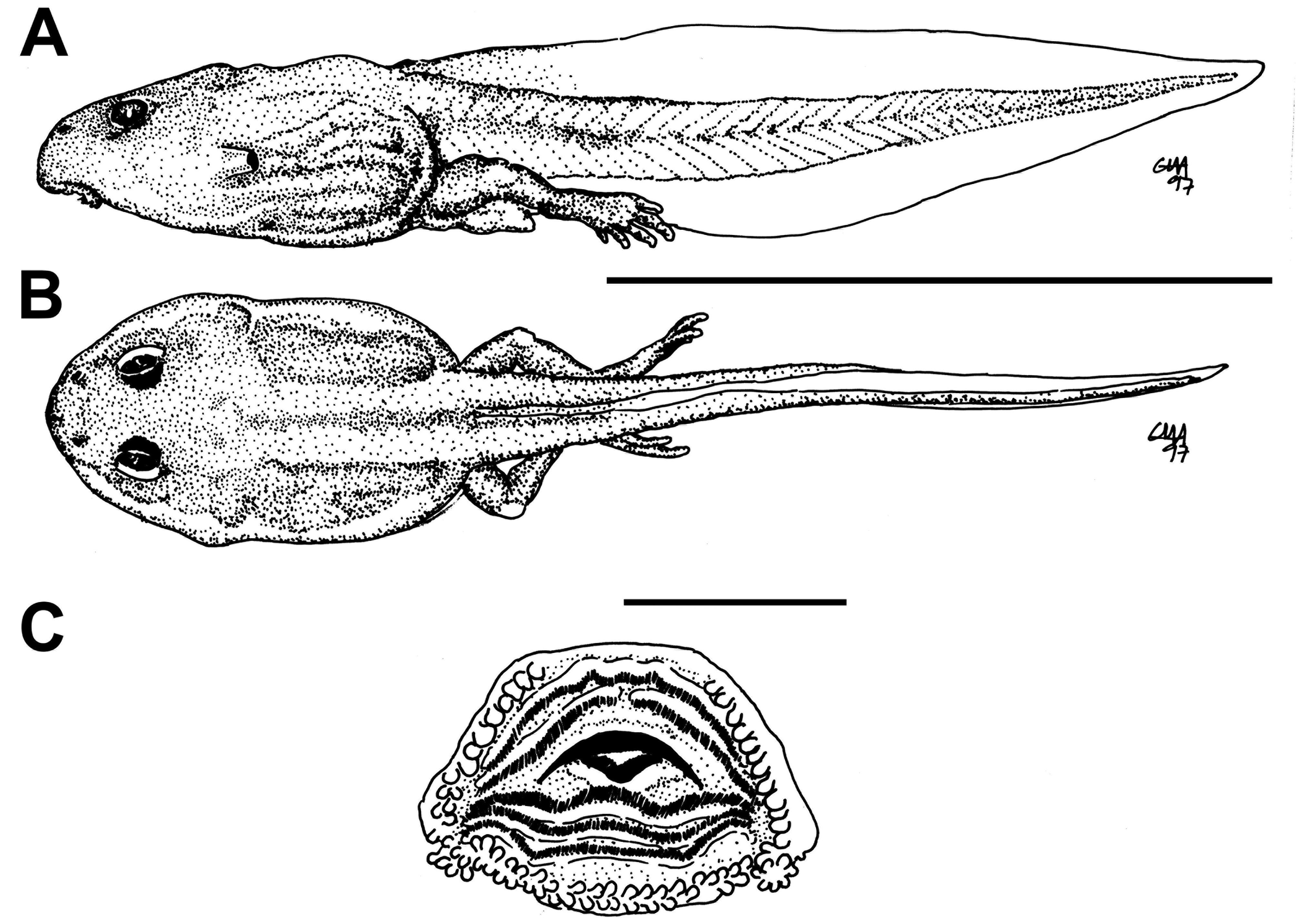
External morphology. Description based on one tadpole at Stage 40 (MNRJ 7953). Total length 26.2 mm. Body oval in dorsal view and globular/depressed in lateral view ( Fig. 59A, B View FIGURE 59 ). Snout rounded in dorsal view and truncate in lateral view. Eyes small, dorsally positioned and dorsolaterally directed. Nostrils small, oval, dorsolaterally positioned near to snout, with opening anterolaterally directed, without a projection on the marginal rim. Oral disc ( Fig. 59C View FIGURE 59 ) ventral, non-emarginate; marginal papillae conical, biseriate, with a dorsal gap. Submarginal papillae absent. LTRF 2(2)/3; A1 and A2 of the same length; P1, P2 and P3 of the same length. Jaw sheaths moderately wide, finely serrated; anterior jaw sheath arch-shaped, posterior jaw sheath V-shaped. Spiracle single, lateroventral, conical, short and wide, posteriorly directed, opening in the medial third of the body, with the centripetal wall fused to the body wall and longer than the external wall. Vent tube medial, fused to the ventral fin, with a medial opening. Caudal musculature of moderate width; in lateral view gradually tapering to a pointed tip. Dorsal fin shallow, originating at the tail-body junction, convex; ventral fin shallow, convex. Tail tip pointed.
Colour. In preservative body and caudal musculature brown, venter lighter; fins translucent. In life body dark brown / black; tail dark brown / black but becoming lighter towards the tip of the tail (as Leptodactylus wagneri / podicipinus in Hero 1990). Gut perfectly visible through ventral skin.
Natural history. Gravid females of L. petersii from Peru contain between 465 and 655 ovarian eggs ( Aichinger 1992). Foam nests are deposited in a depression excavated by the male under fallen leaves at the edge of isolated forest ponds in terra-firme forests ( Lima et al. 2012). Tadpoles were collected in the dry season. In experiments tadpoles were found to be preyed upon by fish ( Hero 1991).
Comments. Tadpoles of L. petersii from Central Amazonia were illustrated as Leptodactylus wagneri / podicipinus by Hero (1990) and described from Peru by Heyer (1994) and Duellman (2005). The tadpole illustrated by Hero (1990, Plate 32) is, according to Heyer (1994), most certainly L. petersii , since it is the only member of the complex that occurs within the forests in the areas sampled by Hero (1990). The only morphologically differences between the tadpoles illustrated by Hero (1990) and those herein characterized is the e tail tip rounded, the sloped snout in lateral viewand the seemingly anteroventral oral disc (Hero 1990). Tadpoles from Peru differ from those herein characterized by having nostril just nearer to eye or midway between eye and snout, oral disc subterminal, marginal papillae uniseriate anteriorly and biseriate lateroventrally, or uniseriate anteriorly and posteriorly and biseriate laterally ( Heyer 1994); and by presenting snout bluntly rounded in lateral view, spiracle directed posterodorsally, vent tube dextral, tail tip rounded, oral disc directed anteroventrally, row of marginal papillae uniseriate, LTRF 2(2)/2(1) or 2(2)/3[1] ( Duellman 2005).
No known copyright restrictions apply. See Agosti, D., Egloff, W., 2009. Taxonomic information exchange and copyright: the Plazi approach. BMC Research Notes 2009, 2:53 for further explanation.