
Pilumnus dofleini Balss, 1933
|
publication ID |
https://doi.org/ 10.5281/zenodo.210231 |
|
DOI |
https://doi.org/10.5281/zenodo.6177466 |
|
persistent identifier |
https://treatment.plazi.org/id/B16B8792-FFB5-483B-CCA1-FEEFE6D41B96 |
|
treatment provided by |
Plazi |
|
scientific name |
Pilumnus dofleini Balss, 1933 |
| status |
|
Pilumnus dofleini Balss, 1933 View in CoL
( Figs 1–5 View FIGURE 1 View FIGURE 2 View FIGURE 3 View FIGURE 4 View FIGURE 5 )
Pilumnus dofleini Balss, 1933: 29 View in CoL , pl. 6, fig. 29. — Sakai 1939: 538; 1965: 169, pl. 70, fig. 1; 1976: 489 (English text), pl. 175, fig. 2. — Takeda & Manuel 2000: 157 (part); Marumura & Kosaka 2003: 57. –– Takeda et al. 2006: 204.
Not Pilumnus dofleini View in CoL . — Miyake 1983: 134, pl. 45, fig. 5. [see Discussion].
Not Pilumnus dofleini View in CoL .— Ng 2000: 301, figs 1, 2. [= Pilumnus curvipenis View in CoL n. sp.]
Not Pilumnus dofleini View in CoL . — Ng et al. 2008: fig. 112. [= Pilumnus bohol View in CoL n. sp.]
Not Pilumnus dofleini View in CoL . — Takeda & Manuel 2000: 157 (part), figs 3F, 5. [= Pilumnus armatus View in CoL n. sp.]
Material examined. Holotype: female (13.0 x 15.0 mm), ZSM A20110199, off Boshu (= Boso Peninsula), Sagami Sea, Japan, 180 m, 10 September 1904, coll. F. Doflein. Photographs provided by S. Friedrich examined.
Additional material: 1 male (10.2 x 12.5 mm), NSMT-CrR 2507, Kan’non-zuka-dashi, Sagami Bay, Japan, 60– 80 m, 18 July 1957; 1 male (12.5 x 15.3 mm), CBM-ZC 10621, off Hashidate, Ishikawa Prefecture, Sea of Japan, 68 m, sandy mud, 17 October 2008, commercial gill net.
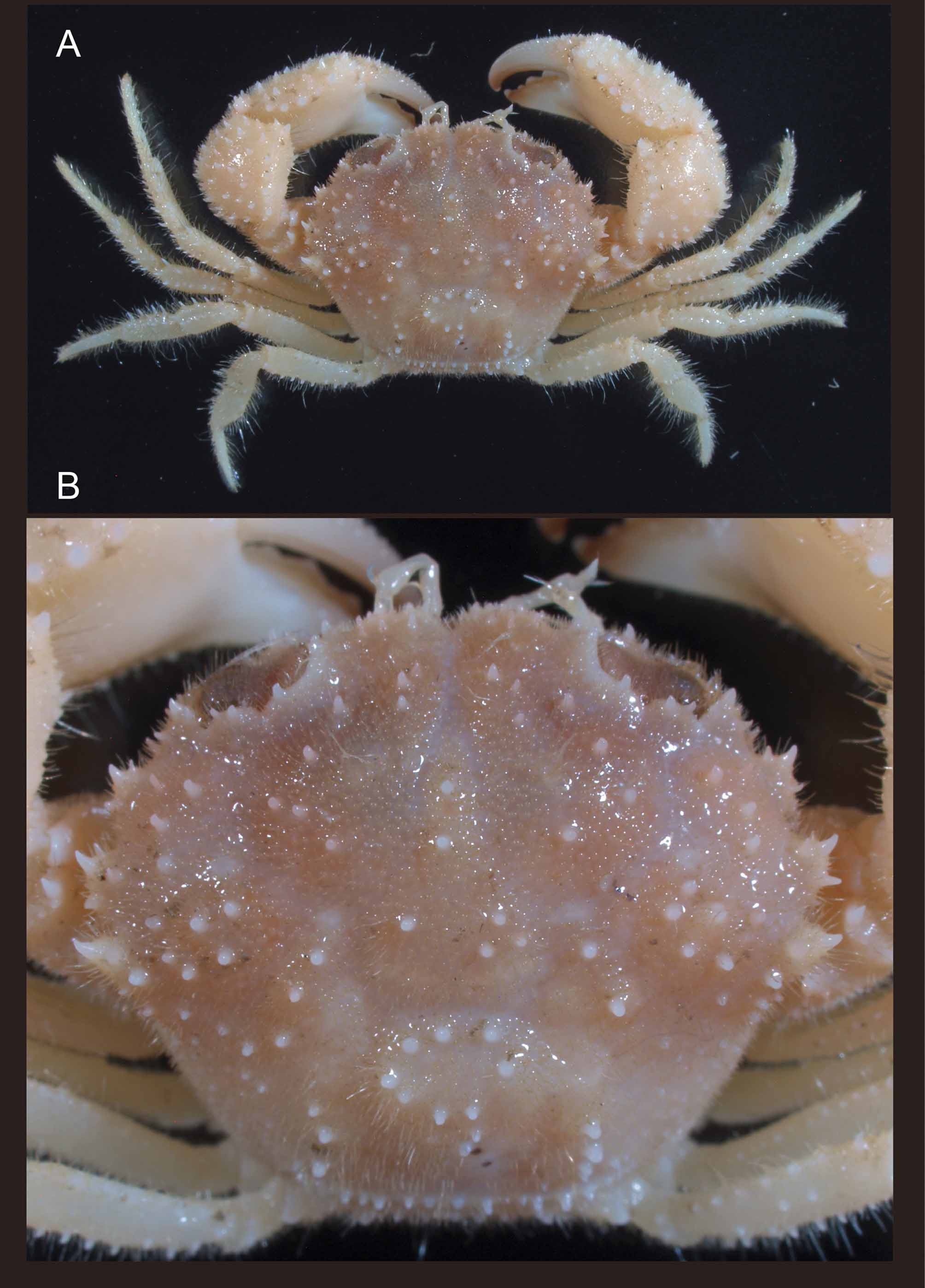
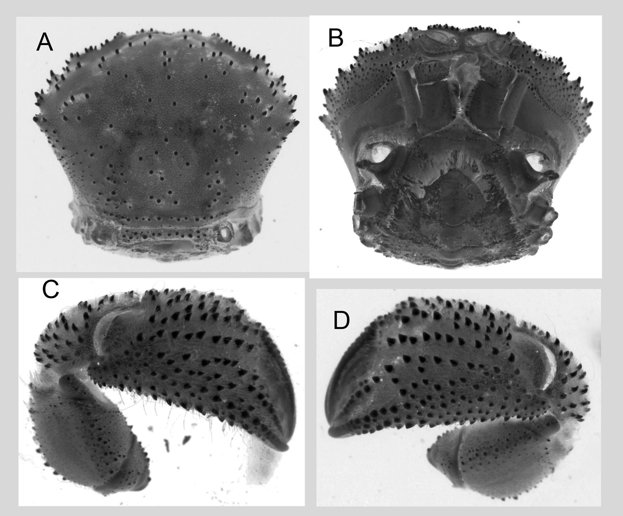
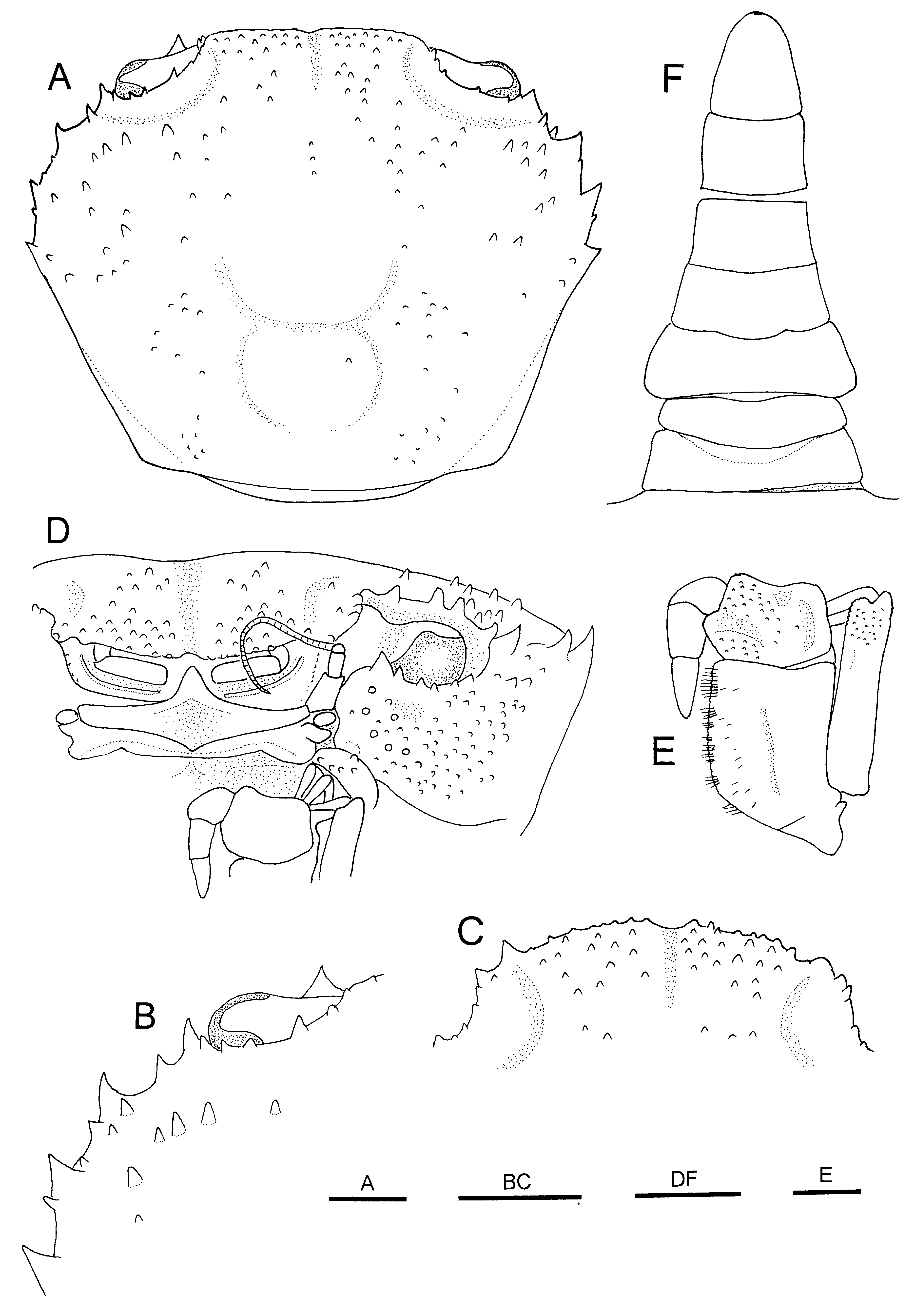
Description. Carapace ( Figs 1 View FIGURE 1 A, B, 3A–D, 5A) fairly vaulted, transversely ovoid, surface covered with numerous short setae; greatest width across fourth anterolateral spines, 1.2 times of length; dorsal surface somewhat convex longitudinally, slightly convex transversely; regions well defined, surface of regions with several conical spines or spiniform tubercles; cardiac region slightly elevated, with about 10 spiniform tubercles; grooves between regions smooth, glabrous. Frontal margin not markedly produced, separated medially by wide, V-shaped notch; each frontal lobe divided in 2 parts by broad hiatus, mesial lobe with 7 small tubercles (innermost tubercle minute), lateral lobes each consisting of single conical spine slightly larger than spines on mesial lobe; inner supraorbital angle with small spine; supraorbital margin with row of small spines, with 2 shallow cleft (one at middle, one near external orbital angle). Anterolateral margin markedly spinose, with 4 main spines (including spinose external orbital angle), second to fourth anterolateral spines subequal in size, obliquely erect, each bearing several accessory spines or spinules. Posterolateral margin almost straight, converging towards posterior margin. Posterior part of carapace with submarginal row of distinct tubercles; posterior margin slightly convex. Suborbital region with covering of numerous spinules or spinulose granules; pterygostomial groove lined with granular rows dorsally and ventrally. Dorsolateral portion of buccal frame with cluster of spines.
Orbits ( Fig. 3 View FIGURE 3 A, D) moderately large, slightly obliquely transversal in dorsal view, transversal in anterior view; eyes filling entire orbits; suborbital margin with row of small spines, innermost spine largest.
Basal segment of antennular peduncle ( Fig. 3 View FIGURE 3 D) with 2 obliquely transversal ridges on outer surface. Antennal peduncle just entering orbital hiatus; second segment with 1 small tubercle distally on outer surface.
Epistome ( Fig. 3 View FIGURE 3 D) divided into 4 crested lobes. Endostomal ridges indistinct.
Third maxillipeds ( Fig. 3 View FIGURE 3 E) completely covering buccal cavity when closed; outer surfaces of ischium, merus exopods granular. Ischium subrectangular, longer than wide, with shallow oblique median sulcus, mesial margin weakly denticulate. Merus subrectangular, wider than long, margins granular, mesial margin somewhat produced in rounded lobe. Carpus to dactylus smooth on surfaces. Exopod moderately stout, distal margin just reaching distal margin of merus, inner subdistal spine prominent.
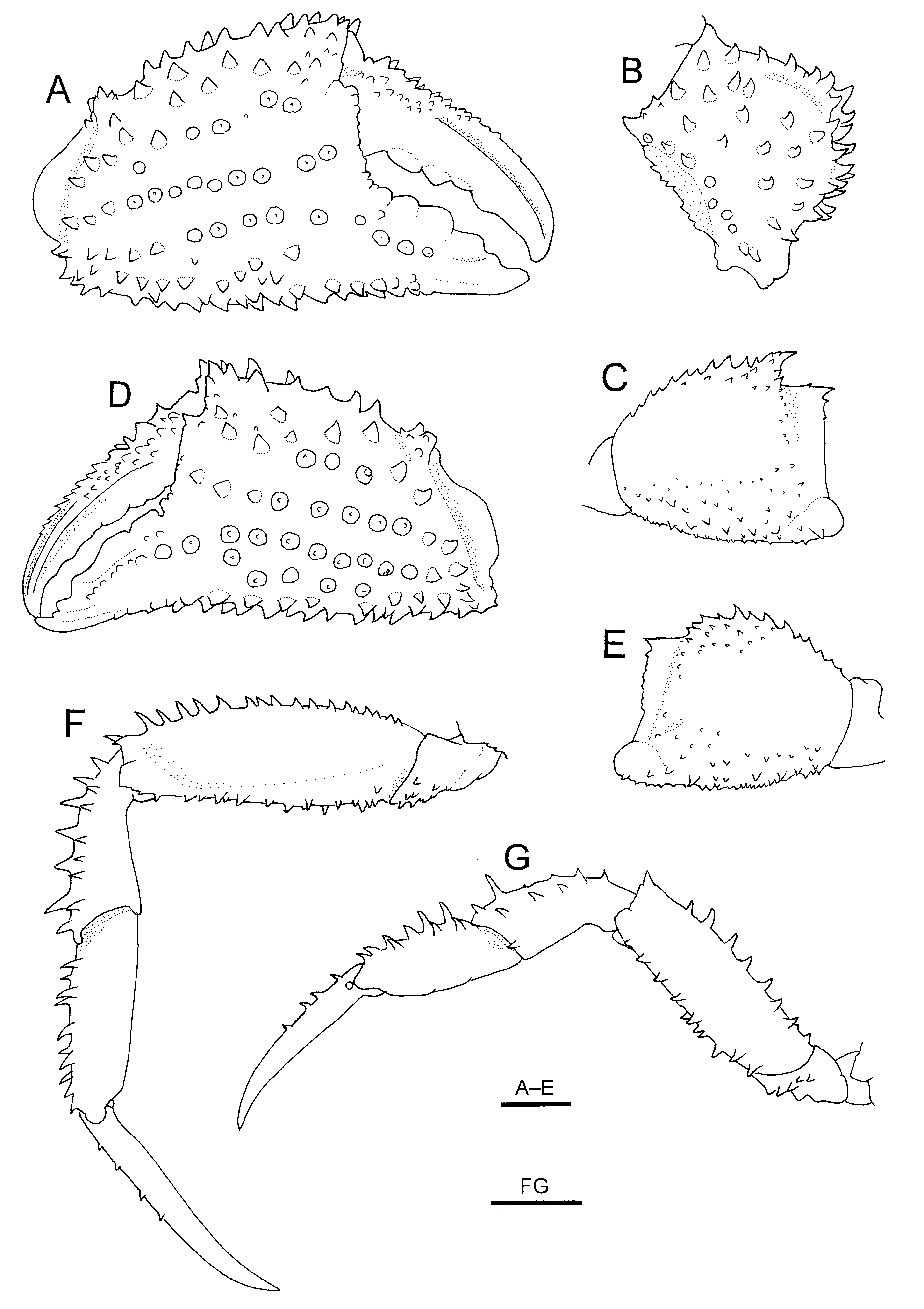
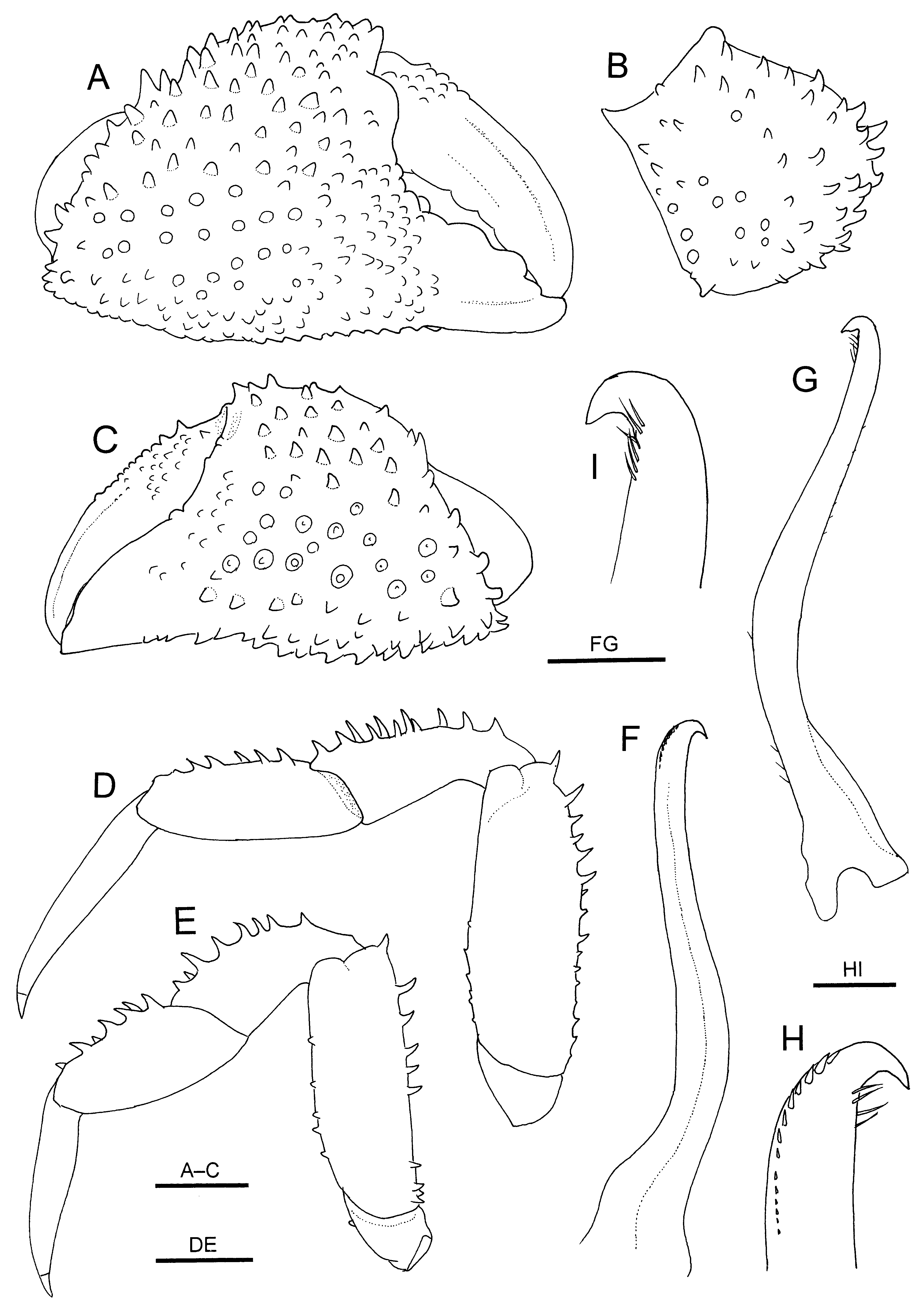
Chelipeds ( Figs 1 View FIGURE 1 A, 2A, 4A–E; 5C, D) slightly unequal, slightly dissimilar with right larger than left; outer surfaces with covering of short setae. Anterior margin of fused basis-ischium with row of small tubercles. Merus nearly as long as high; posterodorsal margin crested, with row of spines (2 distal spines prominent); outer (posterior) surface granular adjacent to margins, otherwise nearly smooth; lower surface bluntly carinate, with rows of acute, minute to small spines. Carpus numerous, small, occasionally curved spines on dorsal to outer (lateral) surface, inner distal angle with prominent spine. Palm with numerous spines on outer surface, arranged in irregular longitudinal rows (ventral rows extending onto fixed finger), inner dorsal margin bluntly carinate, with row of small tubercles or spinules; ventral surface spinulose, bluntly carinate; inner surface nearly smooth. Fixed finger of major cheliped not deflexed, with row of rounded teeth on cutting edge; outer surface flat medially. Fixed finger of minor cheliped slightly deflexed, with row of broadly triangular, sharply edged teeth on cutting edge. Dactylus with multiple rows of small spines or spinulose tubercles on dorsal surface extending beyond midlength, with 1 (right) or 2 (left) distinct longitudinal grooves on outer surfaces.
Ambulatory legs ( Figs 1 View FIGURE 1 A, 2A, 4F, G) moderately long, relatively slender for genus; surfaces with covering of short setae, dorsal (extensor) and ventral (flexor) surfaces numerous moderately short to moderately long, stiff setae; first leg (second pereopod) longest, about 1.5 times of carapace width. Coxa with minutely granular ventrodistal margin. Basis-ischium fused segment each with sharp granules on ventral surface, that of fourth leg (fifth pereopod) with small spinulose tubercles on outer surface along distal margin. Merus with single row of slender, weakly curved spines on dorsal (extensor) margin; flexor surface flanked by distinctly delimited margins each bearing row of small acute spines or tubercles; outer (posterior) surface unarmed (first to third legs) or with some small spines (fourth leg). Carpus with row of long spines on extensor margin; outer (posterior) surface with median row of spines plus few additional spines. Propodus with 2 or 3 rows of spines on extensor surface; flexor margin unarmed (first to third legs) or with 1 or 2 small spines (fourth leg). Dactylus slender, slightly curved distally, terminating in small corneous claw, longer than respective propodus; extensor margin with 0–4 spinules or tiny tubercles.
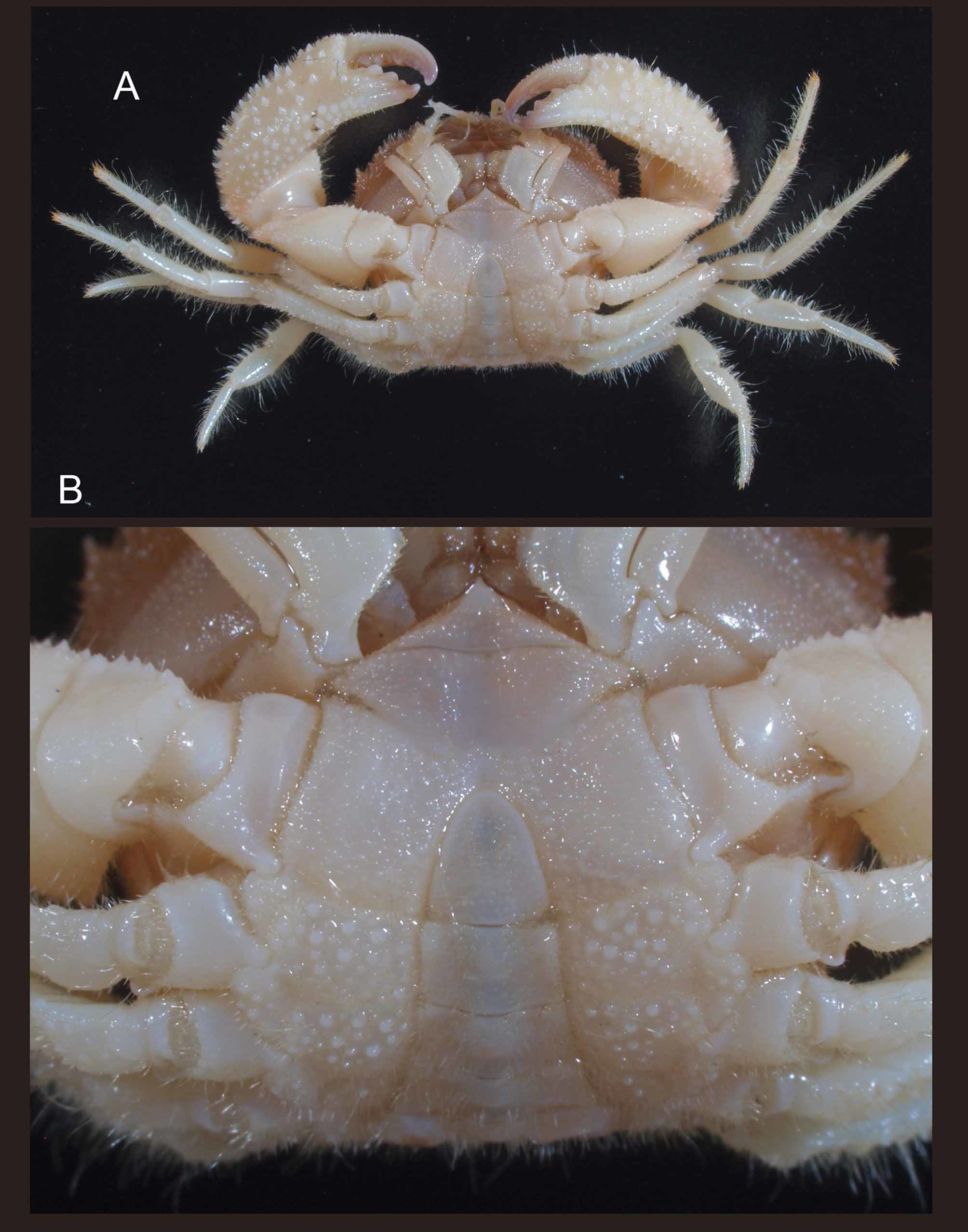
Thoracic sternum ( Fig. 2 View FIGURE 2 B) with covering of granules; these granules becoming more conspicuous in posterior sternites. Sternites 2/3 separated by distinct, straight groove; sternites 3/4 separated by suture sloping just anterior to abdominal cavity, suture becoming obsolete medially.
Male abdomen ( Fig. 3 View FIGURE 3 F) moderately narrow, somites 1 and 3 widest. Somite 1 transversely trapezoidal, armed with transversal row of small tubercles. Somite 2 subtrapezoidal, narrower than flanking somites, with paired tubercles laterally. Somites 3–6 unarmed; somite 3 broadly trapezoidal, narrowed distally, lateral margin gently convex, unarmed; somite 4 trapezoidal, lateral margins slightly concave; somites 5 and 6 subrectangular, with straight lateral margins. Telson ( Fig. 3 View FIGURE 3 F) triangular with rounded tip, 1.1 times longer than wide, lateral margins nearly straight.
Female abdomen ( Fig. 5 View FIGURE 5 B) moderately broad. Somite 1 with several tubercles on outer surface. Somite 2 with pair of tiny tubercle laterally on outer surface.
First gonopod ( Fig. 3 View FIGURE 3 G–K) sinuous; distal 0.2 noticeably arcuate in lateral or mesial view, slightly directed laterally in ventral view, gradually tapering, bearing 2 rows of minute stiff setae not extending to apex. Second gonopod ( Fig. 3 View FIGURE 3 L) short, sigmoidal, distal part spatulate, tip slightly recurved, sharply pointed.
Coloration. Carapace generally brown, whitish grooves between regions, cardiac and posterolateral regions. Chelipeds light brown on dorsal surface; outer surface of palm white. Ambulatory legs whitish with brown tinge on basal parts of dactyli, propodi, carpi and distal parts of meri. Thoracic sternum generally white.
Distribution. Presently known only from Japan (Sagami Sea and Ishikawa Prefecture, Sea of Japan); 60– 180 m.
Remarks. The identity of Pilumnus dofleini is here established. The present male specimens, including one specimen studied by Sakai (1965; 1976), agree well with the holotype female in every non-sexual diagnostic character, including the presence of a submarginal row of distinct tubercles along the posterior margin of the carapace and spines arranged in irregular rows on the outer surfaces of the chelae. These male specimens enable us to provide male diagnostic characters.
The material referred to Pilumnus dofleini by Ng (2000) is here described as P. curvipenis n. sp. Pilumnus dofleini is readily distinguished from this new species by the following characters: spines or tubercles on the dorsal surface and margins on the carapace are more conspicuous in P. dofleini than in P. curvipenis n. sp., in particular, the cardiac region is armed with about 10 distinct spines or tubercles in P. d o f l e i n i, whereas there are only a few indistinct tubercles on that region in P. curvipenis n. sp. ( Fig. 1 View FIGURE 1 B versus Fig. 12 View FIGURE 12 A); grooves defining the regions on the dorsal surface of the carapace are more distinctly marked in P. dofleini than in P. curvipenis n. sp. ( Fig. 1 View FIGURE 1 B versus Fig. 12 View FIGURE 12 A); there is a submarginal row of conspicuous tubercles adjacent to the posterior margin of the carapace in P. dofleini , but such tubercles are absent in P. c u r v i p e n i s n. sp. ( Fig. 3 View FIGURE 3 C versus Fig. 12 View FIGURE 12 A); the upper orbital margin has two small clefts in P. dofleini , whereas no distinct clefts are discernible in P. curvipenis n. sp. ( Fig. 3 View FIGURE 3 A versus Fig. 12 View FIGURE 12 B); the pterygostomial groove of the carapace is flanked by rows of distinct tubercles in P. dofleini , whereas such rows of tubercles are absent or poorly developed in P. curvipenis n. sp. ( Fig. 3 View FIGURE 3 D versus Fig. 12 View FIGURE 12 D); spines on the palms of the chelae are relatively less numerous and stronger in P. dofleini than in P. curvipenis n. sp., with those on the median area are arranged in irregular rows in P. dofleini rather than scattered in P. curvipenis n. sp. ( Fig. 4 View FIGURE 4 A, D versus Fig. 13 View FIGURE 13 A, C); the ambulatory legs are relatively more slender in P. d o f l e i n i than in P. c u r v i - penis n. sp. ( Fig. 4 View FIGURE 4 F, G versus Fig. 13 View FIGURE 13 D, E); the extensor margins of the dactyli of the ambulatory legs are armed with small spines or tubercles in P. dofleini , but unarmed in P. curvipenis n. sp. ( Fig. 4 View FIGURE 4 F, G versus Fig. 13 View FIGURE 13 D, E); the thoracic sternum is distinctly granular in P. dofleini , rather than nearly smooth in P. curvipenis n. sp. ( Fig. 2 View FIGURE 2 B versus Fig. 1 View FIGURE 1 g of Ng 2000); the first abdominal somite is armed with several distinct tubercles in P. dofleini , but is unarmed in P. curvipenis n. sp. ( Fig. 3 View FIGURE 3 C, F versus Fig. 12 View FIGURE 12 F); the first gonopod is more strongly sinuous in P. dofleini than in P. curvipenis n. sp., with its distal part arcuate in the distal 0.2 in P. d o f l e i n i, but strongly curved near the apex in P. curvipenis n. sp. ( Fig. 3 View FIGURE 3 G–K versus Fig. 13 View FIGURE 13 F–I).
Sakai (1965; 1976) reported P. dofleini on the basis of material from Sagami Bay made by the Showa Emperor of Japan (now housed in the collection of the Showa Memorial Institute of Tsukuba Research Center, National Science Museum, Tokyo). Takeda & Manuel (2000) presented a precise list of those specimens, in which the presence of ten lots was clarified. During this study, a single lot (NSMT-Cr R2507) was reexamined, of which the identification has been verified.
Miyake (1983) reported P. dofleini based on a single male specimen from Tosa Bay, Kochi Prefecture, Japan. However, this specimen seems to be different from the present specimens of P. dofleini in the dorsally naked carapace, the weaker armature on the cheliped, the lack of long dorsal or extensor spines on the ambulatory meri to propodi, and the purplish body color. There is little doubt that the specimen used by Miyake (1983) does not represent P. dofleini . Our efforts to locate Miyake’s specimen has not been successful, and the determination of the specific identity of Miyake’s (1983) specimen is not possible at this time.
Takeda & Manuel (2000) identified one female specimen from Balicasag Island, the Philippines, with P. dofleini . Although this specimen was not reexamined, it is assumed that it actually represents P. a r m a t u s n. sp. based on examination of the given photograph ( Takeda & Manuel 2000: Fig. 3 View FIGURE 3 F). The photograph clearly shows the absence of tubercles on the cardiac region of the carapace and the presence of a row of conspicuous tubercles along the posterior margin of the carapace and on the first abdominal somite, representing diagnostic features of P. armatus n. sp.
The record of P. dofleini by Marumura & Kosaka (2003) based on two specimens from Tosa Bay needs to be verified. Ng et al. (2008) published a color photograph of an ovigerous female specimen from the Philippines, referred to as P. dofleini , although P. dofleini was inadvertently omitted from their species list (Ng et al. 2008: 141– 142). This specimen is referred to P. bohol n. sp. because of the weak armature on the carapace, although it was not available for this study.

| ZSM |
Bavarian State Collection of Zoology |
No known copyright restrictions apply. See Agosti, D., Egloff, W., 2009. Taxonomic information exchange and copyright: the Plazi approach. BMC Research Notes 2009, 2:53 for further explanation.
|
Kingdom |
|
|
Phylum |
|
|
Class |
|
|
Order |
|
|
Family |
|
|
Genus |
Pilumnus dofleini Balss, 1933
| Komai, Tomoyuki & Motoh, Hiroshi 2012 |
Pilumnus dofleini
| Takeda 2000: 157 |
Pilumnus dofleini
| Miyake 1983: 134 |
Pilumnus dofleini
| Marumura 2003: 57 |
| Takeda 2000: 157 |
| Sakai 1939: 538 |
| Balss 1933: 29 |