
Hypopleuron caninum Smith & Radcliffe in Radcliffe 1913
|
publication ID |
https://doi.org/10.11646/zootaxa.4521.4.2 |
|
publication LSID |
lsid:zoobank.org:pub:CCCE0C72-7F61-4F25-AA2A-217E6D496929 |
|
DOI |
https://doi.org/10.5281/zenodo.5971214 |
|
persistent identifier |
https://treatment.plazi.org/id/B16AB72F-BE62-EC26-7787-3AFC641CFD90 |
|
treatment provided by |
Plazi |
|
scientific name |
Hypopleuron caninum Smith & Radcliffe in Radcliffe 1913 |
| status |
|
Hypopleuron caninum Smith & Radcliffe in Radcliffe 1913 View in CoL
Meristics. Dorsal-fin rays 141–151; anal-fin rays 110–118; pectoral-fin rays 22–25; pelvic-fin ray 1; caudal-fin rays 7–8; branchiostegal rays 8; abdominal vertebrae 22–24; total vertebrae 97–101; developed gill rakers on first arch 3; median basibranchial tooth patch 1.
Measurements (%SL). Head 15.8–20.2; snout 3.5–4.8; upper jaw 6.8–8.4; horizontal eye 2.9–3.3; interorbital 2.1–4.1; body depth 9.7–13.4; predorsal 16.0–19.8; pectoral fin 11.7–13.3; pelvic fin 2.3–3.4; preanal 40.6–46.2; caudal fin 2.5–4.2.
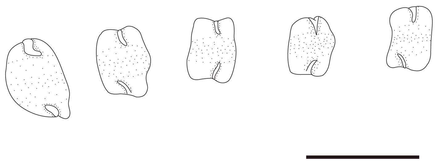
Body elongate, tapering posteriorly. Head relatively slender, about six times longer than eye diameter. Snout slightly pointed, longer than eye. Nostrils in two pairs: anterior nostril about midway between snout tip and anterior margin of eye; posterior nostril about midway between anterior nostril and anterior margin of eye. Mouth terminal, somewhat short; posterior end of upper jaw extending slightly beyond posterior margin of eye. Eye somewhat large, elliptical. Scales on head and body small; lateral line scales considerably larger, covered with skin to form a canal-like structure ( Fig. 4 View FIGURE 4 ). Lateral line well developed, originating above opercle, becoming obscure posteriorly on body. Bases of dorsal and anal fins extremely long, continuous with caudal fin posteriorly. Dorsal-fin origin above pectoral-fin base. Anal-fin origin below ca. 30th dorsal-fin ray. Pelvic fin below hind margin of preopercle. Caudal fin slender, pointed posteriorly.
Color when fresh (photographed just after capture) ( Fig. 1 View FIGURE 1 ). Head and body pale brown dorsally, whitish laterally and ventrally. Pectoral fin brown with whitish base. Dorsal, anal and caudal fins uniformly black.
Color after formalin fixation. Head and body dark dorsally. Infraorbital, cheek, and branchiostegal membrane pale. Abdominal area whitish, becoming yellowish posteriorly. All fins dark, dorsal and anal fins darker posteriorly.

No known copyright restrictions apply. See Agosti, D., Egloff, W., 2009. Taxonomic information exchange and copyright: the Plazi approach. BMC Research Notes 2009, 2:53 for further explanation.
|
Kingdom |
|
|
Phylum |
|
|
Class |
|
|
Order |
|
|
Family |
|
|
Genus |