
Deinonychus antirrhopus (Ostrom, 1969)
|
publication ID |
https://doi.org/ 10.1093/zoolinnean/zlaa048 |
|
persistent identifier |
https://treatment.plazi.org/id/B14487F2-FFCF-FFD4-FF3C-FB19FBEEB8C4 |
|
treatment provided by |
Felipe |
|
scientific name |
Deinonychus antirrhopus |
| status |
|
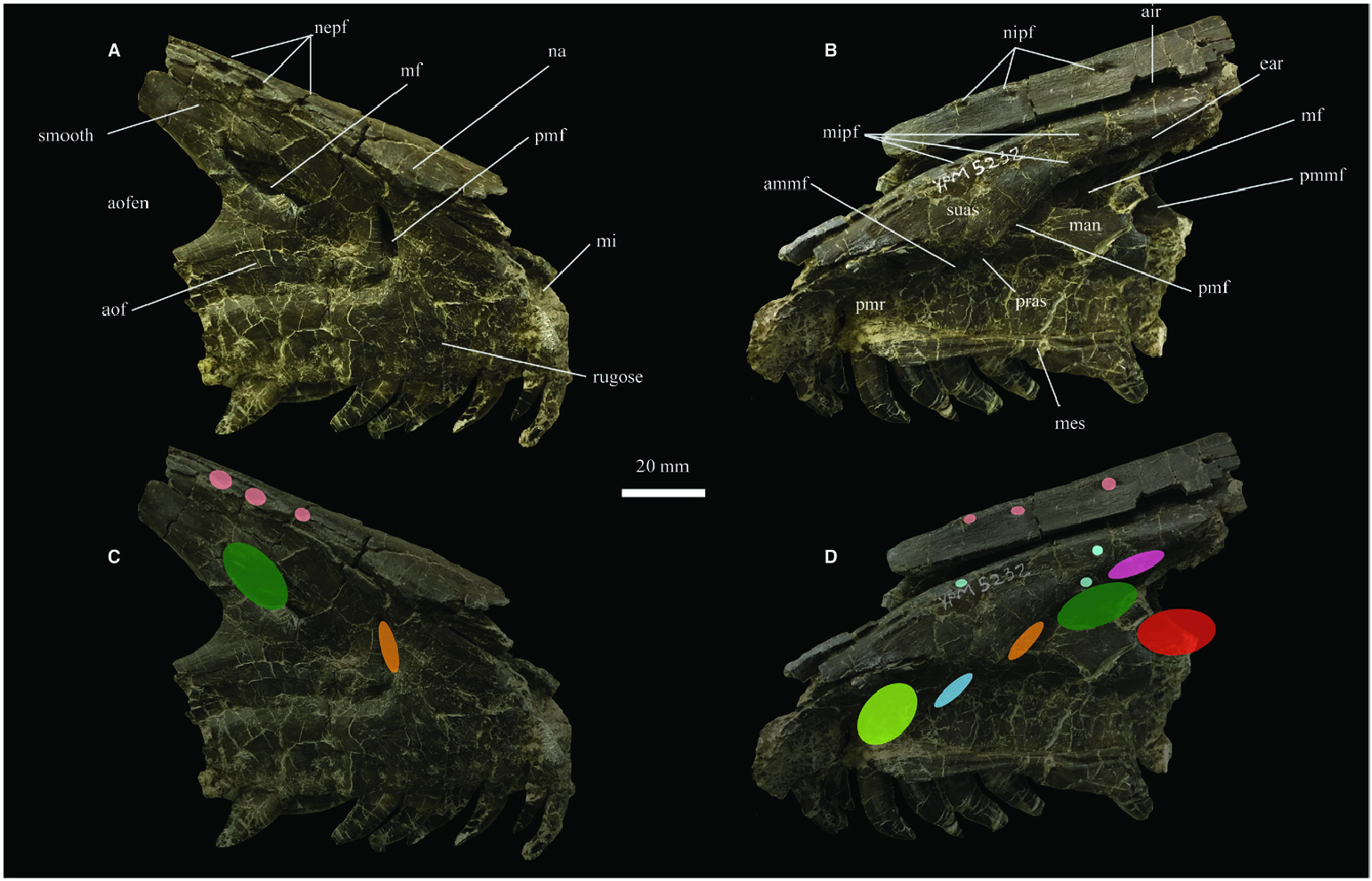
The right maxilla of Deinonychus antirrhopus (YPM VP5232) is well preserved, nearly complete and shows details of its surface texture ( Fig. 2A View Figure 2 ). Many small, ovoid maxillary alveolar foramina sit on the lateral surface of the maxillary body, as in other dromaeosaurids and theropods (e.g. Turner et al., 2012; Hendrickx & Mateus, 2014; Lautenschlager et al., 2014; Barker et al., 2017). Several fenestrae also sit within the antorbital fossa. Among them, two larger pneumatic fenestrae are present. These are the promaxillary and maxillary fenestrae ( Fig. 2A View Figure 2 ). The promaxillary fenestra is dorsoventrally teardrop-shaped and bordered by a developed medial wall. This pneumatic fenestra is deeper than the maxillary fenestra and sits just dorsal to two of the pneumatic foramina. The enlarged, deepened nature of the promaxillary fenestra may indicate an enlarged promaxillary diverticulum (e.g. Witmer, 1997; Witmer & Ridgely, 2008; Tahara & Larsson, 2011). Between the promaxillary and maxillary fenestrae, there is a concave portion of bone that represents the surface on which the antorbital sinus sat. The maxillary fenestra is ovoid and larger than the promaxillary fenestra, sitting just above the anterior end of the dorsal border of the antorbital fenestra. As in tyrannosauroids and other theropods, the presence of the maxillary and promaxillary fenestrae on the maxilla of Deinonychus antirrhopus indicates the presence of the promaxillary sinus and promaxillary antrum in this taxon ( Witmer, 1997; Tahara & Larsson, 2011; Gold et al., 2013). The broken anterior edge of the maxilla of Deinonychus shows this bone is hollow anteriorly. The antorbital fossa is dorsally strongly bordered by a shelf. However, the ventral border of this fossa is not deeply invaginated and lacks a shelf-like border.
Medially, the maxilla of Deinonychus antirrhopus possesses many pneumatic features, including large ovoid to circular pneumatic foramina along the medial surface of the ascending ramus just ventral to the contact with the nasal ( Fig. 1B View Figure 1 ). Some of these pneumatic foramina on the distal end of the medial surface of the enlarged suprantral strut appear as extremely elongate groove-like structures. These are the impressions of diverticula of the maxillary antrum sinus. At the posterodorsal end of the preserved portion of the maxilla, a large concavity is identifiable as the epiantral recess. Anteroventral to this feature, two pneumatic features, the maxillary fenestra and the posteromedial maxillary fenestra, are present and separated by the postantral strut ( Fig. 1B View Figure 1 ). Ventral to these two features, the maxillary antrum appears as a large recess that anteriorly leads into the promaxillary fenestra. The preantral strut is broken; however, it is clear this feature would have separated the promaxillary fenestra from the more ventrally placed anteromedial maxillary fenestra ( Fig. 1B, D View Figure 1 ). This latter fenestra is also separated from the promaxillary recess. In total, the medial pneumatic features of the maxilla of D. antirrhopus indicate a complex maxillary antrum morphology in this taxon. In Deinonychus and other theropods, the maxillary antrum and promaxillary sinus form the maxillary sinus (e.g. Witmer, 1997; Witmer & Ridgely, 2008; Gold et al., 2013). Among the pneumatic features of the medial surface of the maxilla, the maxillary antrum and maxillary fenestra would have interacted with the maxillary antrum sinus, whereas the promaxillary fenestra, promaxillary recess and anteromedial maxillary fenestra would have interacted with the promaxillary sinus and other anterior diverticula of the antorbital sinus (e.g. Witmer & Ridgely, 2008; Tahara & Larsson, 2011; Gold et al., 2013).

No known copyright restrictions apply. See Agosti, D., Egloff, W., 2009. Taxonomic information exchange and copyright: the Plazi approach. BMC Research Notes 2009, 2:53 for further explanation.
|
Kingdom |
|
|
Phylum |
|
|
Class |
|
|
Order |
|
|
Family |
|
|
Genus |
|
Kingdom |
|
|
Phylum |
|
|
Class |
|
|
Order |
|
|
Family |
|
|
Genus |