
Xestaspis semengoh Eichenberger, 2012
|
publication ID |
https://doi.org/10.11646/zootaxa.3160.1.1 |
|
DOI |
https://doi.org/10.5281/zenodo.5248342 |
|
persistent identifier |
https://treatment.plazi.org/id/B12087C5-FFAE-FF97-E3DD-35BA04C51C74 |
|
treatment provided by |
Felipe (2021-08-23 23:03:43, last updated 2024-11-27 09:11:45) |
|
scientific name |
Xestaspis semengoh Eichenberger |
| status |
sp. nov. |
Xestaspis semengoh Eichenberger View in CoL n. sp.
( Figs. 41–42 View FIGURE 41 View FIGURE 42 )
Type material: Holotype male ( PBI_OON 00031783 ): West Sarawak , Semengoh Arboretum , lower track , sieving leaflitter , 23 March 1985, coll. Deeleman, leg. C.L. en P.R. Deeleman ( RMNH). Female paratype ( PBI_OON 00031782 ), West Sarawak, Semengoh Arboretum , litter, humus, wood, 27 March 1985, coll. Deeleman, leg. C.L. en P.R. Deeleman ( RMNH).
Etymology: The species epithet refers to the Semengoh wildlife centre in Sarawak which is the type locality.
Diagnosis: Resembles Gamasomorpha taprobanica Simon, 1893 but sternum with rows of large, rather channellike than droplike pits between coxae I-II, II-III and III-IV (fig. 41 D); abdomen scutopedicel region without second more dorsal ridge (fig. 42. A); carapace posterolateral edges with small, laterally located pits (cpp) (figs. 41. F, H); male palpal conductor (co) distal part curved ventrally, distal tip rounded, without ventral excavation (figs. 42. G–H).
Description: MALE: Body length 2.3 mm. Uniformely orange-brown colored species (figs. 41. A–C). Carapace broadly oval in dorsal view; pars cephalica quite strongly elevated in lateral view, without posterolateral spikes, surface sides granulate (figs. 41 E–F), lateral margin straight from dorsal view, cephalic setae u-shaped in double row, transverse row of setae at posterior end of cephalothorax proximally originating from tiny, blunt denticles. Anterior lateral eyes separated from edge of carapace by their radius or more, eye group by less than diameter of anterior lateral eyes narrower than clypeus, all ovoid, subequal (fig. 41. G). Sternum anterior margin with continuous transverse groove (ctg) (fig. 41. D). Abdomen scuto-pedicel region with paired curved scutal ridges (psr) with triangular projections (fig. 42. A), pedicel tube with small, dorsolateral, triangular extensions (pds), (fig. 42. A), booklung covers (boc) small, elliptical, border elevated from surface, anterolateral edge with tubercle (figs. 42. B–C); postepigastric scutum with short, posteriorly directed, lateral apodemes. Legs patella plus tibia shorter than carapace. No detailed description on promarginal chelicerae setae, serrula, grooves on lateral margin of sternum, trichobothria hood structure, tarsal organ and scopula between claws. Male genitalia: Similar to X. kandy n. sp. with a long slender, lamellar embolus (em), adjacent to an embolic accessory appendage (ma) and a lamellar conductor (co) (fig. 42. E–H). Bulbus distally tapering into a strongly pointed conical extension (ce) (figs. 42. E–F).
FEMALE: Body length 2.4 mm. Postepigastric scutum without lateral apodemes; colulus presented only by setae. Female genitalia: Ventral view (fig. 42. D): Without external features.
Distribution: West Sarawak, Semengoh Arboretum.
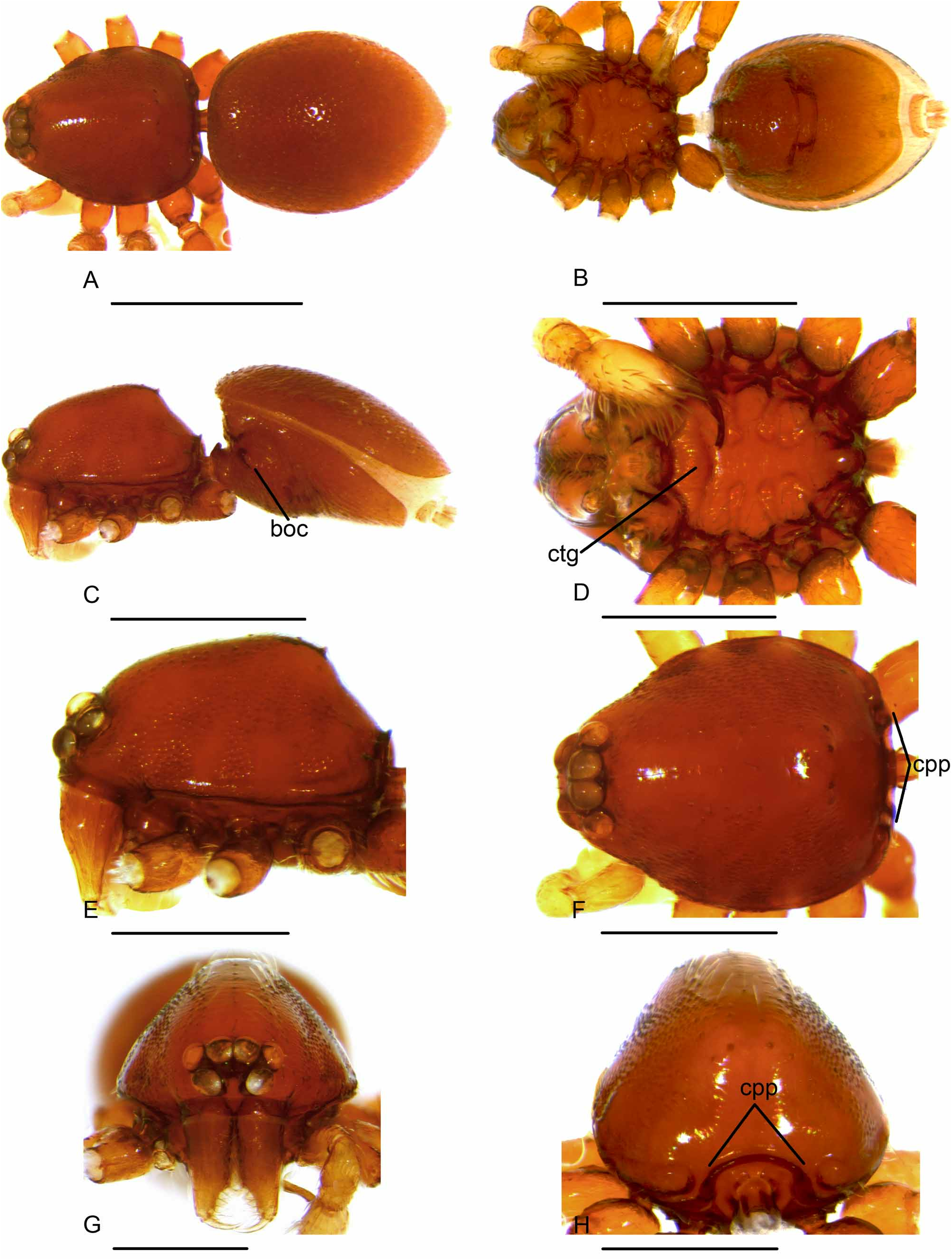
FIGURE 41. Xestaspis semengoh n. sp. Male: A. habitus dorsal view; B. habitus ventral view; C. habitus lateral view; D. sternum ventral view; E. carapace lateral view; F. carapace dorsal view; G. carapace anterior view; H. carapace posterior view. Abbreviations: boc, booklung covers; ctg, continuous transverse groove; cpp, carapace posterolateral pits. Scale bars: A–C 1 mm; D–H 0.5 mm.
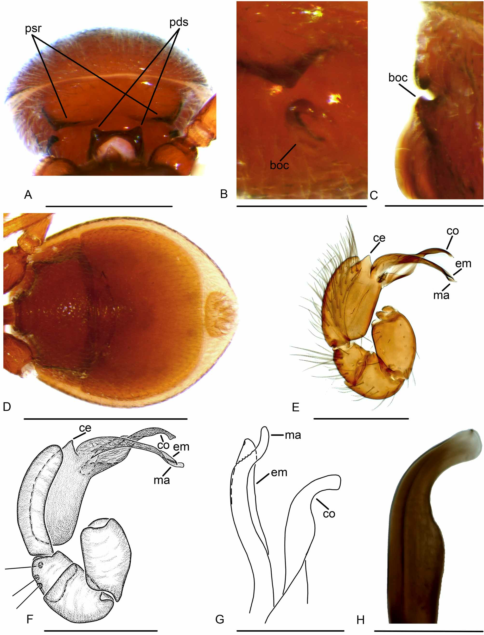
FIGURE 42. Xestaspis semengoh n. sp. Male: A. abdomen anterior view; B. booklung covers lateral view; C. booklung covers posterior view; E–F. right palp, prolateral view; G. right embolus, prolateral-ventral view; H. right palp, ventral appendage, ventral view. Female: D. abdomen ventral view. Abbreviations: psr, paired scutal ridges; pds, pedicel dorsolateral, triangular extensions; boc, booklung covers; ce, conical extension; em, embolus; co, conductor; ma, mesal embolic accessory appendage. Scale bars: A, D 0.5 mm; B–C 0.2 mm; E–F 300 µm; G 100 µm; H 50 µm.
No known copyright restrictions apply. See Agosti, D., Egloff, W., 2009. Taxonomic information exchange and copyright: the Plazi approach. BMC Research Notes 2009, 2:53 for further explanation.
1 (by felipe, 2021-08-23 23:03:43)
2 (by ExternalLinkService, 2021-08-24 17:43:46)
3 (by ExternalLinkService, 2021-08-24 18:41:58)
4 (by ExternalLinkService, 2021-08-24 19:54:16)
5 (by valdenar, 2022-01-25 22:02:27)
6 (by valdenar, 2022-01-27 16:55:20)
7 (by ExternalLinkService, 2022-01-27 17:05:52)
8 (by ExternalLinkService, 2022-01-27 18:31:25)
9 (by carolina, 2022-09-13 16:36:52)
10 (by ExternalLinkService, 2023-08-28 15:22:02)
11 (by plazi, 2023-11-04 14:26:08)