
Lochmanolenellus pentagonalis, Webster, Mark & Bohach, Lisa L., 2014
|
publication ID |
https://doi.org/10.11646/zootaxa.3824.1.1 |
|
publication LSID |
lsid:zoobank.org:pub:023D78D0-4182-48D2-BAEB-CDA6473CF585 |
|
DOI |
https://doi.org/10.5281/zenodo.6129730 |
|
persistent identifier |
https://treatment.plazi.org/id/B10C8793-FFDC-FFA2-61B5-FBD7FB6D85E7 |
|
treatment provided by |
Plazi |
|
scientific name |
Lochmanolenellus pentagonalis |
| status |
sp. nov. |
Lochmanolenellus pentagonalis n. sp.
Figs 11 View FIGURE 11 , 12 View FIGURE 12 , 13 View FIGURE 13
1999 Laudonia bispinata Harrington; Lieberman (part), fig. 20.1 only.
Diagnosis. Cephalon pentagonal in outline; genal spine base transversely opposite lateral margins of L2 or posterior portion of L3; distal portion of posterior cephalic margin oriented anterolaterally at approximately 5° to 25° relative to an exsagittal line when traced toward base of genal spine. Distance (tr.) between genal spine bases 160% to 199% of cephalic length (sag.). Angle between distal portion of posterior cephalic margin and outer margin of proximal portion of intergenal spine 115° to 141°. Length (sag.) of LA 66% to 81% of maximum width of LA. Posterior tip of ocular lobe transversely opposite lateral margins of anterior portion of L1, S1, or posteriormost portion of L2.
Description (mature morphology, phase 5 of cephalic development; sagittal cephalic length> 5 mm)
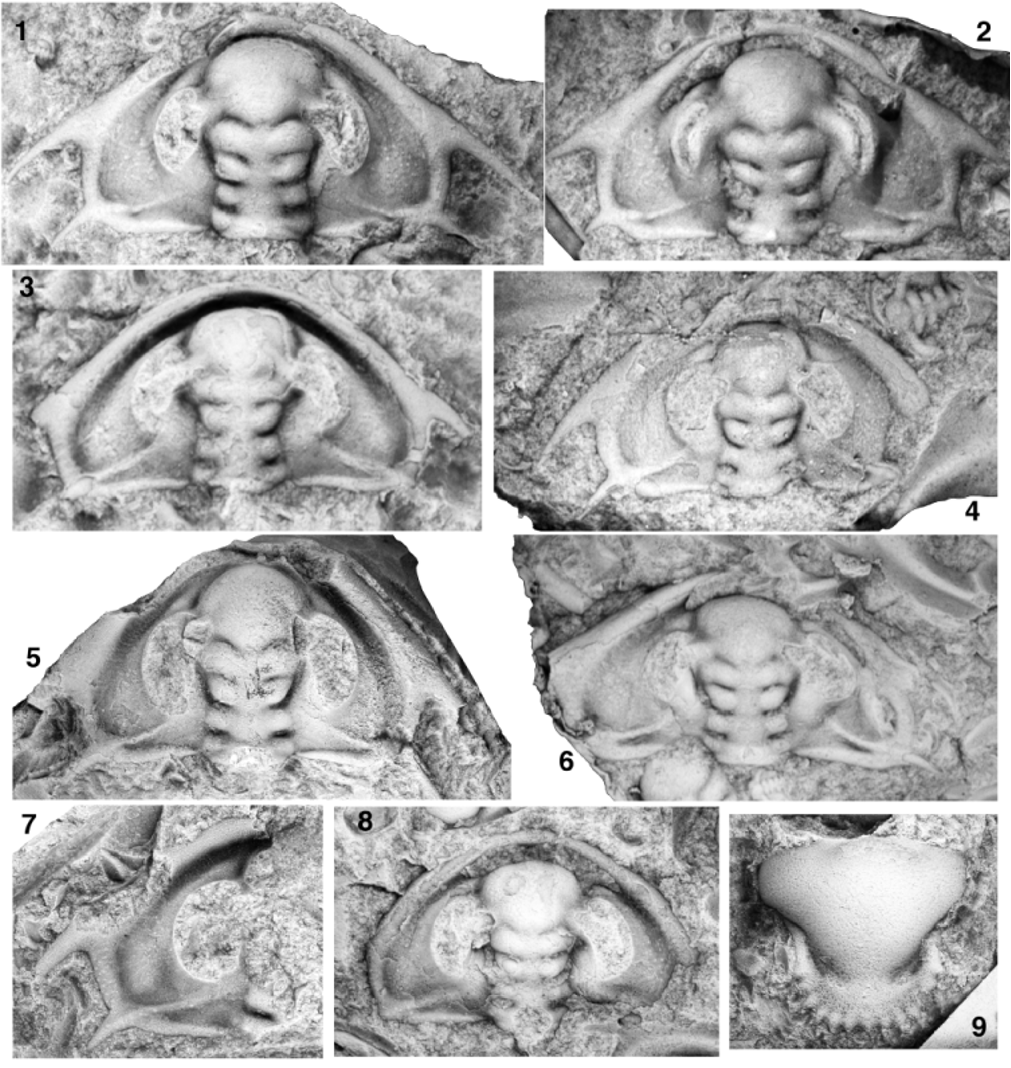
Cephalon pentagonal in outline, widest (tr.) at base of genal spines. Proximal portion of posterior cephalic margin oriented weakly posteriorly by up to 9° relative to a transverse line when traced abaxially, flexing into a roughly transverse or weakly anterior orientation at a weak, rounded adgenal angle located approximately half way along cephalic margin from axial furrow to base of genal spine, flexing anteriorly again at strong intergenal angle so that distal portion of posterior cephalic margin is oriented anterolaterally approximately 16° (range 25° to 5°) relative to an exsagittal line when traced abaxially; sometimes bears slight geniculation along distal portion of posterior cephalic margin (Figs 11.1, 11.2, 11.4, 12.2, 12.4). Genal spine stout, broad-based; angle between distal portion of posterior cephalic margin and inner margin of proximal portion of spine approximately 69° (range 57° to 88°), spines gently curving into slightly more posterior orientation along their length; tips rarely preserved but spine length estimated to be approximately two-fifths cephalic length (sag.); spine base at contact with distal portion of posterior cephalic margin transversely opposite lateral margins of L2 or posterior portion of L3 (more anteriorly located on larger specimens). Distance (tr.) between genal spine bases approximately 174% (range 160% to 199%) of cephalic length (sag.). Intergenal spine slender, broad-based; angle between distal portion of posterior cephalic margin and outer margin of proximal portion of spine approximately 129° (range 115° to 141°); tips rarely preserved but spine length estimated to be approximately one-quarter to one-third cephalic length (sag.); spine base slightly distal to adgenal angle. Cephalic border defined by broad, trough-like border furrow interrupted only by intergenal ridge at it merges with border at base of intergenal spine; border furrow somewhat shallower and narrower along proximal portion of posterior cephalic border. Anterior cephalic border narrowest at sagittal axis, broadens distally, width opposite junction of ocular lobes with LA approximately 15% length (sag.) of glabella (up to 18% on compacted specimens; Fig. 12 View FIGURE 12 ); broadest portion of border located at confluence with genal spines; posterior border tapers to point adaxially, barely touches axial furrow at posterior-most portion of LO. Border well rounded dorsally anterior to LA, somewhat more broadly dorsally arched at and between bases of genal and intergenal spines (entire border of much lower dorsal convexity on compacted specimens; Fig. 12 View FIGURE 12 ). Preglabellar field absent; LA slopes steeply anteriorly into trough-like cephalic border furrow that is slightly narrower than anterior border at sagittal axis. Plectrum not developed. Glabella club- to hourglass-shaped in outline, 81% to 89% of cephalic length (sag.), moderately dorsally convex (tr.), strongly dorsally arched (sag.), summit formed by posterior portion of LA and L3. Maximum width of LA approximately 122% (range 115% to 129%) of basal glabellar width (tr.). Sagittal portion of posterior margin of glabella more–or-less straight, transverse. SO deep only abaxially, abaxial end slightly anterior to adaxial end. LO weakly subtrapezoidal, narrows slightly anteriorly (transverse width across SO approximately 95% basal glabellar width), length (exsag.) approximately 10% to 12% of glabellar length (sag.), strongly dorsally convex (tr.). S1 deepest abaxially, approximately parallel to SO. L1 trapezoidal, narrowing (tr.) anteriorly, transverse width across S1 approximately 82% (range 76% to 90%) of basal glabellar width; length (exsag.) approximately 12% to 15% of glabellar length (sag.). S2 deepest abaxially, connected to axial furrow, arcuate (convex anteriorly) on either side of sagittal axis, adaxial portion slightly posterior to distal portion. L2 broadly V-shaped, lateral margins strongly diverging anteriorly, transverse width across S2 90 % to 110% basal glabellar width, distance (exsag.) between contact of S1 with axial furrow and contact of S2 with axial furrow approximately 10% to 13% of glabellar length (sag.). S3 transglabellar but shallow over axis, oriented anterolaterally away from axis until contact with ocular lobes where it contacts anteriorly converging axial furrow, the two together thus forming a strongly caret-shaped furrow on either side of sagittal axis, deepest at apex of each caret. L3 broadly M-shaped, lateral margins diverging anteriorly until contact with inner margin of ocular lobes, then converging anteriorly until contact with S3; transverse width of glabella at point of contact between axial furrow and inner margin of ocular lobes approximately 117% (range 112% to 132%) of basal glabellar width, distance (exsag.) between contact of S2 with axial furrow and contact of axial furrow with inner margin of ocular lobes approximately 5% to 10% of glabellar length (sag.). LA transversely oblate in outline, anterior margin bluntly rounded and rather bull-nosed; maximum width (tr.) at contact with outer margin of ocular lobes, length (sag.) approximately 74% (range 66% to 81%) of maximum width and 40% to 48% of glabellar length (sag.); moderately dorsally convex (tr.), strongly dorsally convex (sag.); prominently inflated relative to extraocular area, lateral margins clearly defined from anterior portion of extraocular area by axial furrow. Shallow, weakly arcuate (convex posteriorly) preocular furrow runs inwards and anteriorly from contact of LA with outer margin of ocular lobes; not incised over axis. Very shallow transocular furrow runs anterolaterally across ocular lobes from point of contact between S3 and axial furrow to preocular furrow, isolating ocular lobes from LA. Axis of LO bears prominent axial node. Prominent ovoid lateral swellings on L2, more subdued lateral swellings on anterior portion of LO and L1 and sometimes L3. Axial furrow broad, deepest at lateral margins of L2 and L3, shallow adjacent to lateral swellings on LO. Ocular lobes divergent from exsagittal axis by approximately 32° (range 24° to 39°), crescentic; posterior tip transversely opposite lateral margins of anterior portion of L1, S1, or posterior-most portion of L2 (more anteriorly located on larger specimens). Deep ocular furrow runs along length of ocular lobe slightly distal to midline so that inner band of ocular lobe is wider (tr.) than outer band; anteriorly, ocular furrow curves slightly outwards to merge into a “triple junction” of furrows at point where preocular furrow meets the axial furrow at the margin of LA, thus isolating outer band of ocular lobe from LA; posteriorly, ocular furrow curves slightly inwards to meet inner margin of ocular lobe so that posterior tip of ocular lobe is located on outer band. Inner and outer bands of ocular lobe each convex dorsally (tr.). Circumocular suture defines large visual surface (not preserved) that occupied much of length of outer wall of ocular lobe; portion of outer wall of ocular lobe below circumocular suture forms prominent, steep eye socle. Interocular area slopes down from inner margin of ocular lobe to axial furrow; surface sometimes convex upward, giving arched appearance; approximately two-thirds width (tr.) of inner band of ocular lobe opposite S2. Extraocular area climbs gently out of broad border furrow then gently arches to produce a weakly vaulted transverse profile. In vicinity of ocular lobe, extraocular area abruptly changes slope to form a steep-walled, narrow-topped extraocular platform upon which ocular lobe sits; extraocular platform comma-shaped to subcrescentic in outline when viewed from above, platform broadest (tr.) and most obvious in dorsal view adjacent to contact between outer margin of ocular lobe with LA; anteriorly, extraocular platform narrows to a thin, subdued ridge that extends beyond anterior limit of ocular lobe, descending in vertical height and running parallel to lateral margin of LA then fading into anterior border furrow; posteriorly, extraocular platform progressively fades and merges into posterolateral portion of extraocular area just anterior to posterior tip of ocular lobe. Prominent intergenal ridge extends from interocular area opposite lateral margin of L1 to posterior cephalic border at base of intergenal spine; ridge broadens distally and merges smoothly into border, interrupting border furrow. Weak genal ridge rarely present (Fig. 12.1). Weak extraocular caeca sometimes present on extraocular area ( Fig. 11 View FIGURE 11 ), often most clearly expressed in cephalic border furrow. Network of raised ridges forming walls of polygonal, mesh-like prosopon on dorsal surface of extraocular area, intergenal ridge, glabella, cephalic border furrow, and cephalic border (Fig. 11.1, 11.2, 11.7, 11.8). Central raised boss developed in most polygons; central bosses developed into large, pustule-like structures in some polygons on distal portion of extraocular area ( Fig. 11 View FIGURE 11 ) and posterior cephalic border (Fig. 11.7). Subparallel lirae sometimes present on anterior cephalic border and margins of genal spines (Fig. 11.1–11.4). Terrace lines on cephalic doublure and ventral surface of genal spines (Fig. 12.3).
Only known hypostome (Fig. 11.9) approximately 5.65 mm in sagittal length, moderately convex (tr., sag.), subtrapezoidal (widest anteriorly) in outline. Anterior lobe of middle body broadly subtriangular, occupies approximately 78% of sagittal length of hypostome; anterior margin gently curved; extends laterally onto broad, triangular anterior wings, tips of which are located less than one-third distance along hypostomal length (traced from anterior to posterior). Posterior lobe of middle body narrow, <20% of hypostome length (sag.), transversely crescentic in outline, widest anteriorly, divided from anterior lobe over sagittal axis by broad, shallow furrow. Maculae relatively deep. Anterior border not preserved. Posterior border narrow, approximately 6% of hypostome length sagittally, poorly defined by very shallow furrow. Thirteen small, triangular spines project posterolaterally from margin of lateral and posterior border (very short median spine flanked by six spine pairs); spine lengths progressively increase from median spine to second outside pair, second to fourth pairs subequal in size, distalmost two pairs progressively smaller. Network of raised ridges forming walls of polygonal, mesh-like prosopon on entire ventral surface.
Thorax (known from one specimen, Fig. 12.4, 12.5) of at least 20 segments; width (tr.) of axis approximately 94% width (tr.) of inner pleural region on T1, gently tapering posteriorly to at least T14. Axes of T1 to T14 with prominent subtriangular lateral swellings (widening abaxially) in anterolateral corners that deflect axial furrows abaxially; each axial ring therefore trapezoidal in outline, with anteriorly divergent, slightly sigmoidal, lateral margins. Axial portions of axis not well preserved (fractured and often chipped), but axial swelling or node seemingly originally present on at least T2, T3, T7, and T10 to T14 (conceivably originally present on T1 to T14). Inner pleural regions of T1 and T2 transverse, parallel-sided, with straight margins; pleural spines of T1 and T2 slender, divergent, falcate; pleural spine of T1 approximately 95% of width (tr.) of inner pleural region, pleural spine of T2 estimated to have been slightly longer than that of T1 (but spine tip not preserved). T3 weakly macropleural; pleural spine macrospinous with posterior tip located transversely opposite axial ring of at least T13. Inner pleural region of T4 transverse, very slightly tapering distally, with slightly curved anterior margin to accomodate distal expansion of T3; inner pleural regions of T5 to T9 transverse, parallel-sided, with straight margins; those of T10 to T14 increasingly divergent to pendent, with increasingly curved margins; width (tr.) of inner pleural regions relative to width (tr.) of axial rings proportionally decreasing down thorax. Pleural spines of T4 and T5 slender, divergent, weakly falcate; those of T6 and T8 to T14 (that of T7 not preserved) falcate, progressively increasing in length down the thorax to T8 then proportionally decreasing in length on T9 to T14, divergent. Pleural furrows of T1 to T14 relatively broad (exsag.), occupying almost half length (exsag.) of inner pleural region, anterior wall steeper than posterior wall; extending onto pleural spines of T1 to T12, terminating at or just short of base of pleural spine on T13 and T14. Axes of T15 to T20 poorly preserved; that of T15 elongated posteriorly into broad-based axial spine of unknown length; axial furrow of T20 represented by break in slope. Inner pleural regions of T15 to T20 much narrower than axes (tr.), continuing trend in proportional width from T1 to T14; margins curved, converging distally into short, divergent, sentate pleural spines that progressively shorten in length from T15 to T20. Pleural furrows terminate on inner pleural region of T15; not evident on more posterior segments. Rostral plate and pygidium unknown.
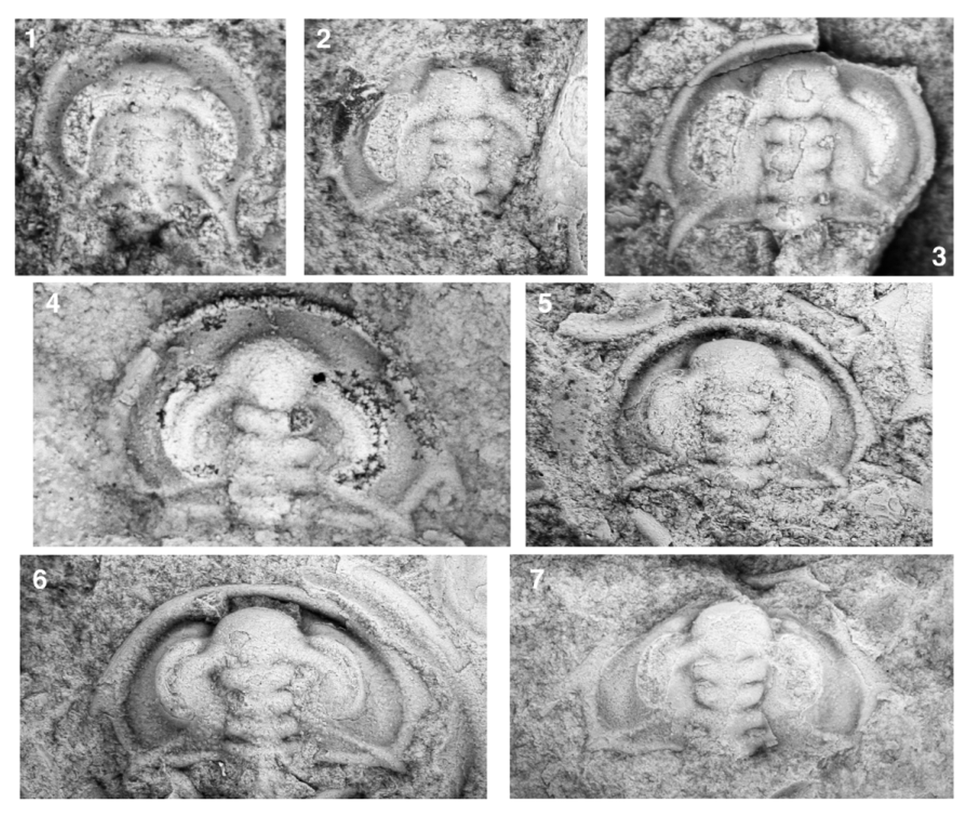
Ontogeny. Well-preserved specimens examined herein range from approximately 1.44 mm to 12.43 mm in cephalic length (sag.). Fragments of much larger cephala ranging up to approximately 20 mm in estimated cephalic length are also known (Fig. 12.3). These specimens together span phases 3 through 5 of cephalic development. Specimens of sagittal cephalic length> 5 mm are essentially morphologically mature and have been described previously.
Assignment of the smallest specimen (Fig. 13.1) to Lochmanolenellus pentagonalis is tentative, because larger, morphologically mature specimens of this species have not been recovered from this horizon (LACMIP locality 17053). Nevertheless, the specimen clearly differs from similarly sized specimens of Lo. subquadratus , with which it co-occurs in LACMIP locality 17053, by having a less strongly anteriorly oriented distal portion of the posterior cephalic margin (between the intergenal and genal spine bases) and thus a proportionally wider (tr.) extraocular area and cephalon, in having less strongly anteriorly advanced genal spines, and in having longer ocular lobes. The morphology of LACMIP 12542 is consistent with an assignment to Lo. pentagonalis based on extrapolation of ontogenetic trends.
Phase 3: Initial morphology: Smallest known specimen (tentatively assigned to this species, see previous) 1.44 mm in cephalic length (sag.; Fig. 13.1). Cephalon broadly horseshoe-shaped in outline. Proximal portion of posterior cephalic margin (between bases of intergenal spines) weakly posterolaterally oriented when traced distally. Broad-based, triangular intergenal spines project posterolaterally from posterolateral corners of cephalon; tips not preserved but spine length at least 30% of cephalic length (sag.); distance (tr.) between inner margins of intergenal spine bases approximately 68% of cephalic length (sag.). Anterior border gently rounded dorsally, sagittal length approximately 8% of glabellar length (sag.), slightly broader around anterolateral margin of cephalon, narrows posterior to genal spines; border extends around lateral cephalic margin to base of intergenal spines. Tiny, thorn-like genal spine projects posterolaterally from lateral margin at widest point of cephalon (tr.), approximately transversely opposite anterior-most portion of lateral margin of L1. Slight geniculation of cephalic margin at anterolateral corner of cephalon where short procranidial spines project anteriorly, another geniculation of cephalic margin midway between base of procranidial spines and genal spines; cephalic margins between genal and intergenal spines divergent anteriorly, each side forming approximately a 32° angle with an exsagittal line. Glabella evenly tapers anteriorly from SO to S3; approximately 86% of cephalic length (sag.); defined by very shallow axial furrow. SO, S1, S2, and S3 extremely shallow, deepest abaxially, straight; SO oriented slightly anterolaterally when traced abaxially, all other glabellar furrows more-or-less transverse. Preocular furrow not present. LO rectangular in outline; sagittal length approximately 11% of glabellar length (sag.); lateral margins poorly differentiated from posterior cephalic border and posterior portion of interocular area; posterior margin convex posteriorly. L1, L2, and L3 together occupy approximately 52% of glabellar length (sag.); each slightly trapezoidal in outline (narrower anteriorly). LA transversely oblate, widest (tr.) at point of contact with outer margin of ocular lobes; clearly delimited from preglabellar field by break in slope. Maximum width (tr.) of LA 147% of basal glabellar width. Presence or absence of axial nodes on LO unknown (axial region chipped); no axial structures on other glabellar segments. Ocular lobes crescentic in dorsal view, clearly differentiated from interocular area by furrow; strongly divergent proximally, bending to run almost parallel with exsagittal axis; posterior tips more widely separated than anterior tips and located transversely opposite posterior portion of lateral margin of L1; convex in cross-section (tr.); dorsal summit slightly higher than highest points of interocular area and anterior portion of glabella. Interocular area more-or-less flat-topped and shelf-like (tr.); interocular furrows not visible. Prominent intergenal ridge originates adjacent to lateral margin of L1, runs onto adaxial portion of intergenal spines; much weaker posterior ocular line extends to abaxial margin of intergenal spines. Narrow (tr.), crescent-shaped extraocular area extends posteriorly to posterior ocular line; sagittally developed as broad, troughlike border furrow (or preglabellar field?) approximately as broad (sag.) as anterior cephalic border.
Subsequent morphological change: Two larger specimens (Fig. 13.2, 13.3; sagittal cephalic lengths estimated to be approximately 1.61 mm and 1.81 mm, respectively) are also in phase 3 of cephalic development and, by comparison to the previous specimen, offer insight into some of the morphological changes that occurred during that phase of cephalic development. On the glabella, all glabellar furrows deepened, especially distally. LA proportionally elongated (sag.) to occupy 36% of sagittal glabellar length and inflated in dorsal relief. The sagittal elongation of LA resulted in a slight posterior deflection of the sagittal portions of S3, which became oriented slightly anterolaterally on either side of the sagittal axis until its contact with the inner margins of the ocular lobes. LO proportionally widened (tr.) so that it became trapezoidal in outline, tapering anteriorly; glabellar width (tr.) across SO became approximately 96% of basal glabellar width (tr.), and the maximum width (tr.) across LA became approximately 117% of basal glabellar width (tr.). Widening (tr.) of the glabella resulted in an increasing area of contact between the anterolateral corners of L3 and the inner margin of the ocular lobes, and S3 became slightly caret-shaped on either side of the sagittal axis. The transocular furrow became visible, albeit very faintly. A prominent axial node is evident on LO of the larger specimen (Fig. 13.3). The posterior ocular line became fainter.
The ocular lobes proportionally shortened and their posterior tips migrated slightly anteriorly to a position transversely opposite the midlength of the lateral margins of L1. An ocular furrow developed. The extraocular area proportionally widened (tr.), with a corresponding proportional elongation (tr.) of the posterior cephalic margin between the axial furrow and the base of the intergenal spine and an increased separation (tr.) between the outer margin of the ocular lobe and the cephalic border. The intergenal spine bases, which were initially located more-orless behind (exsag.) the posterior tips of the ocular lobes on the smallest phase 3 cephalon, migrated to positions outside the abaxial-most points of the ocular lobes. The distal portion of the posterior cephalic margin rotated slightly to form approximately a 30° angle with an exsagittal line when traced toward the genal spine base. The genal spine proportionally elongated (although the tip is not preserved any specimen, so the full length of the spine is unknown). The strip of exoskeleton between LA and the anterior cephalic border deepened into a broad, troughlike anterior border furrow. Weak geniculations are still evident in the cephalic margin slightly anterior to the genal spines and at the site of the procranidial spines, although the procranidial spines themselves are reduced to tiny nubbins or may be entirely absent on the larger phase 3 cephala.
Phase 4: Three specimens ranging from 2.22 mm to 2.89 mm in sagittal cephalic length (Fig. 13.4–13.6) are assigned to phase 4 of cephalic development and offer insight into morphological changes during that portion of ontogeny. As is diagnostic of that phase of development, L3 proportionally widened (tr.) relative to L2 so that the glabella became hourglass-shaped and most constricted at L2; glabellar width (tr.) across S3 increased from approximately 90% to more than 100% of basal glabellar width (tr.), while glabellar width (tr.) across S1 remained at approximately 85% of basal glabellar width (tr.); the area of contact between the anterolateral corners of L3 and the inner margins of the ocular lobes increased. LA continued its proportional elongation (sag.) from phase 3 and occupied almost 40% of glabellar length (sag.) by late phase 4. Lateral swellings became evident, first on L2 (Fig. 13.5) and subsequently also on LO, L1, and L3 (Fig. 13.6). The ocular lobes continued to shorten relative to glabellar length; the posterior tips are transversely opposite the anterior portion of the lateral margins of L1 on a late phase 4 cephalon (Fig. 13.6). The interocular area tilted to slope down from the inner margin of the ocular lobe to the axial furrow. The extraocular area continued to proportionally widen, especially posteriorly. This resulted in the base of the intergenal spines migrating to a position well outside (exsag.) the distal-most part of the ocular lobe, the distal portions of the posterior cephalic margins rotating to form approximately a 24° angle with an exsagittal line when traced toward the genal spine base, and the intergenal ridges becoming more strongly divergent posteriorly. A very weak intergenal angle developed along the posterior cephalic margin slightly distal to the midway point between the axial furrow and the inner margin of the intergenal spine base. The intergenal spines became more strongly laterally flared. The genal spines continued to proportionally elongate into slender spines (although their full length is unknown) and migrated slightly anteriorly (by the end of phase 4 the genal spine bases were located transversely opposite the lateral margins of S1). The anterior cephalic border proportionally broadened relative to the exsagittal length of L1 and L2, and the anterior cephalic border furrow deepened. Procranidial spines and posterior ocular lines are absent on phase 4 cephala. A narrow extraocular platform was well developed around each ocular lobe by the end of phase 4.
Phase 5: Several specimens ranging from approximately 3.5 mm to 4.84 mm in sagittal cephalic length are assigned to phase 5 of cephalic development based on exhibiting an L2 that widens (tr.) anteriorly (with the glabella being narrowest [tr.] at S1), indicating that L2 had initiated the pronounced lateral widening (tr.) that defines entry into that phase (Fig. 13.7). However, these specimens differ from the mature morphology (sagittal cephalic length> 5 mm; described previously) in several respects, and thus reveal several ontogenetic changes that occurred during early phase 5 of cephalic development. During early phase 5 of development, LA continued to increase in proportional length (sag.) relative to both glabellar length (sag.; Fig. 14.1) and to the maximum width of LA (tr.; Fig. 14.2). As a result, the length (exsag.) of L1 and L2 relative to glabellar length (sag.) proportionally decreased (Fig. 14.3, 14.4). The length of the ocular lobes continued to shorten relative to glabellar length (sag.; Fig. 14.5). The genal spines continued to migrate anteriorly (Fig. 14.6). The extraocular area continued to proportionally widen, especially posteriorly. This resulted in the proximal portion of the posterior cephalic margin (between the axial furrow and the intergenal spine base) becoming proportionally longer (Fig. 14.7) and becoming less strongly posterolaterally orientated when traced abaxially (Fig. 14.8). The transverse expansion of the extraocular area also led to a proportional increase in the transverse distance between the bases of the genal spines relative to glabellar length (sag.; Fig. 15.1). However, the proportional widening near the genal spine bases was less dramatic than was the proportional widening along the posterior cephalic margin, so the distal portion of the posterior cephalic margin rotated into a more strongly anteriorly directed orientation (when traced towards genal spine base; Fig. 15.2, 15.3). The genal spines became more strongly laterally flared relative to the distal portion of the posterior cephalic margin (Fig. 15.4).
Etymology. Named for the pentagonal cephalic outline.
Holotype. LACMIP 12550a and LACMIP 12550b (part and counterpart; Fig. 11.1, 11.2), from LACMIP locality 17052, Slate Ridge, Esmeralda County, Nevada.
Other material. Quantitative morphometric and/or qualitative descriptive data were recorded from first-hand examination of the holotype (see previous) and the following 36 specimens: 12 additional cephala from LACMIP locality 17052 (LACMIP 12547, LACMIP 12548 [seven cephala], LACMIP 12549 [2 cephala], LACMIP 12550/2, and LACMIP 12551.2); one tentatively assigned morphologically immature cephalon from LACMIP locality 17053 (LACMIP 12542); 12 cephala from ICS-10241 (including FMNH PE 58483 to FMNH PE 58486); 10 cephala from GSC 95190 (including GSC 137492 and GSC 137493); and MCZ 110679, an almost complete dorsal exoskeleton from Mumm Peak. This last specimen was figured by Lieberman (1999, fig. 20.1) and assigned to Laudonia bispinata (see also Lieberman, 1998, p. 73). However, MCZ 110679 is herein reassigned to Lochmanolenellus pentagonalis . One hypostome from LACMIP locality 17052 is also assigned to this species.
Occurrence. CANADA: Cassiar Mountains, British Columbia: GSC locality 95190, limestone approximately12.5 metres above the base of Unit 4 of the Rosella Formation, in Section 2 of Fritz (1978, text-fig. 3.1a), on the north bank of the Dease River, approximately 2.7 km ( 1.7 miles) east of McDame. Mount Robson area, British Columbia and Alberta: Talus on the flanks of Mumm Peak near the Mural Glacier, Alberta, sourced from the middle member of the Mural Formation. U.S.A.: Montezuma Range, Esmeralda County , Nevada: ICS- 10241, fossiliferous shale collected from talus from the interval 131.2 metres to 135.1 metres above the base of the middle member of the Poleta Formation (47.6 metres to 46.9 metres above the local base of the Dyeran), Indian Springs South section, Indian Springs Canyon (unpublished data). Slate Ridge, Esmeralda County, Nevada: LACMIP locality 17052, brown packstone to grainstone approximately 138 metres above the base of the section (122 metres above the base and 67 metres below the top of the middle member of the Poleta Formation), and?LACMIP locality 17053 (a tentative occurrence), light grey packstone approximately 150 metres above the base of the section (approximately 134 metres above the base and 55 metres below the top of the middle member of the Poleta Formation), both in the Gold Point section of Bohach (1997, text-fig. 62, p. 374).
Discussion. Lochmanolenellus pentagonalis is easily distinguished from Lo. trapezoidalis and Lo. subquadratus by its pentagonal rather than subquadrate or trapezoidal cephalic outline (Figs 9.1, 9.2, 18.8) and by its less strongly anteriorly advanced genal spine bases (Fig. 9.3). Differences between Lo. pentagonalis and Lo. primus are listed in the discussion of the latter species. In addition to these differences, the glabella of Lo. pentagonalis is proportionally narrower across LA (tr.) relative to basal glabellar width (tr.) than all other species (Fig. 9.6).

No known copyright restrictions apply. See Agosti, D., Egloff, W., 2009. Taxonomic information exchange and copyright: the Plazi approach. BMC Research Notes 2009, 2:53 for further explanation.
|
Kingdom |
|
|
Phylum |
|
|
Class |
|
|
Order |
|
|
SubOrder |
Olenellina |
|
Family |
|
|
Genus |