
Pleurostomum flabellatum Ruinen 1938
|
publication ID |
https://doi.org/ 10.4467/16890027AP.22.008.17111 |
|
DOI |
https://doi.org/10.5281/zenodo.11152089 |
|
persistent identifier |
https://treatment.plazi.org/id/AF4987FE-FF90-033A-806D-E351FED24ECB |
|
treatment provided by |
Felipe |
|
scientific name |
Pleurostomum flabellatum Ruinen 1938 |
| status |
|
Pleurostomum flabellatum Ruinen 1938
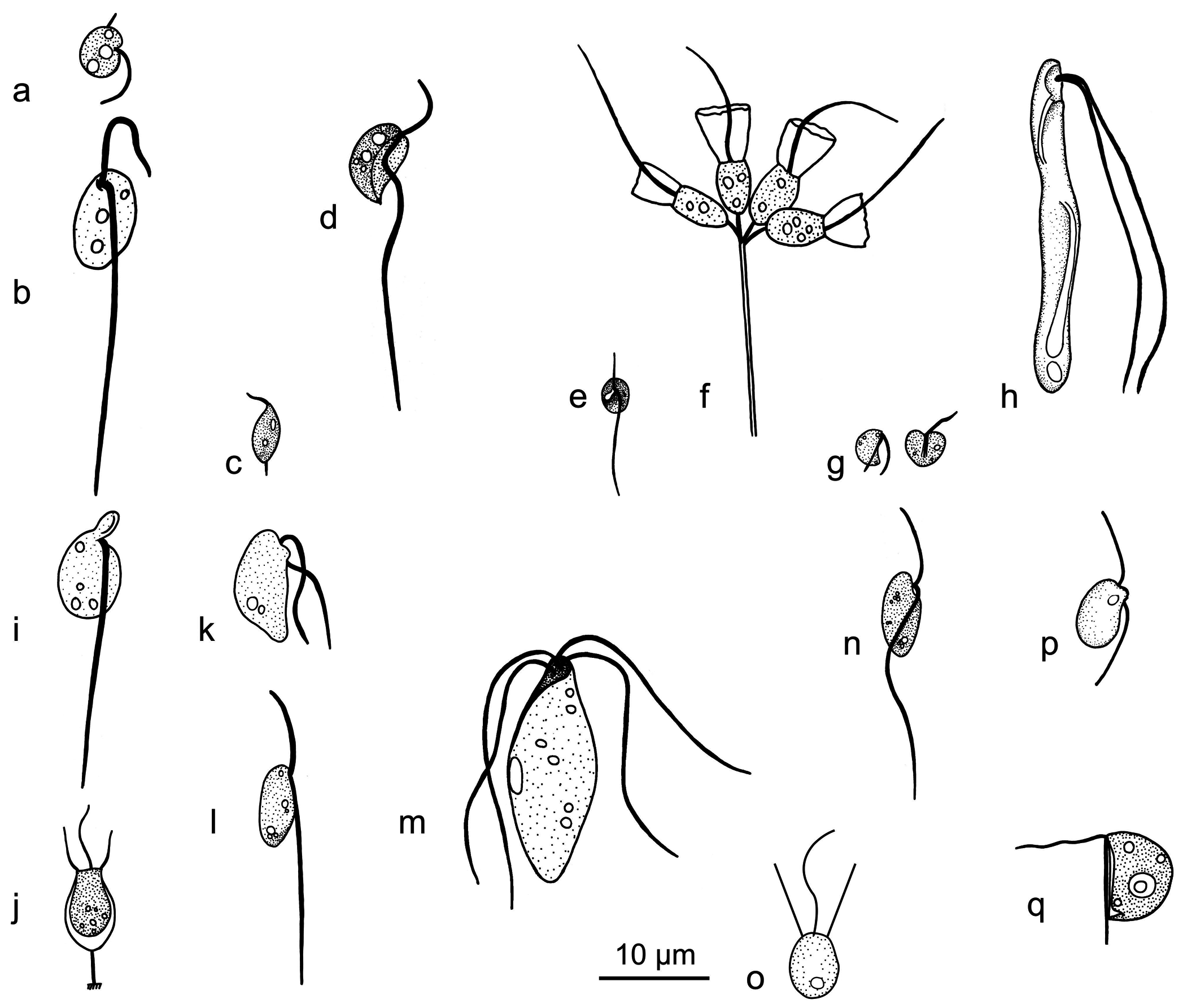
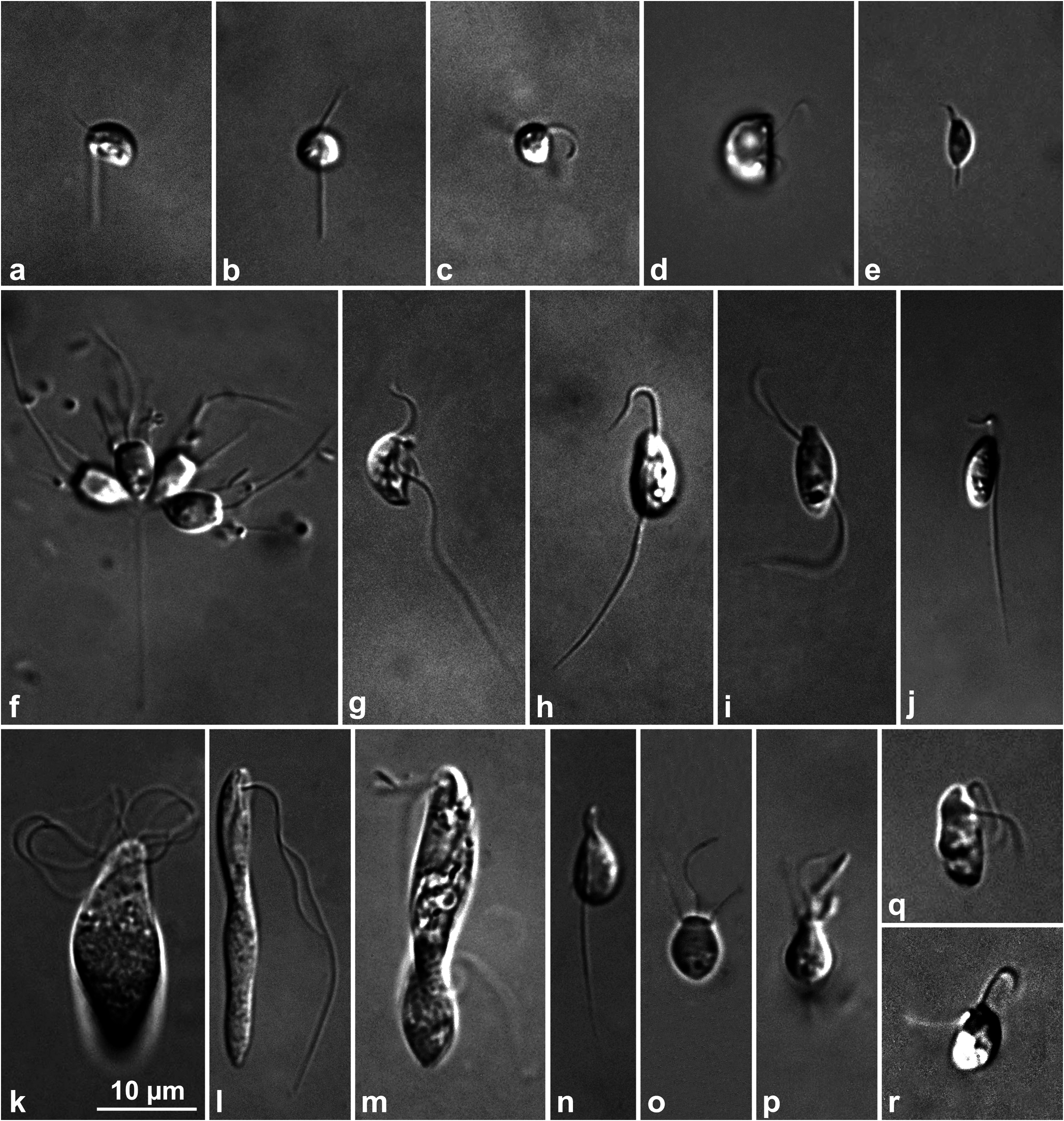
( Figs 2h View Fig , 3 View Fig l-m)
Cells are 18–30 µm long, flattened, somewhat elongate and narrower posteriorly. The synchronously moving two flagella are 1.5–2 times the cell length and emerge apically beneath the rostrum. Because of the rostrum, flagella seemed to emerge subapically. Both flagella move in close contact with lamella. The cytotomal structure originates near the point of flagellar insertion and extends in a spiral to the posterior end of the cell. The extension of the cytostomal structure gives the cell its spiral shape. A vacuole is located posteriorly. The cells swim slowly by making spirals. Description based on observations of 16 cells. Occurance: every month with the exception of July and August at Meke Lake, temperature 0–21 °C, salinity 107.5–215 psu, dissolved oxygen 0.25–5.18 mg /L.
Remarks: The genus Pleurostomum contains 5 species and is characterized by two parallel homodynamic subapically (and also apically in regard to former descriptions) emerging flagella and by its cytostomal groove. Generally, our observations are in accordance with the original description of Ruinen (1938) with respect to cell length and general appearance of the cell. The only difference is the point of flagellar insertion. According to Ruinen (1938) both flagella emerge apically, whereas in our cells the flagella emerged subapically. Pleurostomum flabellatum is similar to P. salinum , which was reported with the cell length of 20–22 µm and with a cytostomal groove extending 2/3 of the cell ( Namyslowski 1913, Ruinen 1938), but can be distinguished because the cytostomal groove of P. flabellatum extends almost to the end of the cell and is relatively longer. The cells of Pleurostomum flabellatum reported by Patterson and Simpson (1996) and Park et al. (2007) are smaller than the original cells (16–30 µm) of Ruinen (1938) and the cells (18–30 µm) observed here: Patterson and Simpson, 11–14 µm; Park et al., 10–14 µm. Their cells resemble P. salinum Namyslowski 1913 because of the cell length and general appearance. Further studies are required to establish the identities of these taxa.
Park et al. (2009) reported a new amoeboid species Tulamoeba peronaphora , a close relative of Pleurostomum flabellatum in the 18S rRNA gene tree, but can be easily distinguished because of their morphology and because Tulamoeba peronaphora does not have a flagellated stage. Pleurostomum flabellatum and Tulamoeba bucina Kirby et al. 2014 ’s flagellated stage resemble each other, but can be distinguished by the paths of their cytostome: P. flabellatum has a cytostome extending along the body spirally and T. bucina has a cytostome curling around the lateral axis. Pleurostomum flabellatum has been reported from hyposaline habitats in Australia, India, Korea and Poland ( Namyslowski 1913, Ruinen 1938, Patterson and Simpson 1996, Park et al. 2007).
No known copyright restrictions apply. See Agosti, D., Egloff, W., 2009. Taxonomic information exchange and copyright: the Plazi approach. BMC Research Notes 2009, 2:53 for further explanation.
|
Kingdom |
|
|
Phylum |
|
|
Order |
|
|
Family |
|
|
Genus |