
Antespathius Belokobylskij, 1995
|
publication ID |
https://doi.org/ 10.5852/ejt.2021.741.1289 |
|
publication LSID |
lsid:zoobank.org:pub:932D3C8F-6F22-4103-ABCE-47F1E4E8FF43 |
|
DOI |
https://doi.org/10.5281/zenodo.4651642 |
|
persistent identifier |
https://treatment.plazi.org/id/AF1D4E27-AD7E-5F1A-FD91-E12F3ADE3FC4 |
|
treatment provided by |
Plazi |
|
scientific name |
Antespathius Belokobylskij, 1995 |
| status |
|
Genus Antespathius Belokobylskij, 1995
Spathius (Antespathius) Belokobylskij, 1995: 49 .
Spathius (Antespathius) – Long & Belokobylskij 2011: 24.
Antespathius – Zaldívar-Riverón et al 2008: 358 (as genus in Rhaconotini ). — Jasso-Martínez et al. 2019: 165.
Type species
Spathius (Antespathius) buonluoicus Belokobylskij, 1995 View in CoL , by original designation.
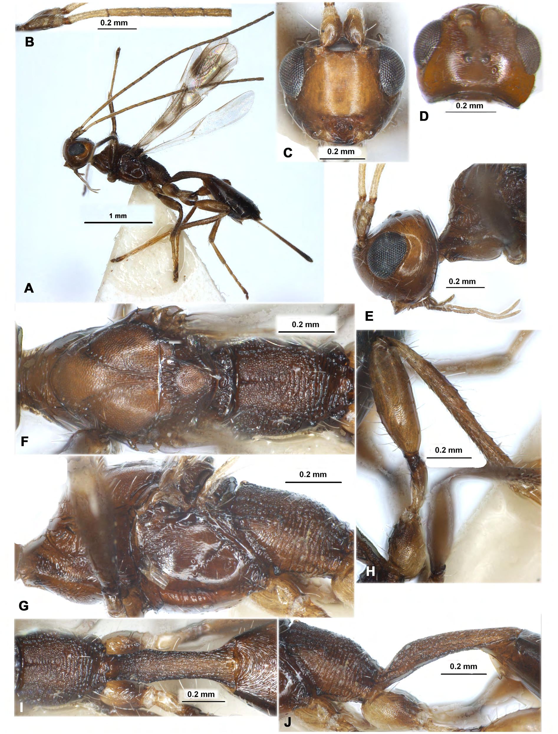
Description ( Figs 1–2 View Fig View Fig )
HEAD. Head not depressed, high, weakly transverse, face with very dense, even and slender transverse aciculation as on CD disc. Ocelli arranged in triangle with base larger than its sides. Frons flat, without median carina, with shallow longitudinal furrow.Antennal socket situated weakly lower than upper level of eyes (front view). Eyes glabrous. Occipital carina distinct, complete dorsally, ventrally obliterated at short distance of upper base of mandible. Malar suture very shallow, almost indistinct. Clypeus very narrow, with short lower flange. Hypoclypeal depression medium-sized, suboval. Postgenal bridge narrow. Maxillary palps long, 6-segmented, sixth (apical) segment 1.3 times as long as fourth segment; labial palps short, 4-segmented, second segment thickened, third segment weakly shortened, about 0.7 times as long as fourth segment. Scapus of antenna rather narrow and long, without apical lobe and basal constriction, its ventral margin (lateral view) shorter than dorsal margin, apically with distinct excavation in outer side. First flagellar segment subcylindrical, weakly curved, longer than second segment. Apical segment obtuse and without apical “spine”.
MESOSOMA. Mesosoma not depressed. Neck of prothorax rather long, without pronope. Pronotum dorsally flat (lateral view); pronotal carina fine and medially fused with posterior margin of pronotum at short distance. Propleural lateroposterior flange short and wide. Mesonotum highly and roundly elevated above pronotum. Median lobe of mesonotum weakly protruding forwards (lateral view), without median longitudinal furrow and anterolateral corners (dorsal view). Notauli complete, crenulate at least in anterior half, deep in anterior half and shallow in posterior half. Scuto-scutellar suture present and distinct. Prescutellar depression evenly long, rather deep, with several distinct carinae. Lateral longitudinal flanges on the level of prescutellar depression lower. Scutellum convex, with distinct lateral carinae. Metanotum medially without median carina and with short high lateral carinae (dorsal view); with short wide tooth (lateral view). Mesopleural pit transformed in narrow, long and oblique furrow running to median part of mesopleuron. Sternaulus (precoxal furrow) deep, wide, short. Prepectal carina complete, wide ventrally. Postpectal carinae absent. Metapleural flange short, wide, rounded apically. Propodeum without areas delineated by carinae, without depressions and lateral tubercles; propodeal bridge absent. Propodeal spiracles small and round, situated in basal 0.4 of propodeum.
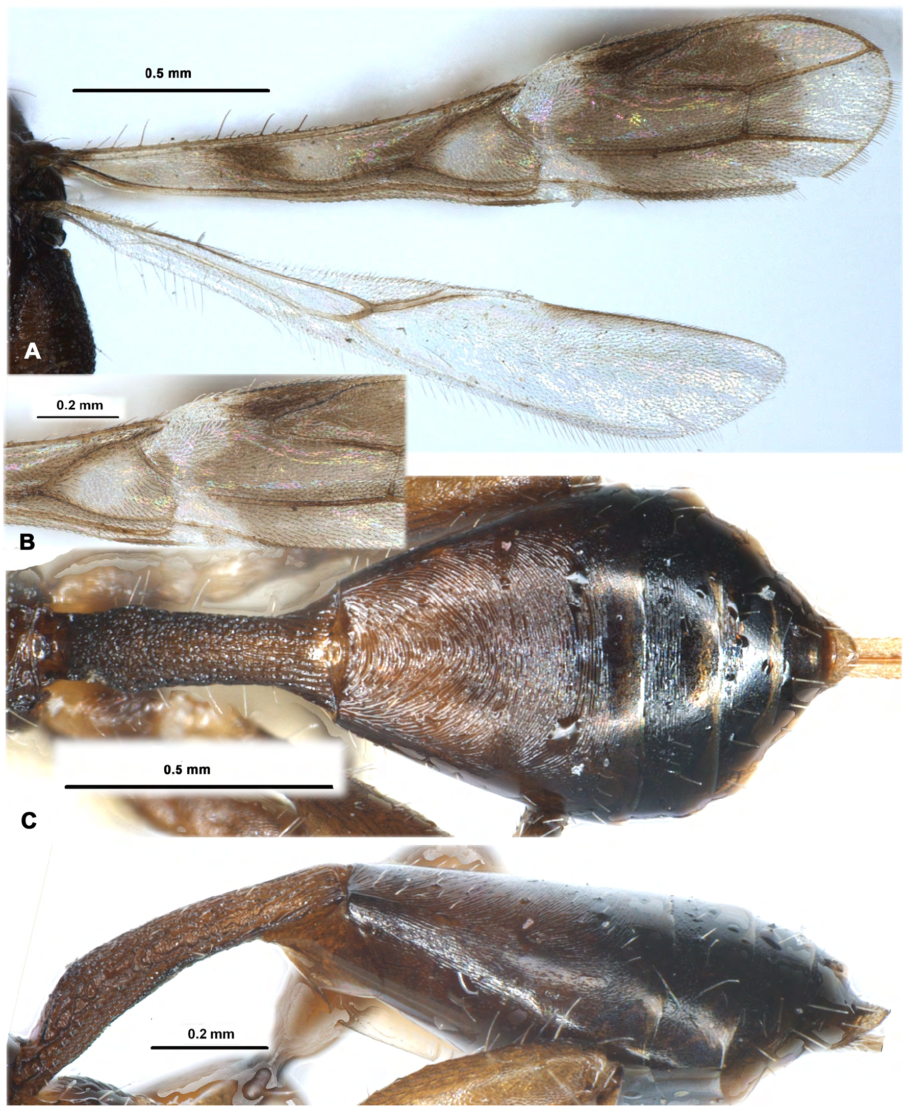
WINGS. Pterostigma of fore wing narrow and long. Radial vein (r) arising from middle of pterostigma. Radial (marginal) cell not shortened. First radiomedial vein (2-SR) absent, second radiomedial vein (r-m) present and sclerotised. Nervulus (cu-a) present and strongly antefurcal. Discoidal (discal) cell petiolate anteriorly, petiole (1-SR) short. Parallel vein (CU1a) interstitial. Mediocubital vein (M+CU1) distinctly sinuate. Brachial (subdiscal) cell closed postero-apically by evenly curved brachial vein (CU1b). Transverse anal veins (2A, a) absent. Hind wing with three hamuli. First abscissa of costal vein (C+SC+R) 0.8 times as long as second abscissa (1-SC+R); first abscissa (C+SC+R) not divided apically on two abscissae. Radial vein (SR) almost indistinct, arising from costal vein (2-SC+R) far from basal vein (1r-m). Radial (marginal) cell weakly and evenly widened toward apex, without additional transverse vein (r). Medial (basal) cell narrow, weakly widened towards apex from its base. Nervellus (cu-a) present and declivous. Submedial (subbasal) cell short. First abscissa of mediocubital vein (M+CU) 0.45 times as long as second abscissa (1-M). Recurrent vein (m-cu) rather short, strongly oblique toward base of wing, straight, hyaline.
LEGS. Fore tibia with several short and rather thick spines situated almost in single line. All tibiae slender. Middle tarsal segments rather long. Hind coxa rather short, wide, without basoventral corner and tubercle. Fore and middle femora with very low and wide dorsal protuberance. Hind femur wide, elongate-oval, without dorsal protuberance. Hind basitarsus short, about 0.6 times as long as second– fifth segments combined.
METASOMA. First metasomal tergite petiolate, long and narrow. Acrosternite of first segment 0.7 times as long as first tergite, its apical margin situated significantly behind spiracles. Dorsope of first tergite small; basolateral lobes absent. Spiracular tubercles present, distinct, situated in basal 0.3 of tergite; dorsal carinae distinctly only basally. Second and third tergites without any furrows and areas, its lateral parts fused. Second suture very shallow. Only second tergite with separate laterotergites. Fourth–sixth tergites with single submedian transverse line of short, sparse and semi-erect pale setae. Fifth tergite not enlarged, 0.9 times as long as fourth tergite; apical segments weakly protruding behind fifth tergite. Hypopygium in apical margin obtuse and rounded apically. Ovipositor sheath weakly widened towards subapex, not longer than metasoma.
Diagnosis
Antespathius was originally described as a subgenus of Spathius within the tribe Spathiini ( Belokobylskij 1995) . However, recent molecular phylogenetic studies that have included this taxon ( Zaldívar-Riverón et al. 2008; Jasso-Martínez et al. 2019) have confirmed its placement within the tribe Rhaconotini (sensu nova) as a separate genus. Antespathius distinctly differs from the remaining Rhaconotini genera by having a considerable petiolate first metasomal tergite with strongly elongated acrosternite, equal to 0.6–0.7 times length of tergite, face with very dense, even and slender transverse aciculation (as on CD disc), and nervulus (cu-a) of fore wing distinctly antefurcal. This genus distinctly differs from the Spathius species by the antefurcal position of nervulus (cu-a) in fore wing together with the loss of first radiomedial (2-SR) vein.
Composition
Antespathius buonluoicus ( Belokobylskij, 1995) (OR) , and an undescribed species from Madagascar ( Zaldívar-Riverón et al. 2008).
Hosts
Unknown.
Distribution
Oriental and Afrotropical regions.
Remarks
This genus remains monotypic with its single described species from Vietnam. However, a second, undescribed species of this genus has been recorded from Madagascar ( Zaldívar-Riverón et al. 2008: 347).
No known copyright restrictions apply. See Agosti, D., Egloff, W., 2009. Taxonomic information exchange and copyright: the Plazi approach. BMC Research Notes 2009, 2:53 for further explanation.
|
Kingdom |
|
|
Phylum |
|
|
Class |
|
|
Order |
|
|
Family |
Antespathius Belokobylskij, 1995
| Belokobylskij, Sergey A. & Zaldívar-Riverón, Alejandro 2021 |
Spathius (Antespathius)
| Long K. D. & Belokobylskij S. A. 2011: 24 |
Antespathius
| Jasso-Martinez J. M. & Belokobylskij S. A. & Zaldivar-Riveron A. 2019: 165 |
| Zaldivar-Riveron A. & Belokobylskij S. A. & Leon-Regagnon V. & Briceno R. & Quicke D. L. J. 2008: 358 |
Spathius (Antespathius)
| Belokobylskij S. A. 1995: 49 |