
Leptohyphes cornutillus, Nascimento, Jeane M. C., Molineri, Carlos & Salles, Frederico F., 2014
|
publication ID |
https://doi.org/ 10.11646/zootaxa.3893.3.5 |
|
publication LSID |
lsid:zoobank.org:pub:0B0E56EE-295C-46C7-A680-D2D9F959F6CA |
|
DOI |
https://doi.org/10.5281/zenodo.5662834 |
|
persistent identifier |
https://treatment.plazi.org/id/AD09B31E-FFEE-9E15-FC8E-549EFBD0FC5A |
|
treatment provided by |
Plazi |
|
scientific name |
Leptohyphes cornutillus |
| status |
sp. nov. |
Leptohyphes cornutillus sp. nov.
( Figs. 6–9 View FIGURE 6 View FIGURES 7 View FIGURES 8 View FIGURES 9 and 14 View FIGURE 14 )
Material examined. HOLOTYPE, male mature nymph, BRAZIL, Espírito Santo, Domingos Martins, Megalcinho, 30/iii/2010, S 20˚ 15’ 0.6” / W 40˚ 43’ 26.9”, 622m, Salles FF leg. ( INPA). PARATYPE, 1 female mature nymph (parts on slide), same data as holotype ( CZNC).
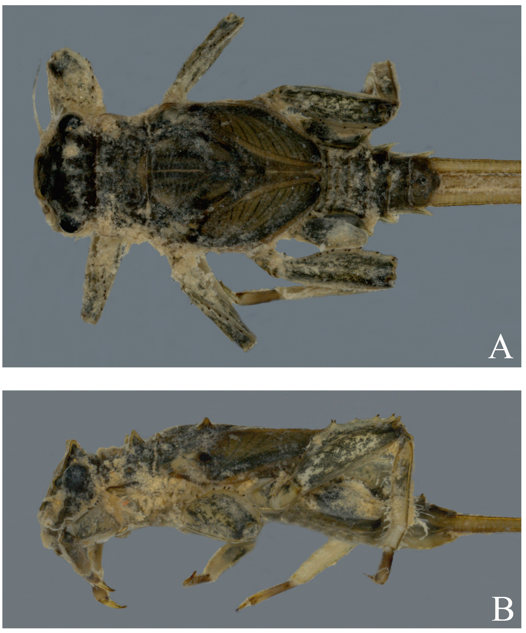
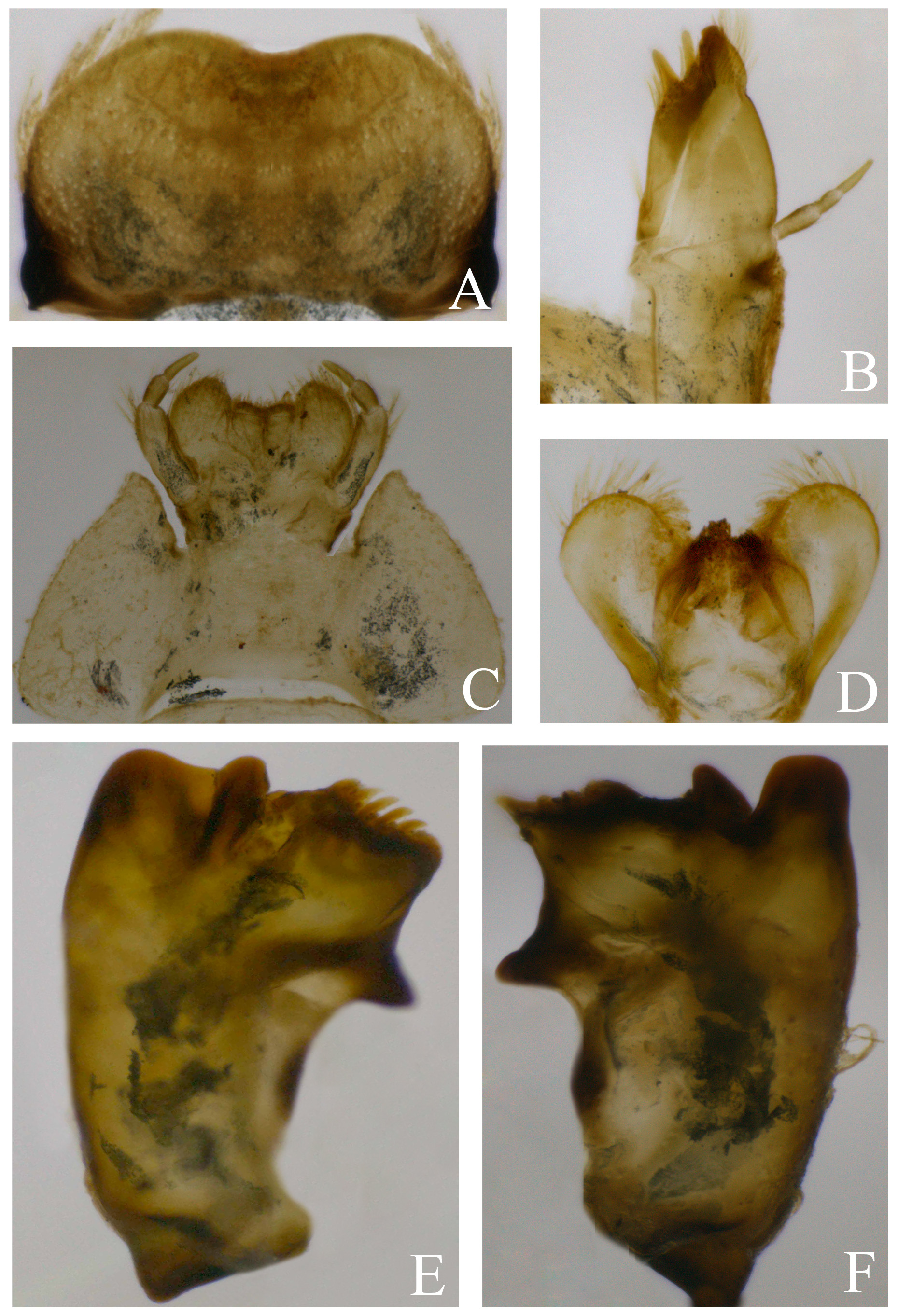
Mature nymph. Length of male: body, 5.0 mm; hind femur, 1.5 mm; caudal filaments, 4.0 mm. Length of female (mm): body, 8.2 mm; hind femur, 2.0 mm; caudal filaments, 4.5 mm. General coloration brownish, almost completely shaded with black ( Figs 6 View FIGURE 6 , 7 View FIGURES 7 ). Head ( Fig. 7 View FIGURES 7 A) with two pairs of tubercles, a submedian smaller and sublateral longer and pointed; clypeus well developed and subquadrate. Gena ( Fig. 7 View FIGURES 7 A) strongly projected. Mouth parts ( Fig. 8 View FIGURES 8 ). Labrum ( Fig. 8 View FIGURES 8 A): with an anteromedial marked indentation on fore margin. Labium ( Fig. 8 View FIGURES 8 C): submentum with pointed and projected anterolateral corner; glossae shorter than paraglossae; labial palp with basal segment three times the length of segment two, apical segment subequal in length to segment two. Hypopharynx ( Fig. 8 View FIGURES 8 D): lingua with short and strong setae. Maxillae ( Fig. 8 View FIGURES 8 B): maxillary palp three-segmented with setae at joints, suture on galea and lacinea complete. Mandibles ( Fig. 8 View FIGURES 8 E, F): left mandible with fused and strong incisives, both mandibles with a dense row of setae on dorsolateral surface; prostheca reduced (left), or absent (right). Thorax. Pronotum subrectangular with two pairs of sublateral tubercles, a large and pointed tubercle near hind margin and a smaller blunt anterolateral tubercle. Mesonotum with three pairs of tubercles: a short sublateral pair on fore margin, a longer and acute sublateral pair at 1/3 from anterior margin and a submedian pair of tubercles at apex of wing pads. Mesonotum with blunt anterolateral projection. Hind wing pad present in both sexes, small. Thoracic sterna paler than terga. Legs: brownish yellow shaded with black, except on apex of tibia and tarsi, tarsi brownish. Fore leg ( Fig. 9 View FIGURES 9 A): femur ratio length/maximum width 2.1–2.3; transversal row of seven stout spines at 1/3 from base; fore margin bare, hind margin with five stout spines (on elevated sockets) distally to transversal row; apex of femur medially and dorsally projected. Tibia subequal in length to femur with row of setae on inner margin; tarsus half the length of tibia with row of setae on inner margin, dorsum with elevated ridge; tarsal claw ( Fig. 9 View FIGURES 9 C) apically curved with six marginal denticles and one subapical submarginal denticle. Middle and hind legs ( Figs. 9 View FIGURES 9 B): coxae with short semicircular dorsal projection; hind femur ratio length/maximum width 2.2; with dorsally row of six blunt spines at base, fore margin bare, hind margin with ten stout spines on elevated sockets, dorsal surface with three stout spines at the middle; dorsomedial projection on apex of femur more pronounced than fore leg. Tibia 1.2x the length of femur, with dorsal ridge, inner margin with strong setae; hind margin with 2–3 strong setae basally and weaker setae distally. Tarsus 0.4x the length of femur, inner margin with setae, tarsal claw as in fore leg. Abdomen ( Fig. 7 View FIGURES 7 B) with strong spines and weak setae on dorsum. Lateral margins of segments III–VI expanded forming pointed flanges; segment VII and VIII strongly expanded laterally. Terga with a pair of blunt submedian projection on hind margin. Posterolateral spines on segments V–IX (flange on IV is also somewhat pointed resembling a posterolateral spine). Abdominal sternum IX with distal V-shaped indentation (female). Gills: operculate gill on abdominal segment II yellowish translucent almost completely shaded with black on dorsum, except medially and along distal margin, ventrally with strong basal spine and two long and pointed lamellae; other gills whitish almost completely shaded with black. Gill formula 3/11/9 /9/6. Caudal filaments completely covered with blunt scattered setae, and with a whorl of strong spines at the segment joint after every fourth segment.
Adults. unknown.
Diagnosis ( Table 1 View TABLE 1 ). i) paired tubercles on head (2 pairs), pronotum (2 pairs, anterior pair very small), and mesonotum (3 pairs), abdominal terga with remnants of paired tubercles on hind margin (blunt and short undulations in dorsal view) ( Figs. 6 View FIGURE 6 , 7 View FIGURES 7 ); ii) fore femur length/maximum width, 2.1–2.3 ( Fig. 9 View FIGURES 9 A); iii) fore margin of middle and hind femora without spines, hind margin with 5 rounded and stout spine-like setae on elevated sockets ( Figs. 9 View FIGURES 9 B, C); iv) tarsal claws denticulation 6+1 ( Fig. 9 View FIGURES 9 C); v) middle and hind coxa only with short semicircular projection on dorsum ( Figs. 9 View FIGURES 9 B, C); vi) hind wing pads present in females; vii) gill formula 3/11/9 /9/6, gill V without ventral extension on dorsal lamella.
Etymology. From cornutus , given the similarity with L. cornutus , and the diminutive suffix ilus. An allusion to the smaller size of the tubercles of the new species.
Distribution. Brazil (Espírito Santo).
Discussion. L. cornutillus is similar to L. cornutus , but they can be differentiated by the presence of three paired tubercles on mesonotum of the first species ( L. cornutus shows two pairs), and by the absence of coxal projection in L. cornutillus (character shared with L. airuoca ) ( Table 1 View TABLE 1 ). The form of mandibles in L. cornutillus , with very short canines ( Figs. 8 View FIGURES 8 E, F), somewhat resembles those of L. mandibulus Baumgardner (2007) , but this Central American species does not present tubercles on body, among other important differences. As stated by Baumgardner (2007), the short canines could be as a result of wear and tear, but we discard this probability because other mouthparts do not show any mark related with wear and tear ( Arens 1990).


| INPA |
Instituto Nacional de Pesquisas da Amazonia |
No known copyright restrictions apply. See Agosti, D., Egloff, W., 2009. Taxonomic information exchange and copyright: the Plazi approach. BMC Research Notes 2009, 2:53 for further explanation.