
Schistopeltis lizeri Rehn 1928
|
publication ID |
https://doi.org/ 10.5281/zenodo.210491 |
|
DOI |
https://doi.org/10.5281/zenodo.6172399 |
|
persistent identifier |
https://treatment.plazi.org/id/AB3C87DE-FFE6-FFAE-FF24-FE30FCF47C6D |
|
treatment provided by |
Plazi |
|
scientific name |
Schistopeltis lizeri Rehn 1928 |
| status |
|
Schistopeltis lizeri Rehn 1928 View in CoL
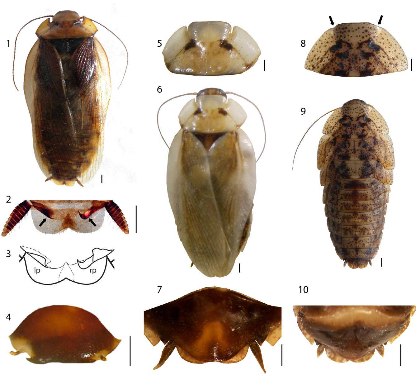
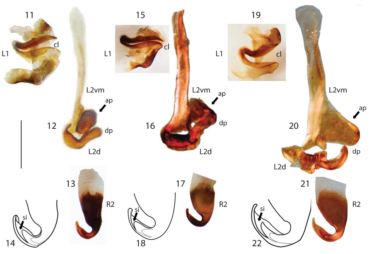
( Figs. 1–10 View FIGURES 1 – 10 , 15–18 View FIGURES 11 – 22. S ).
The genital sclerites of male. Supra-anal plate transverse with the distal margin bilobated. In the genital chamber the right-side paraproct hook-shaped with its apex directed apically ( Figs. 2–3 View FIGURES 1 – 10 ). Subgenital plate slightly asymmetrical with simple styles ( Fig. 4 View FIGURES 1 – 10 ).
Left phallomeres, subdivided in L1 and L2. The first genital sclerite L1 ( Fig. 15 View FIGURES 11 – 22. S ) very well developed and various areas darkly sclerotized. L1 presenting a cleft with a sigmoid shape and a narrow aperture.
L2 placed medially with two intimately articulated articles. The ventromedial article L2vm ( Fig. 16 View FIGURES 11 – 22. S ) with a well developed posterior apophysis, which extends dorsally (laterally in the photograph). Its posterior apophysis with a wide base that tappers distally. The dorsal article L2d ( Fig. 16 View FIGURES 11 – 22. S ) with curved dorsal process under L2vm apophysis.
The right phallomere R2 a hooked sclerite retracted in a soft cuticular tube. The apical third ( Figs. 17–18 View FIGURES 11 – 22. S ) sclerotized, with a globulous subconic base and a hooked apex with a narrow subapical incision that forms a free blade shaped lamina with a rounded apex.
External morphology of the female ( Fig. 6 View FIGURES 1 – 10 ). Size similar to male ( Table 1). Female pronotal fissures ( Fig. 5 View FIGURES 1 – 10 ) narrower than those of male. Front leg type D, with ventral anterior and posterior margins unarmed and genicular spine absent. Tibial formula with the same distribution as those in the male and nymph [5–6*2*1] [10*2*4] [13*2*8]. The cerci tapering distally with color similar to male. Supranal plate darker than male. Subgenital plate with different shape with a clear subapical area ( Fig. 7 View FIGURES 1 – 10 ).
Mature nymph ( Fig. 9 View FIGURES 1 – 10 ). Body length 23 mm; pronotum length 4.45 mm; pronotum width 9 mm; interocular distance 2 mm; interantennal distance 2 mm; interocelar distance 1.55 mm.
The nymph with a pronotum not bifissate, but the fissures feeble, insinuated on the pronotal anterior border ( Fig. 8 View FIGURES 1 – 10 ). The mature nymph is likely to develop into a female, a deduction based on the absence of the styli ( Fig. 10 View FIGURES 1 – 10 ) and the measurements similar to those of the female. The pharate adult can be observed by transparent cuticle; its pronotal lateral wings and fissures are evident.
Species S. lizeri S. peculiaris S. microschistos Country Argentina Bolivia Brasil Colombia
Sex M F M M M F Body length 20.80–25.90 23.00 20.00 26.80 25.00 27.00–28.50 Pronotum length 3.80–4.20 4.60 4.90 6.30 5.30–5.50 5.80 Pronotum width 8.00–9.35 8.90 8.60 11.40 10.80–11.00 12.00–12.50 Tegmen length 19.30–20.40 19.90 19.40 23.00 23.5–24.00 27.00 Tegmen width 5.30–6.00 6.00 6.90 8.20 8.00 9.00 Inter ocular distance 1.70–1.85 2.00 1.58 1.51 1.50 1.70–1.80 Inter antenal distance 2.00–2.15 2.50 2.16 2.15 2.00 2.10–2.20 Inter ocelar distance 1.35–1.45 1.50 1.34 1.45
No known copyright restrictions apply. See Agosti, D., Egloff, W., 2009. Taxonomic information exchange and copyright: the Plazi approach. BMC Research Notes 2009, 2:53 for further explanation.
|
Kingdom |
|
|
Phylum |
|
|
Class |
|
|
Order |
|
|
Family |
|
|
SubFamily |
Zetoborinae |
|
Genus |
|
Kingdom |
|
|
Phylum |
|
|
Class |
|
|
Order |
|
|
Family |
|
|
SubFamily |
Zetoborinae |
|
Genus |
|
Kingdom |
|
|
Phylum |
|
|
Class |
|
|
Order |
|
|
Family |
|
|
SubFamily |
Zetoborinae |
|
Genus |
|
Kingdom |
|
|
Phylum |
|
|
Class |
|
|
Order |
|
|
Family |
|
|
SubFamily |
Zetoborinae |
|
Kingdom |
|
|
Phylum |
|
|
Class |
|
|
Order |
|
|
Family |
|
|
SubFamily |
Zetoborinae |
|
Genus |