
ACROTHORACICA Gruvel, 1905
|
publication ID |
https://doi.org/ 10.1080/00222933.2014.896488 |
|
persistent identifier |
https://treatment.plazi.org/id/A6708794-FF9B-FFE1-3263-FA7EFE7F9BC0 |
|
treatment provided by |
Felipe |
|
scientific name |
ACROTHORACICA Gruvel, 1905 |
| status |
|
Superorder ACROTHORACICA Gruvel, 1905
Order LITHOGLYPTIDA Kolbasov, Newman and Høeg (cited in Kolbasov 2009) Family LITHOGLYPTIDAE Aurivillius, 1892 View in CoL Subfamily BERNDTIINAE Utinomi, 1950b Genus Berndtia Utinomi, 1950b Berndtia denticulata sp. nov.
( Figures 2A View Figure 2 , 3A, B View Figure 3 , 4–7 View Figure 4 View Figure 5 View Figure 6 View Figure 7 , 22A View Figure 22 )
Material examined
Holotype. RUMF-ZC-02827. One specimen in coral Psammocora superficialis, Odo , Okinawa, 26 July 2010. Paratype. RUMF-ZC-02828 – three specimens, same data as holotype . Paratype. ASIZCR000326 – three specimens, same data as holotype . Paratype. ZMMSU – three specimens, same data as holotype . Other materials. CEL-Virac-001. Three specimens, Virac, the Philippines, May 2011 .
Comparative material
Berndtia nodosa Tomlinson, 1967 . Paratype. 1971.2.25.26. Date det 28 Feb. 1967. Raffles Museum of Biodiversity Research Department of Biological Sciences Faculty of Science National University of Singapore.
Diagnosis
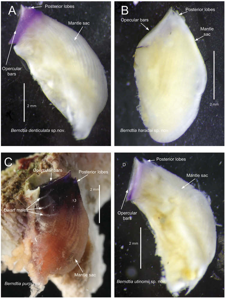
Female. Operculum wide oval shaped, composed of a pair of massive opercular bars. When alive, opercular bars have a pair of symmetrical pale white areas at the middle portions and a pair of small pale white spots at anterior and posterior ends. The remaining parts are dark brown in colour. Mantle sac with large multifid (tertra- and pentafid) teeth and the lateral margin of the opercular bars has simple, bi- and trifid sharp teeth. Outer surfaces of the opercular bars with numerous rosette nodules. Posterior lobes of operculum small, spherical and slightly swollen. Thorax with two small ventral conical processes. Live in Psammocora corals.
Description
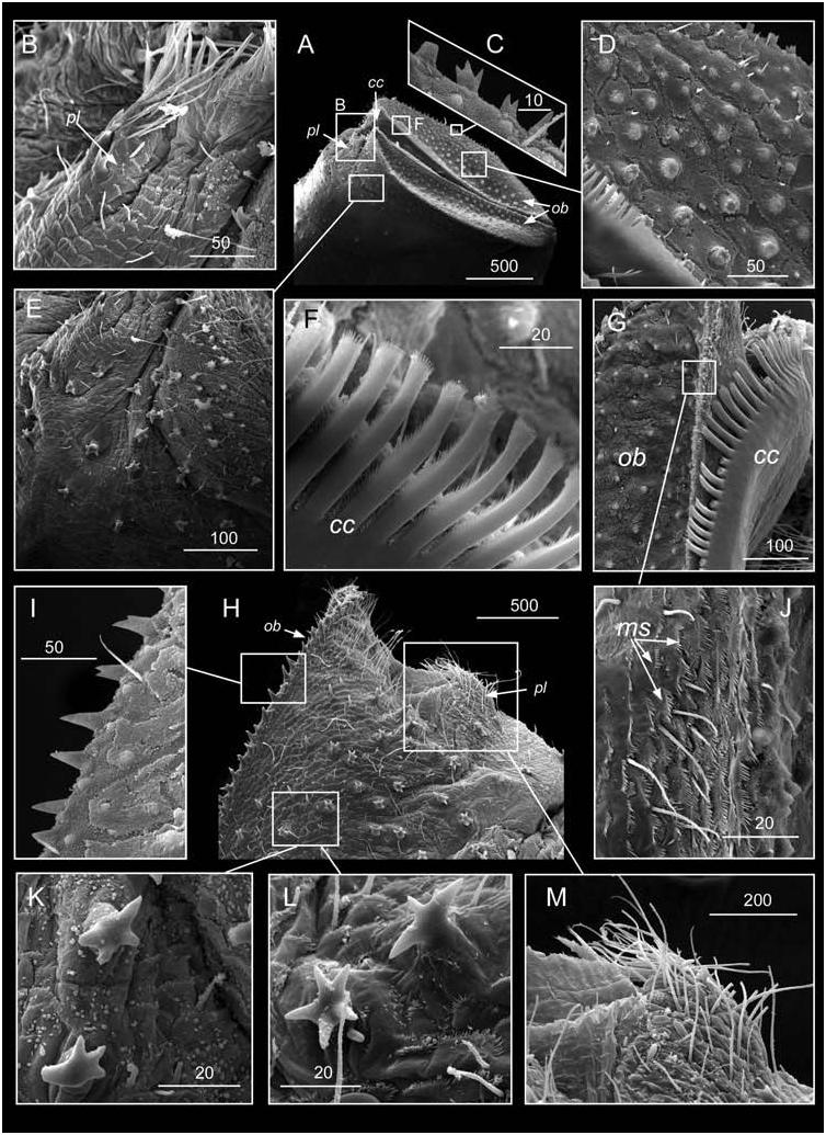
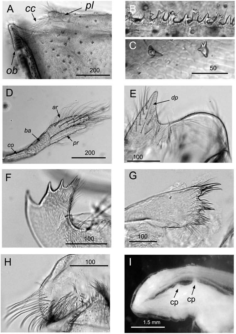
Female. Burrow opening wide oval shaped, well fitted with the operculum shape. Operculum wide oval shaped, with a pair of massive opercular bars, separated by an occludent slit ( Figures 2A View Figure 2 , 3A, B View Figure 3 , 4A View Figure 4 ). When alive, the opercular bars with a pair of large pale white areas at the middle region and a pair of small pale white spots at anterior and posterior ends, the remaining parts are brown ( Figure 3A, B View Figure 3 ). Posterior lobes of operculum small, spherical and slightly swollen, with sparse setae along outer (posterior) surface and tip ( Figures 4B, M View Figure 4 , 5A View Figure 5 , 23A View Figure 23 ), outer surface at basal part with bifid and trifid teeth ( Figure 4B View Figure 4 ). Outer surface of the opercular bars concaved, anterior and posterior ends separated by deep notches from the mantle sac ( Figure 4A, H View Figure 4 ). Lateral margins of opercular bars equipped with simple, bifid and trifid sharp teeth ( Figures 4H, I View Figure 4 , 5B View Figure 5 ). Outer surface of the opercular bars with numerous large rosette nodules approximate 20 μm in diameter, but decreased toward the lateral margin ( Figure 4D View Figure 4 ). The occludent margins of the opercular bars have numerous simple smooth setae ( Figure 4G, J View Figure 4 ) and the comb collar comprises villous setiform protrusions, extending towards the posterior region and terminated at the posterior lobes of operculum ( Figures 4A, F, G View Figure 4 , 5A View Figure 5 ). Lateral surfaces of the operculum area bear numerous trifid, quadrifid and pentafid teeth, dense simple setae and dense rows of massive multifid (ctenoid) scales ( Figures 4A, E, H, K, L View Figure 4 , 5A, C View Figure 5 ). Mantle sac without orifice knob and lateral bars, yellowish and with densely distributed bifid, trifid, quadrifid and pentafid teeth ( Figure 4K, L View Figure 4 ).
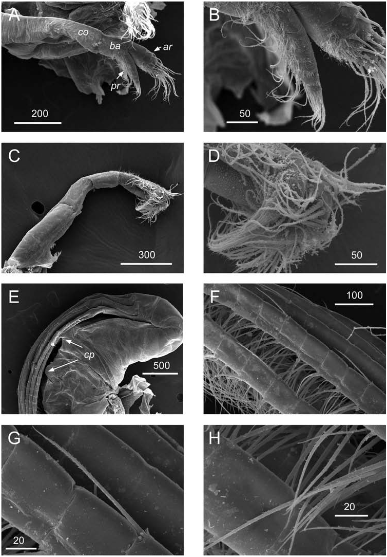
Mouth cirri with short rami about one-quarter of total length of the mouth appendage ( Figures 5D View Figure 5 , 6A View Figure 6 , 7A–D View Figure 7 ). Anterior ramus (inner) 6 segmented ( Figures 5D View Figure 5 , 7A View Figure 7 ), posterior (outer) ramus 5 segmented ( Figures 5D View Figure 5 , 6A View Figure 6 , 7 View Figure 7 A-D). Both rami with dense, long setulated and serrate setae ( Figure 7B, D View Figure 7 ).
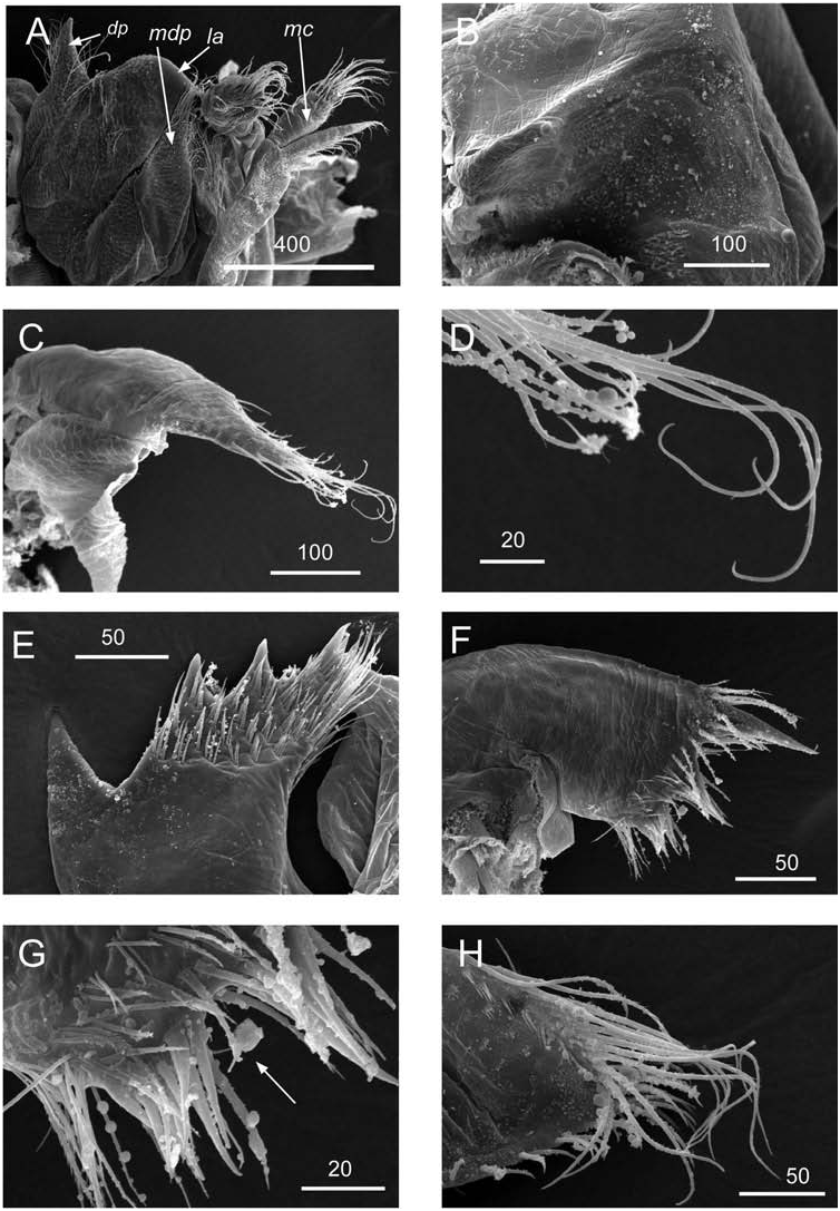
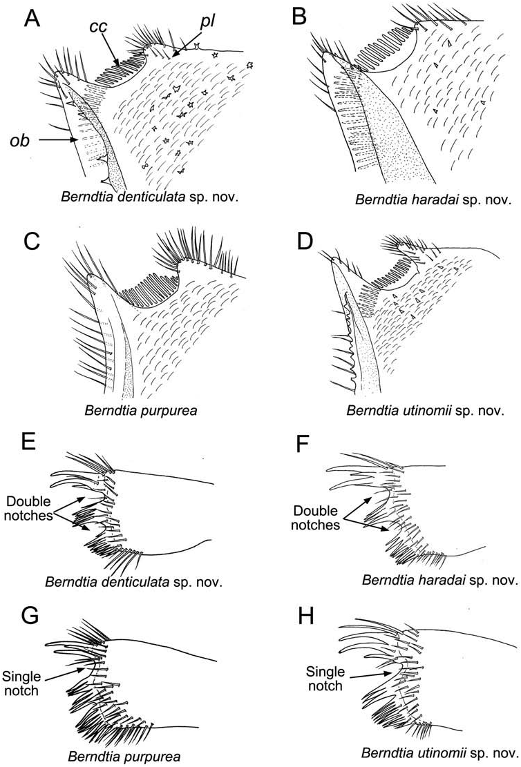
Labrum saddle-shaped ( Figures 5E View Figure 5 , 6A View Figure 6 ), bullate, with convex upper edge armed with developed dorsal process, horseshoe-shaped anterior edge smooth ( Figure 6A, B View Figure 6 ), dense smooth setae on dorsal process and a few smooth setae in upper lateral side, lateral sides and dorsal process with numerous multifid ctenoid scales ( Figures 5E View Figure 5 , 6A View Figure 6 ). Distal part of mandibular palp trapezoid, elongated, narrow, proximal part swollen ( Figure 6A, C View Figure 6 ), with dense simple setae and setae with tiny sparse setules on tip and sparse simple setae on both anterior and posterior margins ( Figure 6C, D View Figure 6 ). Mandible with three teeth excluding the inferior angle, first biggest upper tooth separated from the other lower teeth, lower margin beneath 3 rd tooth with 3–4 small sharp denticles and inferior angle ended in three large massive denticles, lateral surfaces in lower half of blade with tufts of dense short biserrated setae ( Figures 5F View Figure 5 , 6E View Figure 6 ). Maxillule double notched, with a larger upper notch (one-quarter to one-third of total length of the cutting edge) and a smaller lower notch (one-sixth of total length of the cutting edge), both notches lack denticles ( Figures 5G View Figure 5 , 6F View Figure 6 , 23E View Figure 23 ). Two large cuspidate and one short setae above the large notch ( Figures 5G View Figure 5 , 6F View Figure 6 , 23E View Figure 23 ), eight sharp setae in cluster upper the smaller notch and bundles of serrate setae at the cutting edge below the smaller notch ( Figures 5G View Figure 5 , 6F, G View Figure 6 , 23E View Figure 23 ). Lateral surfaces of maxillule with dense biserrated setae ( Figure 6F, G View Figure 6 ). Maxilla sub-triangular, with long, dense simple setae on tip and short sparse serrate setae on inferior margin ( Figures 5H View Figure 5 , 6H View Figure 6 ).
Thorax with two small conical processes on ventral side ( Figures 5I View Figure 5 , 7E View Figure 7 ). Terminal cirri five pairs. Annuli of terminal cirri normally with single distal serrate seta on posterior margin (on each third or fourth annulus) and paired long distal and small middle setae with fine setules on anterior margin ( Figure 7F–H View Figure 7 ). Caudal appendages absent.
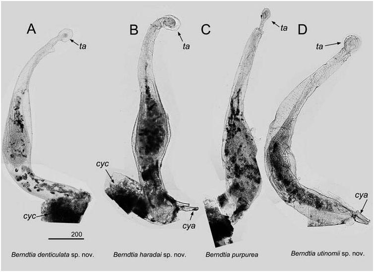
Dwarf males
One to three dwarf males can be occasionally found on the mantle sac of the female and on wall of the burrow. Males elongated, tadpole shaped, and with a terminal ampulla on developed stalk at the posterior end. Some opaque reddish purple granules were found at the middle of the elongated body, believed to be remnants of the cypris eyes ( Figure 22A View Figure 22 ), attachment of cypris antennules without stalk.
Etymology
The specific name originates from Latin ‘ denticulatus ’ meaning having a lot of teeth referring to numerous multifid teeth on the lateral surfaces of the opercular area of mantle sac.
Distribution
At present, only recorded in Okinawa ( Japan) and Virac ( Philippines).
Comparison ( Table 1)
Berndtia denticulata is similar to B. nodosa Tomlinson, 1967 ( Figure 8 View Figure 8 ) identified from Psammocora contigua in Singapore. Both of the species have noticeable nodular teeth on the opercular bars ( B. denticulata : Figure 4A, D; B View Figure 4 . nodosa: Figure 8B, C, I, J View Figure 8 ), but these nodules are more dense in B. denticulata . Berndtia denticulata also differs from B. nodosa in having multifid teeth on the lateral margins of the opecular bars ( Figure 4C View Figure 4 ), whilst B. nodosa has simple sharp or bifid teeth on the lateral margins of the opercular bars ( Figure 8B, I, L View Figure 8 ; see description in Tomlinson 1967). Lateral surfaces in posterior parts of opercular area of B. denticulata have dense multifid teeth ( Figure 4E, H View Figure 4 ), while B. nodosa has simple teeth in this area ( Figure 8H View Figure 8 ). The teeth on the mantle sac of B. denticulata can be pentafid, while Tomlinson (1967) described the teeth on the mantle sac of B. nodosa as at most quadrifid ( Figure 8E– G View Figure 8 ). Tomlinson (1967) did not find ventral conical processes of the thorax in B. nodosa . In the present study, we have recorded two small conical processes of thorax of B. denticulata (see Table 1 for details). Berndtia denticulata has differently patterned opercular bar colourations (absent blue coloration) in comparison with B. purpurea and B. utinomii . This species also differs from B. fossata , B. purpurea and B. utinomii by presence of rosette nodules on outer surface of the opercular bars and two notches on the cutting edge of maxillule. Berndtia denticulata differs form B. haradai in having bigger rosette nodules on the opercular bars and in presence of the ventral conical processes of the thorax.


No known copyright restrictions apply. See Agosti, D., Egloff, W., 2009. Taxonomic information exchange and copyright: the Plazi approach. BMC Research Notes 2009, 2:53 for further explanation.
|
Kingdom |
|
|
Phylum |
|
|
Class |
ACROTHORACICA Gruvel, 1905
| Chan, Benny K. K., Kolbasov, Gregory A., Hirose, Mamiko, Mezaki, T. & Suwa, R. 2014 |
Berndtia denticulata
| Chan & Kolbasov & Hirose & Mezaki & Suwa 2014 |
BERNDTIINAE
| Utinomi 1950 |
Berndtia
| Utinomi 1950 |
LITHOGLYPTIDAE
| Aurivillius 1892 |