
Chrysapace merimbunensis, Yamada & Lin & Eguchi, 2019
|
publication ID |
https://doi.org/ 10.2478/aemnp-2019-0036 |
|
publication LSID |
lsid:zoobank.org:pub:4CDE0401-572B-45CD-B7FE-5CC7F9C5606B |
|
DOI |
https://doi.org/10.5281/zenodo.6473150 |
|
persistent identifier |
https://treatment.plazi.org/id/A2686266-FF84-FFB7-FC33-BCC0FBF9FEAB |
|
treatment provided by |
Marcus |
|
scientific name |
Chrysapace merimbunensis |
| status |
sp. nov. |
Chrysapace merimbunensis Yamada & Eguchi sp. nov.
( Figs 1–19 View Figs 1–8 View Figs 9–14 View Figs 15–19 )
Type material examined. HOLOTYPE: ♀, Tasek Merimbun , Tutong, Brunei, K.Eguchi leg., 17.viii.1999, colony: Eg99-BOR-590 ( MBD). PARATYPES: 1♀, 3♂♂ from the same colony as the holotype ( MBD; MHNG).
Diagnosis. In the worker, cranium subtrapezoidal in fullface view; dorsum of mesosoma longitudinally costate; abdominal tergite III with costae which are transversely arched around center of posterior margin in dorsal view ( Fig. 5 View Figs 1–8 ); abdominal sternite III longitudinally costate; cinctus of abdominal segment IV smooth without any costae; abdominal tergite and sternites IV smooth with sparse hair-bearing foveae.
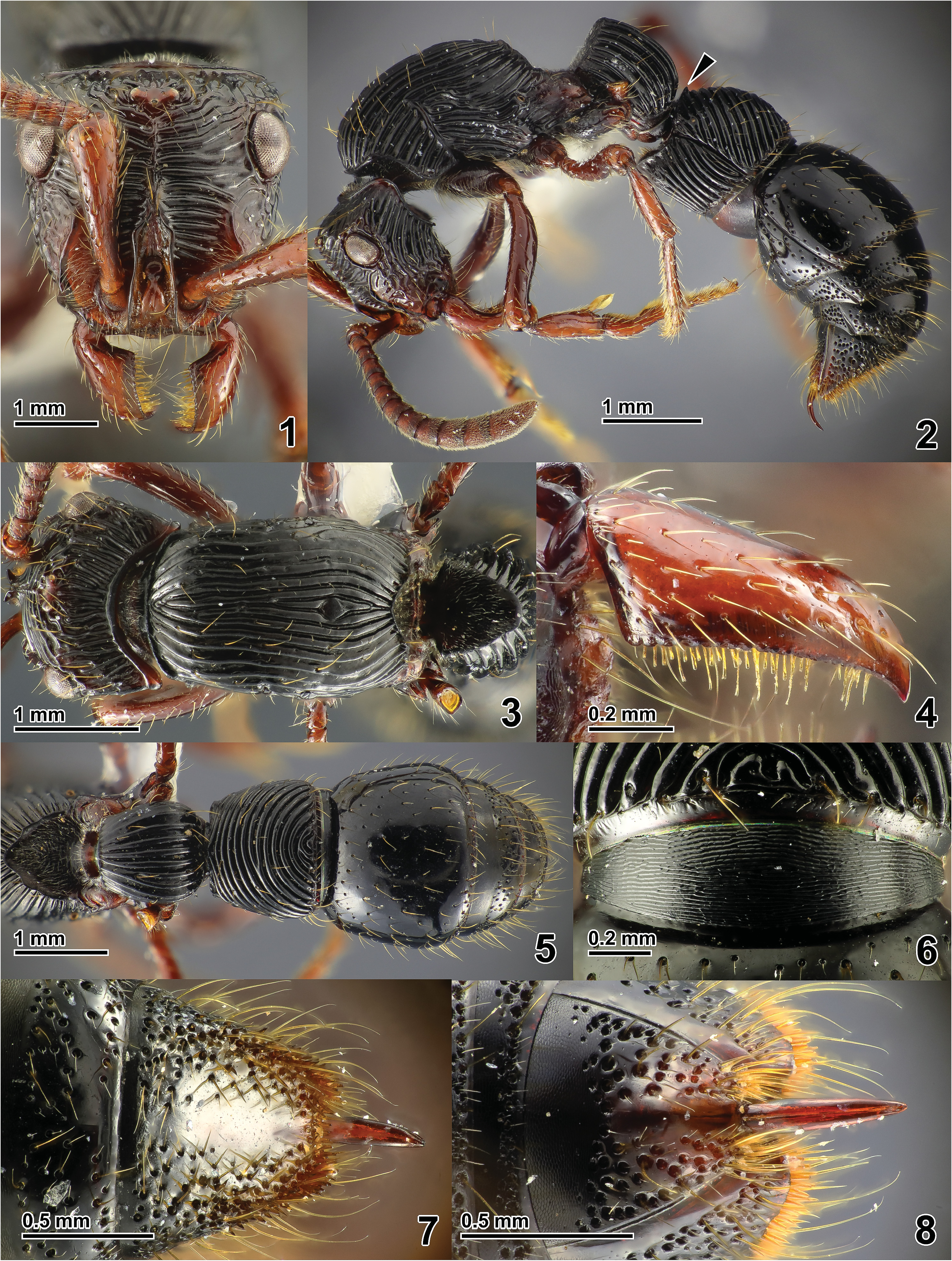
Description. Worker ( Figs 1–8 View Figs 1–8 ). Body black; antennae, anterior part of cranium, mandibles, and legs reddish brown. Body pilosity relatively sparse.
Cranium in full-face view subtrapezoidal, wider posteriorly than anteriorly, faintlylonger than wide (CI, 95–96); posterior part of cranium above eyes relatively short (EPI, 16–17), with PEHL much shorter than EL. Median ocellus located close to level of posteromost points of eyes in fullface view. Torulo-posttorular complex in full-face view relativelybroad and partially concealing antennal socket; width, when measured at level of antennal socket, distinctly greater than length of major axis of antennal socket; lateral margin almost straight but slightly undulate, slightly converging posteriad; anterior protrusion of anterolateral lobe encircling antennal socket weak, not distinctly protruding beyond anterior margin of lateral portion of clypeus in full-face view. Median portion of anterior clypeal margin weakly convexin full-face view, protruding beyond anterior margin of lateral portion of clypeus. Antenna 12-segmented. Maxillary palp 5-segmented. Labial palp 3-segmented. Mandible elongate, with relatively acute apex; masticatory margin feebly dentate, in dorsal view longer than twice as long as basal margin; ventral face of mandible with a series of modified setae along masticatory margin. Mesosoma in dorsal view relatively stout (DMI, 73), with evenly rounded lateral margins. Dorsal margin of propodeal declivity in dorsal view strongly arched anteriad and margined by strong edge. Petiole indorsal view subtrapezoidal, wider posteriorly than anteriorly, with strongly convex lateral margin. Anterodorsal corner of abdominal tergite III forming sharp edge (arrow in Fig. 2 View Figs 1–8 ). Anteroventral corner of abdominal sternite III (prora) in lateral view stronglyangulate with acute apex directed ventrad. Posterior margin of pygidium in dorsal view weakly concave medially.
Costae on body smoothly running, without overlaying microsculpture. Frons and vertex transversely costate ( Fig. 1 View Figs 1–8 ). Posterolateral face of cranium and posterior face above preoccipital carina longitudinallycostate ( Figs 2–3 View Figs 1–8 ). Torulo-posttorular complex and clypeus largely smooth. Gena largely smooth on face inside parafrontal ridge, whereas weakly coarsely rugose on face outside the ridge. Outer face of antennal scape longitudinally rugulate-striate on about basiposterior diagonal half, whereas smooth on remaining face. Outer face of mandible smooth ( Fig. 4 View Figs 1–8 ). Promesonotum, dorsal and lateral face of propodeum longitudinally costate. Mesopleuron with oblique-longitudinal costae which run parallelly with anteroventral margin of mesopleuron. Metapleuron partlycostate weakly and longitudinally. Posterior declivity of propodeum smooth. Legs largely smooth, with each coxa partly shagreened.Anterior faceof petiole smooth; dorsal andlateral faces of petiolar tergite longitudinally costate. Posterodorsal face of helcium imbricate, without any costae. Costae on abdominal tergite III with costae which are transversely arched around centerof posterior margin in dorsal view (see Figs 2, 5 View Figs 1–8 ). Abdominal sternite III longitudinallycostate. Pretergite and presternite of abdominal segment IV transversely striate (but partly imbricate, Fig. 6 View Figs 1–8 ). Cinctus of abdominal segment IV (boundary between pre- and postsclerites) smooth without any costae. Abdominal posttergite and poststernite IV smooth, with sparse hair-bearing foveae. Abdominal tergites and sternites V–VI imbricate on anterior marginal areas, whereas smooth, with relatively dense hair-bearing foveae on remaining face. Pygidium and hypopygium imbricate on anterior marginal area, whereas smooth submedially with dense hair-bearing foveae on remaining face ( Figs 7–8 View Figs 1–8 ).
Holotype worker (paratype worker in the parentheses). HL 1.63 (1.57) mm; HW 1.56 (1.50) mm; EL 0.39 (0.38) mm; EW 0.27 (0.27) mm; ES 0.33 (0.32) mm; PEHL 0.28 (0.24) mm; OL 0.05 (0.04) mm; SL 1.12 (1.05) mm; WL 2.63 (2.51) mm; DML 1.96 (1.85) mm; MW 1.43 (1.35) mm; MFL 1.70 (1.63) mm; PH 1.15 (1.12) mm; PTL 1.13 (1.11) mm; PTW 1.06 (0.97) mm; A3L 1.21 (1.27) mm; A3 W 1.43 (1.37) mm; CI 96 (95); SI 72 (70); EI 21 (22); EPI 17 (16); OI 3 (3); DMI 73 (73); DMI2 74 (74); LMI 59 (60); MFI 109 (109); PTI 94 (87); A3I 119 (107).
Male ( Figs 9–19 View Figs 9–14 View Figs 15–19 ). Body black; antennae, mandibles, and legs dark reddish brown. Body pilosity relatively dense.
Cranium in full-face view bulb-shaped, just faintly longer than wide (CI, 98–99); posterolateral corner somewhat angulate; vertex not strongly raised, without concealing dorsal edge of preoccipital collar in full-face view. Eye and ocelli relatively small (EI, 32–33; OI, 10); median ocellus located close to level of posteromost points of eyes in full-face view. Torulo-posttorular complex in full-face view relatively broad, partially concealing antennal socket; width,when measured at level of antennal socket, distinctly greater than length of major axis of antennal socket; lateral margin parallel in short anterior part, but linearly converging posteriorly; anterior protrusion ofanterolateral lobe encircling antennal socket weak. Anterior clypeal margin in full-face view strongly convex.Antenna 13-segmented. Maxillary palp 5-segmented. Labial palp 3-segmented. Mandible elongate, with relatively acute apex; masticatory margin feebly dentate, in dorsal view about twice as long as basal margin; ventral face of mandible with a series of modified setae along masticatory margin. Mesoscutum subpentagonal, a little wider than long (MSI, 118–119), with faintly convex anterolateral margin; notaulus completely absent; parapsidal line present as very weak and thin furrow. Scuto-scutellar suture fairly deep and broad, strongly scrobiculate. Posterior margin of mesoscutellum in dorsal view strongly convex. Dorsal margin of propodeal declivity in dorsal view broadly and shallowly arched anteriad, and forming strong edge. Wing venation as generic redescription in BOROWIEC (2016) ( Figs 13–14 View Figs 9–14 ).Petiole in dorsal view subtrapezoidal, wider posteriorly than anteriorly, with weakly convex lateral margin. Dorsal outline of abdominal tergite III in lateral viewstrongly rounded convex, without anterodorsal angle. Anteroventral corner of abdominal sternite III (prora) in lateral view strongly produced, with acute apex directed ventrad.
Frons weakly transversely costate. Vertex, lateral and posterior face of cranium coarsely reticulate. Torulo-posttorular complex and clypeus largely smooth. Outer face of antennal scape largely longitudinally rugulate-striate, with smooth apical face. Outer face of mandible smooth. Pronotum coarsely reticulate on dorsal face, whereas weakly and coarsely longitudinally rugose on lateral face. Mesopleuron, metapleuron, and lateral face of propodeum coarsely and irregularly rugoso-reticulate. Mesoscutum coarsely foveolate-reticulate. Mesoscutellum coarsely reticulate. Dorsum of propodeum longitudinally costate. Posterior declivity of propodeum smooth. Legs largely smooth, with meso- and metacoxae partly coarsely shagreened. Anterior face of petiole smooth; dorsal and lateral faces of petiolar tergite coarsely rugoso-reticulate. Posterodorsal face of helcium imbricate, without any costae. Abdominal tergite and sternites III largely weakly coarsely rugoso-reticulate. Pretergite and presternite of abdominal segment IV transversely striate (but partly imbricate). Cinctus of abdominal segment IV (the boundary between pre- and postsclerites) smooth, without any costae. Abdominal posttergite and poststernite IV smooth, with sparse hair- -bearing foveae. Abdominal tergites and sternites V–VII imbricate on anterior marginal areas, whereas smooth with dense hair-bearing foveae on remaining face. Pygidium with dense hair-bearing foveae.
Spiculum (anterior apophysis) of abdominal sternite IX ( Fig. 15 View Figs 15–19 ) 0.30–0.31 times as longas entirelength of sternite IX when spiculum length measured from transverse line spanning posteromost points of each anterolateral margin; outer margin of posterior spine of sternite IX in ventral view weakly concave; posteromedian flange between basesof posterior spines narrow, with some large irregular foveae along anteroventral margin of flange. Posterodorsal part of basimere in lateral view weakly broadly produced (arrow in Fig. 16 View Figs 15–19 ); articulation of basimere to teromere have thickened margin followed by membranous articulation ( Figs 16–17 View Figs 15–19 ). Telomere in lateral view subtriangular, 1.1–1.3 times as long as wide. Cuspis absent. Digitus in lateral view claw-shaped, entirely hooked ventrad with relatively acute apex ( Fig. 18 View Figs 15–19 ). Posterior apex of valviceps in lateral view strongly hooked ventrad, withlarge acute apical denticle; posterior half of ventral margin having 10–12 broad denticles (including apical denticle, Fig. 19 View Figs 15–19 ); anteroventral part of valviceps reduced, in lateral view concealed by lateral apodeme.
Paratype males (n = 3). HL 1.31–1.33 mm; HW 1.29–1.32 mm; EL 0.47–0.50 mm; EW 0.34–0.38 mm; ES 0.41–0.44 mm; PEHL 0.36–0.37 mm; OL 0.12–0.13 mm; SL 0.54–0.57 mm; WL 2.67–2.80 mm; DML 2.32–2.38 mm; MW 1.70–1.76 mm; MSL 1.09–1.14 mm; MSW 1.30–1.35 mm; MFL 1.50–1.55 mm; PTL 0.91–0.96 mm; PTW 0.82–0.87 mm; A3L 1.06–1.12 mm; A3 W 1.20 –1.26 mm; CI 98–99; SI 42–44; EI 32–33; EPI 27–28; OI 10; DMI 73–74; DMI2 85–87; MSI 118–119; MFI 117–119; PTI 88–90; A3I 113–114.
Remarks. Chrysapacemerimbunensis sp. nov. is relatively similar to C. sauteri and its sibling species C. costatus by sharing the following characteristics of the worker: i) dorsum of mesosoma, petiolar tergite, and abdominal sternite III longitudinally costate; ii) abdominal tergite IV–VI lacks any costation. However, the worker of the former is easily distinguished from those of the latter two species by subtrapezoidal shape of cranium in full-face view, non-costate posterodorsal face of helcium, abdominal tergite III with costae which are transversely arched around center of posterior margin in dorsal view, andnon-costate cinctus of abdominal segment IV. A worker and a male of C. merimbunensis from the same colony as the type series, whose images are provided in Antweb, were once misidentified as C. sauteri (https://www.antweb.org/specimen/ CASENT0179562; CASENT0179567 [Date accessed: 30 January 2019]). The queen of C. merimbunensis is currently unknown.
In this genus the male was previously described only for C. sauteri by TERAYAMA et al. (1998). Our comparison of the male morphology of C. merimbunensis and C. sauteri highlights that the male genitalia are highly differentiated between these species, especially in the shape of posterior spines and posteromedian flange of abdominal sternite IX, shape of posterodorsal projection of basimere, thickness of articulation of basimere and telomere, shape of telomere, shape of digitus, and shape of valviceps. These characters are likely to be useful for taxonomy at the species level.
Etymology. This species is named after the type locality, Tasek Merimbun ( Brunei, Tutong), adjective.
Distribution. Only known from the type locality, Tasek Merimbun ( Brunei, Tutong).


| MHNG |
Museum d'Histoire Naturelle |
No known copyright restrictions apply. See Agosti, D., Egloff, W., 2009. Taxonomic information exchange and copyright: the Plazi approach. BMC Research Notes 2009, 2:53 for further explanation.
|
Kingdom |
|
|
Order |
|
|
Family |
|
|
SubFamily |
Dorylinae |
|
Genus |