
Parapercis rubromaculata, Ho & Chang & Shao, 2012
|
publication ID |
https://doi.org/10.5281/zenodo.5347185 |
|
persistent identifier |
https://treatment.plazi.org/id/A145D130-2A6B-FFAC-FF03-FE8AFE0EFCA3 |
|
treatment provided by |
Tatiana |
|
scientific name |
Parapercis rubromaculata |
| status |
sp. nov. |
Parapercis rubromaculata View in CoL , new species
(Fig. 5A–D, 6A–C) New English name: redspot sandperch
Material examined. — Holotype: NMMB-P12635 (97.2 mm), Hengchun , Pingtung, southern Taiwan, northern South China Sea, hook and line, ca. 50–80 m, 10 Oct.2010, purchased from Hengchun market by H.-C. Ho.
Paratype: QM I. 38836 (1 ex., 89.5 mm), near type locality, 29 Sep.2010 ; NMMB-P12637 (1 ex., 78.1 mm), near type locality, 11 Oct.2010 , otolith taken before preservation; both purchased from Hengchun market by H.-C. Ho . NMMB-P12636 (1 ex., 114.0 mm), Hengchun , Pingtung, southern Taiwan, 24 May 2008 , purchased from Houbihu fish market by C.-W. Chang .
Diagnosis. — Many irregular red spots on caudal fin; five reddish-brown blotches on dorsal body surface; five reddish patches on midline of lateral body, each connected to an overlying dorsal blotches; a diagonal reddish-yellow bar below eye crossing cheek; a series of irregular yellowish patches below midline of lateral body; dorsal fin with medial row of yellowish spots and a row of reddish spots on base of soft rays; anal fin yellowish with a whitish base and some irregular pinkish lines; eye yellowish with two horizontal bar, one above and other below iris; and a combination of the following characters: dorsal-fin rays V, 21; anal-fin rays I, 17; pectoral-fin rays 17; pored lateral-line scales 52–53; gill rakers on 1st gill arch 13–14; pseudobranches 15–17; three pairs of canine teeth anteriorly in lower jaw; no palatine teeth; vomerine teeth stout, in a single curved row; cycloid scales on cheek and in predorsal, prepectoral and prepelvic areas; margin of preopercle smoothly indented; 4 th dorsal spine longest; ventral half of caudal fin slightly rounded, dorsal half truncate with a prolongation on upper corner; appressed pelvic fin extends well beyond anus.
Description. — Morphometric and meristic data of type series are provided in Table 1. The following data are provided for the holotype, followed by the range for all types in parentheses, if different from the holotype.
Dorsal-fin rays V, 21; anal-fin rays I, 17; all dorsal and anal soft rays branched, last soft ray branched to base; pectoralfin rays 17, all branched except uppermost; pelvic-fin rays I, 5; principal caudal-fin rays 17, uppermost and lowermost unbranched; upper procurrent caudal-fin rays nine, lower procurrent caudal-fin rays seven (seven to eight); lateral-line scales 53 (52–53, not including three smaller pored scales on base of caudal fin); scale rows above first lateral-line scale to origin of dorsal fin six; scale rows above highest part of lateral line to base of dorsal fin 4.5; scale rows below lateral line postero-ventrally to origin of anal fin about 11 (11–12); median predorsal scales eight; circumpeduncular scales 24; gill rakers 4+9 (4 + 9–10); pseudobranchial filaments 16 (15–17); branchiostegal rays six; vertebrae 10+19.
Body depth 5.8 (5.8–6.0) times in SL, 1.9(1.7–1.9) in HL; body nearly cylindrical anteriorly, width 5.2 (5.2–5.5) in SL, 1.6 (1.6–1.8) in HL, strongly compressed posteriorly; head length 3.1 (3.1–3.4) in SL; ventral part of head, chest, and abdomen slightly convex; snout length 2.8 (2.8–3.4) in HL; orbit diameter 3.8 (3.1–3.8) in HL; interorbital space
A
B
C
D
E
Fig. 5. A–D: Parapercis rubromaculata n. sp. A, NMMB-P12635, holotype, 97.2 mm SL, fresh; B, preserved holotype; C, QM I.38860, paratype, 89.5 mm SL, fresh; D, NMMB-P12637, paratype, 78.1 mm SL, fresh; E, Parapercis randalli Ho & Shao, 2010 , QM I.38817, paratype, 96.9 mm SL, fresh.
flat, least fleshy width 7.2 (7.2–8.5) in HL; caudal-peduncle depth 3.6 (3.3–3.7) in HL; caudal-peduncle length 3.6 (2.8–3.6) in HL.
Mouth large, maxilla nearly reaching vertical through center of eye, the upper-jaw length 2.5 (2.3–2.7) in HL; mouth oblique, forming an angle of about 20° to horizontal axis of body, the lower jaw projecting; front of upper jaw with two or three pairs of recurved canines, last on each side twice as large as anteriormost; side of upper jaw with one row of about four to six slender conical teeth that curve medially and posteriorly, gradually increasing in size posteriorly; remaining teeth in outer row on side of jaw decreasing in length; broad band of villiform teeth in about 10 rows medial to canines at front of upper jaw, gradually narrowing posteriorly in jaw to narrow band in about three irregular rows; front of lower jaw with three pairs of incurved canines, increasing in length laterally, the third twice as large as second and strongly curving laterally as well as posteriorly; a band of about seven rows of villiform teeth medial to canines at front
A
C
of lower jaw, medial row continuing laterally in jaw posterior to last canine as a row of five to seven increasingly larger and more strongly recurved teeth, followed by a single row of small teeth to end of jaw; vomer with a chevron-shaped row of five to six stout conical teeth, middle largest, lateral teeth progressively smaller; no palatine teeth; lips smooth, their inner surface with large fleshy papillae that interdigitate with anterior teeth; tongue broadly rounded, reaching forward to posterior vomerine teeth.
Gill membranes free from isthmus, with a broad free fold across. Gill rakers short and spinous, longest about one-third length of longest gill filaments. Nostrils small; anterior nostril in front of center of eye (as viewed from side), slightly more than half way to groove at edge of upper lip with a slight anterior rim and a pointed posterior flap that reaches three-fourths internarial distance when laid back; posterior nostril dorso-posterior to anterior nostril, aperture ovate with a slight rim.
B
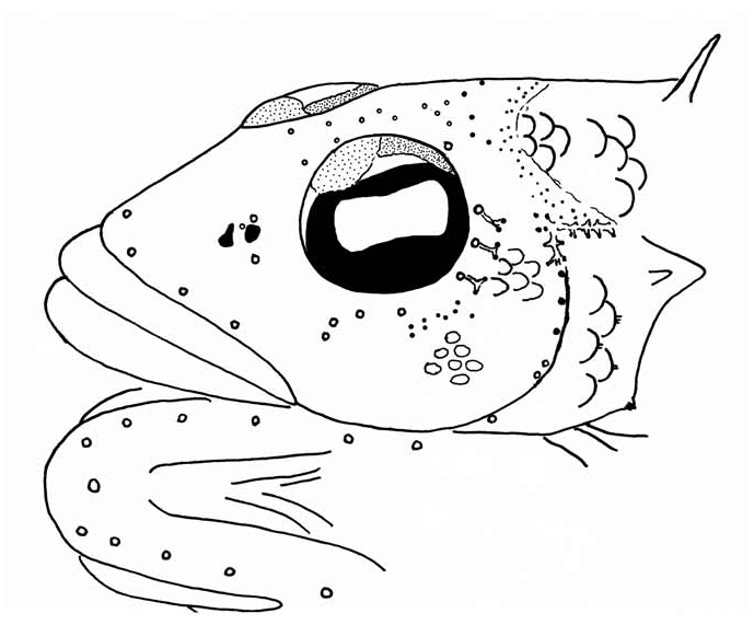
Pores of cephalic sensory system as shown in Fig. 6C. A View Fig row of three large pores on each side of maxilla; two or three median pores near nostrils, one above and one below, one small pore, when present, between both nostrils; two median pores anteriorly in interorbital space; a irregular series of small pores posteriorly in interorbital space, followed by two irregular transverse series of pores posteriorly on occiput, connected by a canal under the skin, divided into three double series, one continuing to ventroposterior margin of eye, one continuing to above free margin of preopercle and one continuing to anterior end of lateral line on body; one pore at dorso-posterior corner of eye; a row of four median pores below anterior half of eye; a series of three pores at posterior margin of eye, each connected by a canal beneath the skin and two terminal pores; a series of eight large pores along the inner margin of preopercle, the central two close together, continuing to a series of four large pores on mandible; a large pore at front of chin.
Opercle with a single sharp spine at level of ventral margin of pupil (when viewed from side); margin of interopercle smooth except for three or four (two to five in paratypes) tiny, close-set serrae on a small bony prominence at upper end; preopercle broadly rounded, its free edge smooth except for slight indentation at pore sites, extending downward and forward from level of ventral edge of orbit to slightly in front of a vertical at posterior edge of orbit.
Scales finely ctenoid on body, becoming cycloid anterior to a line from base of third dorsal spine to anterior end of lateral line, and on pre-pectoral and pre-pelvic areas; scales on opercle cycloid except above spine where a few are very weakly ctenoid; those on subopercle large and weakly ctenoid; those on cheek cycloid, small, mostly non-imbricate, in about 12 irregular horizontal rows, from below center of eye to posterior edge of preopercle, with 8 additional short rows of scales extending dorsally to behind ventral half of orbit; no scales on dorsal, anal, or pelvic fins; progressively smaller scales extending out on basal portion of caudal fin for at least two-thirds length of fin; base of pectoral fin with up to four rows of small cycloid scales; lateral line broadly arched over pectoral fin, then gradually slanting to straight midlateral portion on about posterior fourth of body.
Origin of dorsal fin over second to third lateral-line scale, predorsal length 3.1 (3.1–3.3) in SL, about equal to head length; 1 st dorsal-fin spine 12.4 (10.0–13.6) in HL; second dorsal-fin spine 7.2 (6.0–7.2) in HL; third dorsal-fin spine 5.2 (4.5–5.2) in HL; fourth dorsal-fin spine longest, 4.8 (3.9–4.8) in HL; fifth dorsal-fin spine 5.8 (4.9–5.8) in HL, entirely attached to first soft ray by membrane; last dorsal soft ray longest, 2.3 (1.9–2.7) in HL; origin of anal fin below base of fourth dorsal soft ray, preanal length 2.4 (2.0–2.4) in SL; anal-fin spine 5.4 (4.7–7.0) in HL; last anal soft ray longest, 2.4 (2.3–2.5) in HL; ventral half of caudal fin slightly rounded, a small excisur at middle, dorsal half truncate with a prolonged upper lobe which centered on third branched ray, extending about two-thirds orbit diameter posterior to central margin of fin, total fin length 4.9 (4.3–5.0) in SL, 1.6 (1.3–1.6) in HL; pectoral fins broadly rounded when spread, tenth ray longest, 4.8 (4.8–5.2) in SL, 1.5 in HL; origin of pelvic fins anterior to that of pectoral fin, below base of exposed part of opercular spine, prepelvic length 3.7 (3.5–4.0) in SL, 1.2 (1.1–1.2) in HL; pelvic-fin spine slender, 5.4 (4.2–5.4) in HL; pelvic fins extending beyond anus, fourth soft pelvic ray longest, 4.3 (4.1–4.9) in SL, 1.4 (1.3–1.4) in HL.
Colour when fresh. — See Figs. 5 A, C, D, 6A, B. Dorsal surface reddish, grading to white on ventral portion of lateral body, bright white between pectoral and pelvic fins; anterior portions of both jaws and snout reddish orange; five very broad pale brownish blotches evenly distributed on dorsal surface of body; a row of five reddish patches along midline of lateral body; anterior portion of eye yellowish, followed by a bright red band; eye yellowish, iris black with a horizontal reddish bar both above and below; a reddish-yellow ventroposteriorly directed bar crossing cheek, soft dorsal fin with a horizontal series of yellowish spots centrally and a series of red spots at base; a pale blue line on two-thirds height of soft portion of dorsal fin; spinous portion of dorsal fin reddish yellow anteriorly and dorsally; anal fin yellowish, with a whitish base, a whitish margin and some pinkish lines; about 20–30 irregular bright red spots on caudal fin; prolonged caudal-fin rays reddish; pelvic and anal fins yellowish.
Colour in alcohol. — See Fig. 5B. Creamy white with five broad blackish blotches on dorsal surface of body, becoming progressively smaller from anterior to posterior, first one between posterior margin of neurocranium and origin of soft dorsal fin, middle three below soft dorsal fin, slightly forked ventrally, and the last at caudal peduncle.
Distribution. — Known from the type series collected at Hengchun, southern tip of Taiwan, northern part of South China Sea, at depth ca. 50– 80 m.
Etymology. — ruber – red, and maculate – spot, referring to the characteristic red spots on the caudal fin of this species when fresh.
Remarks. — Parapercis rubromaculata is most similar to a recently described species P. randalli (Fig. 5E) which coexists in the area. The holotype and one paratype of the former were collected together with the type series of P. randalli . However, as these specimens lacked the characteristic black spots on the caudal fin, they were not included in the description of P. randalli . Parapercis rubromaculata differs from P. randalli in having many red spots on caudal fin (vs. two vertical rows of black spots) and a red area on the dorso-posterior corner of the eye (vs. red area absent); in lacking black spots posterior to the eye, above the opercle and the area between the spinous portion of dorsal fin and the pectoral fin (vs. spots present); having a relatively long pelvic fin (23.5–25.0% SL vs. 18.0–20.5% SL), which extends well beyond the origin of anal fin (vs. just reaches); and a different CO I DNA structure.
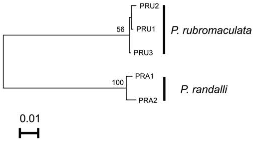
Genetics. — The 594 bp of CO I gene sequences were obtained from three specimens of P. rubromaculata and two specimens of P. randalli . Genetic distances between specimens of P. rubromaculata and P. randalli are 14% K2P genetic distance, whereas those of individuals of each species were not more than 1% ( Fig. 7 View Fig ).
| QM |
Queensland Museum |
No known copyright restrictions apply. See Agosti, D., Egloff, W., 2009. Taxonomic information exchange and copyright: the Plazi approach. BMC Research Notes 2009, 2:53 for further explanation.