
Telmatobius pefauri Veloso & Trueb, 1976
|
publication ID |
https://doi.org/ 10.11646/zootaxa.4250.4.1 |
|
publication LSID |
lsid:zoobank.org:pub:905BFB94-3B5C-44C3-8749-7EFA8EDC2A6D |
|
DOI |
https://doi.org/10.5281/zenodo.5632793 |
|
persistent identifier |
https://treatment.plazi.org/id/A0780A36-881C-A15B-FF27-74FFEF956F84 |
|
treatment provided by |
Plazi |
|
scientific name |
Telmatobius pefauri Veloso & Trueb, 1976 |
| status |
|
Telmatobius pefauri Veloso & Trueb, 1976 (Arica’s water frog)
Topotype: DBGUCH-1501049, adult female collected at Quebrada Murmuntani (18°20’55’’S, 69°34’12’’W, 3300 m elevation), Provincia de Parinacota , Región de Arica y Parinacota, Chile, on 26 January 2015 by H. Salinas, F. Cruz and M. Sallaberry. GoogleMaps
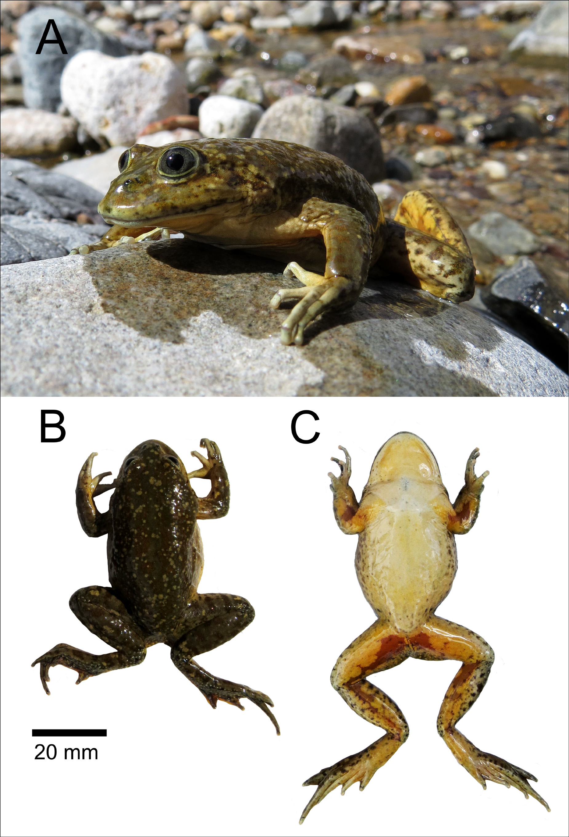
Description ( Figures 2 View FIGURE 2 and 3 View FIGURE 3 ; Table 1 View TABLE 1 ) of the topotype: moderate-sized adult female, SVL 57.13 mm ( Figure 2 View FIGURE 2 D). Large depressed head (HL/SVL=0.26), wider than longer (HL/HW=0.73), with an elliptic dorsal profile of the snout. The lateral profile has a long and sloping snout from the orbital region to the subacuminate end ( Figure 2 View FIGURE 2 A), and flared lips. Small nostrils, barely protuberant, with flanges, located slightly closer to the eyes than to the tip of the snout, with a flat internarial region and a concave loreal zone. Cantus rostralis poorly defined. Tympanic ring is less evident externally on the right side and not distinguishable on the left. Supratympanic fold poorly developed on the right side, extending from the posterior border of the orbit to the shoulder on the left side, granular. Large eyes (diameter = 5.23 mm) positioned in the front of the head with frontal orientation. Developed maxillary and pre-maxillary teeth, curved towards the tongue and embedded in the labial mucosa, only the tip protruding in the buccal cavity. Very reduced dentigerous processes of the prevomers, distinguishing only one vestigial tooth on the left, situated about halfway between the choanae. Large subcircular choanae, widely separated in the middle. Large circular tongue with the posterior border free and unnotched. Forelimbs are robust, without a dermic fold on the wrist, with a relative finger length I>II<III>IV ( Figure 2 View FIGURE 2 B). Palmar webbing is absent and tip of fingers slightly expanded in spherical pads. Lateral margins of digits have differentiated fringes distinguishable along toes II and III. Inner palmar tubercles are large, elliptic and depressed. Outer palmar tubercles are prominent and quadrangular. One large subarticular tubercle is present on each of the first two fingers; two smaller rounded subarticular tubercles are on fingers III and IV. Supernumerary palmar tubercles present and well developed. Hindlimbs and toes are long and thin ( Figure 2 View FIGURE 2 C). Relative toe length I<II<III<IV>V. Webbing of toes is concave, decreasing distally to shape wide fringes along the lateral margins of the toes. Tips of toes are expanded and spherical. Internal metatarsal tubercles are small, elliptic and elevated. Outer metatarsal tubercles are slightly evident, rounded and small. Subarticular tubercles are present, but small in size; distribution of subarticular tubercles in toes I(1)-II(1)- III(2)-IV(3)-V(2). Tiny tubercles are present on the ventral surface of tarsus. Postfemoral fold scarcely developed. Tarsal fold reduced to a fringe along the tarsus and decreasing distally towards the fringe of the internal margin of the first toe. “Bagginess” is absent. Dorsal skin is smooth. Small dispersed tubercles are present in the presacral region. There is profusion of small tubercles on flanks, ventral surface of forearms, knees, external surfaces of tibias and tarsi and posteroventral surfaces of the thigh. Ventral surface is smooth, except for the tubercles in the cloacal region. Cloacal aperture is directed posteriorly at the dorsal level of thighs, and discretely ornamented below with folded skin and small tubercles.
Dorsal surface is olive brown, with brown mottling and small cream-colored dispersed spots ( Figure 3 View FIGURE 3 B). Ventral surface of the body is cream to yellow in color. Dorsal region of arms and legs have brown mottling, that are extended to the ventral surface of the tibia: ventral region is orange-yellow, with apricot-colored blotches ( Figure 3 View FIGURE 3 C), which continue to the tibio-tarsus. Dorsal region of head and face have a color pattern similar to the dorsum. Lips are cream with brown spots. Throat is yellow-cream. Flanks are dark mottling. Iris is grey with dark reticulation ( Figure 3 View FIGURE 3 A). Body measurements are shown in Table 1 View TABLE 1 .

No known copyright restrictions apply. See Agosti, D., Egloff, W., 2009. Taxonomic information exchange and copyright: the Plazi approach. BMC Research Notes 2009, 2:53 for further explanation.
|
Kingdom |
|
|
Phylum |
|
|
Class |
|
|
Order |
|
|
Family |
|
|
Genus |