
Camptoscaphiella paquini Ubick, 2010
|
publication ID |
https://doi.org/10.1206/667.1 |
|
persistent identifier |
https://treatment.plazi.org/id/A01C316A-AB09-FF8A-B321-0DCAFEB8F8C3 |
|
treatment provided by |
Carolina (2021-08-29 08:45:59, last updated 2023-11-15 03:19:33) |
|
scientific name |
Camptoscaphiella paquini Ubick |
| status |
sp. nov. |
Camptoscaphiella paquini Ubick View in CoL , new species
Figures 1–160 View FIGURES 1–8 View FIGURES 9–15 View FIGURES 16–22 View FIGURES 23–28 View FIGURES 29–34 View FIGURES 35–40 View FIGURES 41–47 View FIGURES 48–51 View FIGURES 52–58 View FIGURES 59–66 View FIGURES 67–78 View FIGURES 79–90 View FIGURES 91–102 View FIGURES 103–115 View FIGURES 116–126 View FIGURES 127–138 View FIGURES 139–145 View FIGURES 146–153 View FIGURES 154–160 , 179–181 View FIGURES 179–181 , 196–197 View FIGURES 194–199 , 301–324 View FIGURES 301–310 View FIGURES 311–320 View FIGURES 321–326 ; map 3
TYPES: Male holotype and female allotype, from China, on stable scree slope on soil at Lishadi, 500 m before Shibali Yaku ( Pass #31), 3585 m, 27.21354°N, 98.70021°E, Yunnan Province (7 Aug 2005, P. Paquin, PP2405, CASENT 9022602) deposited in HNU (PBI_OON03638) GoogleMaps .
ETYMOLOGY: The specific name is a patronym in honor of Pierre Paquin, who collected the type series and majority of the known specimens.
DIAGNOSIS: The male of this species differs from all congeners except C. schwendingeri in having a palpal bulb that lacks a distinct conductor, and from C. schwendingeri in having a much smaller embolar region (figs. 139–145, 179–181). The female differs from all other Camptoscaphiella in having genitalia with a sinuous copulatory duct (figs. 154–159, 197, 323, 324).
MALE (PBI_OON 02187, figs. 301–310): Total length 2.00. Carapace yellow-brown, surface and sides of pars cephalica very finely striated, lateral margin undulate. Clypeus straight in front view, sloping forward in lateral view; setae dark. Eyes: ALE largest, ALE oval, PME squared, PLE oval; posterior eye row procurved from both above and front; ALE-PLE touching, PME touching for less than half their length. Sternum as long as wide, yellowish white, not fused to carapace, with weak radial furrows between coxae I–II, II–III, III–IV, furrows wrinkled, surface smooth, anterior margin with continuous transverse groove, posterior margin extending posteriorly beyond anterior edges of coxae IV as single extension, lateral margins unmodified, male sternum with a pair of rounded anterolateral processes; setae abundant, dark, originating from surface. Mouthparts: chelicerae, endites, and labium pale orange. Chelicerae straight; promargin with one small tooth; setae dark, evenly scattered; paturon promargin with row of flattened setae. Labium broadly triangular; with 6 or more setae on anterior margin, subdistal portion with unmodified setae. Endites distally excavated, serrula present in single row, anteromedian margin a truncate lobe bearing dense brush of broad setae. Abdomen: pedicel ribbed. Dorsal scutum moderately sclerotized, yellow-brown, covering about 1 ⁄ 2 of abdomen, between 1 ⁄ 4 and 1 ⁄ 2 abdomen width, fused to epigastric scutum, postepigastric scutum pale orange, short, covering about 1 ⁄ 3 of the abdominal length, posteriorly rounded. Spinneret scutum present, incomplete ring; with fringe of needlelike setae. Spinnerets: colulus represented by 2 setae; ALS with 3 spigots, a larger median one surrounded by 2 smaller ones (pyriform); PMS with 2 subequal spigots; PLS with 4 subequal spigots. Dorsum setae dark. Epigastric area dark. Postepigastric area setae dark. Legs: yellow-brown, tibiae with circular depressions on the cuticle that lack visible pores and probably represent regions of muscle attachment. Tarsi I to IV superior claws examined in detail; all surfaces striated; proclaws and retroclaws I–III each with 3 subapical teeth along outer margin; proclaw IV with 2 basal teeth; retroclaw IV with 4 teeth; teeth actually originate from the ventral surface and bend outwards; inner margins without apparent teeth. Trichobothria examined with SEM; hairs plumose, hood with fine striations, base with longitudinal slit, aperture not gratelike. Tarsal organ of palp round with 1 large and 1 medium-sized sensilla; tarsal organ of legs I–II an irregular oval, with 2 large and 1 small sensilla. Epigastric region with sperm pore large, oval, rebordered; with 1 pair of additional orifices mesad of booklung openings (figs. 48–51). Palp not strongly sclerotized, proximal segments red-brown; trochanter normal size; femur one to two times as long as trochanter; patella greatly elongate, longer than other segments combined, tibia and tarsus rounded, subequal, cymbium orange-brown, ovoid in dorsal view; bulb orange-brown, stout, tapering apically, without distinct ventral conductor.
FEMALE (PBI_OON 3056, figs. 311–324): Total length 2.32. As in male except as noted. Carapace brown, anteriorly narrowed to between 0.50 and 0.75 times its maximum width; marginal setae light. Eyes subequal. Sternum yellow-brown, lacking anterior transverse groove (embolarium), lacking anterolateral process. Endites without distal excavation, anteromedian margin lacking lobe or strongly modified setae. Abdomen oval, dorsal scutum covering less than 1 ⁄ 2 of abdomen, not fused to epigastric scutum. Epigastric scutum strongly sclerotized. Postepigastric scutum strongly sclerotized, widely hexagonal, only around epigastric furrow, with short posteriorly directed lateral apodemes, hollow basally and with external opening (fig. 152). Spinnerets: colulus represented by 2 setae; ALS with 4 spigots, a larger median one surrounded by 3 smaller ones (pyriform); PMS with 5–6 subequal spigots; PLS with 9 subequal spigots. Legs: orange-brown; shorter than in male. Tarsal claws as in male, except that female retroclaw IV has only 2 subapical teeth (4 in male). Trichobothria as in male. Tarsal organ I and II large and rounded, with 2 large and 1 small sensilla; tarsal organ of palp, III, and IV small and oval, with 1 large and 1 small sensilla.
NOTES: The copulatory duct of C. paquini is strongly sinuous and somewhat distinct from the straight to slightly curved duct found in all other Camptoscaphiella . At first glance this suggests that the female may be misplaced and would better fit in Ischnothyreus , which is somatically similar and where sinuous ducts are common. Apart from the fact that C. paquini males and females were collected together, a closer comparison with a true Ischnothyreus , also from western Yunnan Province, has turned up additional differences between the two genera. In C. paquini , the copulatory duct opens externally at its anterior end through a small longitudinal slit (fig. 150), and is similar to other Camptoscaphiella that have been closely examined (as in Brignoli, 1976: figs.1–2). In Ischnothyreus , the copulatory duct opens to the outside at its posterior end through a larger opening and (at least in the Yunnan species) has additional invaginations anteriorly (figs. 325, 326, arrow). Thus, it seems reasonable that the female of C. paquini is, indeed, a Camptoscaphiella , albeit one with a unique morphology. In fact, the morphological uniqueness of the female C. paquini , is also shared by the male that, in contrast to most other species, has a much smaller embolar region and lacks a separate ventral prong (“conductor”). A conductor is also apparently absent in C. schwendingeri , which suggests relationship, a possibility that can be tested when females of that species are discovered.
OTHER MATERIAL EXAMINED: CHINA: Yunnan Province: Lishadi, 500 m before Shibali Yaku ( Pass #31), 3585 m, 27.21354°N, 98.70021°E, stable scree slope on soil, 7 Aug 2005, P. Paquin, PP2405, 2♂ (PBI_OON 02187, CASENT 9022603, CAS) GoogleMaps , 1♀ (PBI_OON 003046, CASENT 9022605, HNU) ; Lishadi , 10 km W Shibali, 3221 m, 27.20055°N, 98.71399°E, mature pine forest with bamboo understory, under rocks and logs, 6 Aug 2005, P. Paquin, PP2305, 1♀ (PBI_OON 003056, CASENT 9022537, HNU) GoogleMaps ; 6.18 km 280° W Shibali , 3100 m, 27.18413°N, 98.72024°E, turning rocks in open meadow along stream, 7 May 2004, C. Griswold, D. Kavanaugh, CGY35, 1♀ (PBI_OON 36310, CASENT 9020666, CAS) GoogleMaps ; 7.41 km 315° WNW Shibali, 36.0 km 325° NNW Fugong , 3336 m, 27.20629°N, 98.72001°E, beneath objects amidst dormant bamboo, along snowfield and avalanche debris, 8 May 2004, C. Griswold, D. Kavanaugh, CGY39, 1♀ (PBI_OON 36312, CASENT 9019944, CAS) GoogleMaps ; 1♀ (PBI_OON 36311, CASENT 9019943, HNU) GoogleMaps ; Lishadi, 1 km before Shibali Yaku ( Pass #31), 3585 m, 27.21447°N, 98.70064°E, talus slope atop alpine meadow, 12 Aug 2005, P. Paquin, PP3405, 1♂, 1 juvenile (PBI_OON 003058, CASENT 9023117, HNU) GoogleMaps ; Lumadeng, Lao Shibali pass (pass #30), 3265– 3060 m, 27.06427°N, 98.75123°E, rock cliffs along the road, 13 Aug 2005, P. Paquin, PP3705, 1♀ (PBI_ OON 003053, CASENT 9022533, CAS) GoogleMaps ; Lishadi , 10.5 km W of Shibali, 3250 m, 27.20192°N, 98.71321°E, rhododendron patch in conifer forest, in wet leaf litter, 17 Aug 2005, P. Paquin, PP4205, 2♀ (PBI_OON 003054, CASENT 9022547, CAS) GoogleMaps ; 1♀ (PBI_OON 003059, CASENT 9022548, HNU) GoogleMaps ; Yakou of Shibali , 3615 m, 27.21234°N, 98.69601°E, 5–7 Aug 2005, T. Guo, TG0503, 1♂ (PBI_OON 003057, CASENT 9025931, CAS) GoogleMaps ; 41 km W Gongshan on Dulong Valley Rd , 3000 m, 27.79655°N, 98.50562°E, 27 Sep–6 Oct 2002, D. Kavanaugh, P. Marek, D. Dong, H. Liang, DHK2002031A, 1♀ (PBI_OON 003049, CASENT 9030686, CAS) GoogleMaps .
DISTRIBUTION: Known only from the Gaoligongshan region in westernmost Yunnan Province, China (map 3).
Brignoli, P. M. 1976. Spinnen aus Nepal, III. Uber einige Spinnen aus dem Himalaya, dazu Revision einiger Arten aus dem Karakorum (Arachnida, Araneae). Ergebnisse der Forschung-Unternehmens Nepal Himalaya 5: 229 - 253.

FIGURES 1–8. Camptoscaphiella paquini, new species, male (PBI_OON 02187). 1. Habitus, ventral view. 2. Same, dorsal view. 3. Same, lateral view. 4. Abdomen, ventral view. 5. Same, dorsal view. 6. Same, lateral view. 7. Dorsal scutum, lateral view. 8. Same, dorsal view.
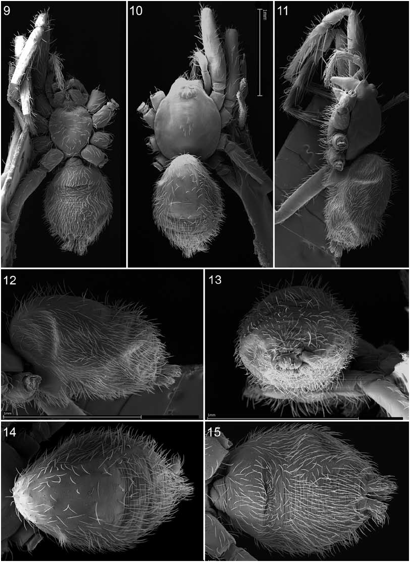
FIGURES 9–15. Camptoscaphiella paquini, new species, female (PBI_OON 03056). 9. Habitus, ventral view. 10. Same, dorsal view. 11. Same, lateral view. 12. Abdomen, lateral view. 13. Same, posterior view. 14. Same, dorsal view. 15. Same, ventral view.
FIGURES 16–22. Camptoscaphiella paquini, new species, male (PBI_OON 02187). 16. Cephalothorax, lateral view. 17. Same, dorsal view. 18. Same, anterior view. 19. Eye region, lateral view. 20. Same, dorsal view. 21. Chelicerae and mouthparts, lateral view. 22. Pedicel region, ventral view.

FIGURES 23–28. Camptoscaphiella paquini, new species, female (PBI_OON 03056). 23. Cephalothorax, anterior view. 24. Same, lateral view. 25. Same, dorsal view. 26. Eye region, anterior view. 27. Same, lateral view. 28. Same, dorsal view.

FIGURES 29–34. Camptoscaphiella paquini, new species, male (PBI_OON 02187). 29. Chelicerae and mouthparts, ventral view. 30. Same, sublateral view. 31. Labium, ventral view. 32. Endite distal end, ventral view. 33. Cephalothorax, ventral view. 34. Same, lateral view.

FIGURES 35–40. Camptoscaphiella paquini, new species, female (PBI_OON 03056: 35–39; PBI_ OON 03053: 40). 35. Chelicerae and mouthparts, ventral view. 36. Endite distal end, ventral view. 37. Cephalothorax, ventral view. 38. Pedicel region, ventral view, with arrow to coxal groove. 39. Chelicerae, anterior view. 40. Left chelicerae, posterior view, with enlargement showing promarginal tooth (arrow).
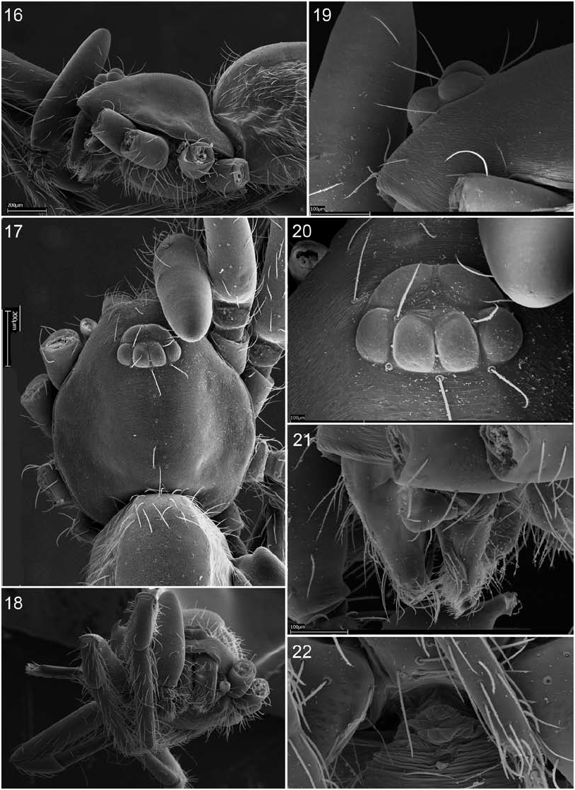
FIGURES 41–47. Camptoscaphiella paquini, new species, female (PBI_OON 03056). 41. Left palp, anterior view. 42. Right palp, prolateral view. 43. Palpal tibia, dorsal view. 44. Palp, dorsal view. 45. Tibia I, dorsal view, showing slit sensillum. 46. Same, magnified view, with arrow to pore. 47. Tibia I, dorsal view, showing probable muscle scars.

FIGURES 48–51. Camptoscaphiella paquini, new species, male (PBI_OON 02187). 48. Epigastric area, ventrolateral view, showing anterior (A) and posterior (P) respiratory spiracles in relation to additional orifice (O). 49. Same, ventral view. 50. Same, lateral view. 51. Same, enlarged view of spiracle region.
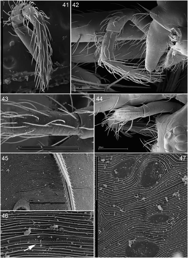
FIGURES 52–58. Camptoscaphiella paquini, new species, male (PBI_OON 02187). 52. Spinnerets, posterior view. 53. ALS, posterior view. 54. PLS, posterior view. 55. PMS, posterior view. 56. Spinnerets, ventral view. 57. Same, dorsal view. 58. Same, lateral view.

FIGURES 59–66. Camptoscaphiella paquini, new species, female (PBI_OON 03056). 59. Spinnerets, posterior view. 60. ALS, posterior view. 61. PLS, posterior view. 62. PMS, posterior view. 63. Spinnerets, ventral view. 64. Same, lateral view. 65. Abdomen, dorsal view, showing posterior edge of dorsal scutum. 66. Same, magnified view of cuticle structure.
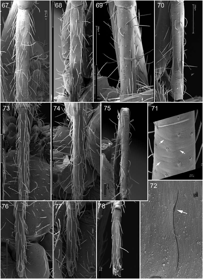
FIGURES 67–78. Camptoscaphiella paquini, new species, male (PBI_OON 02187), leg segments, dorsal views. 67. Tibia I. 68. Tibia II. 69. Tibia III. 70. Tibia IV. 71. Tibia IV, magnified to show slit sensilla (arrows). 72. Same, showing single sensillum (rotated) with arrow to pore. 73. Metatarsus I. 74. Metatarsus II. 75. Metatarsus IV. 76. Tarsus I. 77. Tarsus II. 78. Tarsus IV.
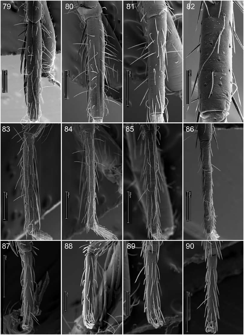
FIGURES 79–90. Camptoscaphiella paquini, new species, female (PBI_OON 03056), leg segments, dorsal views. 79. Tibia I. 90. Tibia II. 81. Tibia III. 82. Tibia IV. 83. Metatarsus I. 84. Metatarsus II. 85. Metatarsus III. 86. Metatarsus IV. 87. Tarsus I. 88. Tarsus II. 89. Tarsus III. 90. Tarsus IV.

FIGURES 91–102. Camptoscaphiella paquini, new species, male (PBI_OON 02187), sensory structures, dorsal view. 91. Tarsal organ, palpal tarsus. 92. Same, tarsus I. 93. Same, tarsus II. 94. Same, tarsus IV. 95. Trichobothrium, palpal tibia. 96. Same, tibia I. 97. Same, tibia II. 98. Same, tibia III. 99. Same, tibia IV. 100. Same, metatarsus I. 101. Same, metatarsus II. 102. Same, metatarsus IV. Scale bar for tarsal organs = 20 µm (vertical), for trichobothria = 10 µm (horizontal).
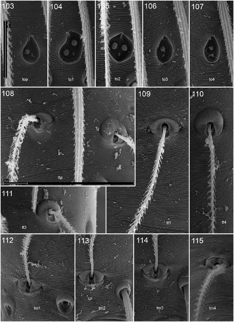
FIGURES 103–115. Camptoscaphiella paquini, new species, female (PBI_OON 03056), sensory structures, dorsal view. 103. Tarsal organ, palpal tarsus. 104. Same, tarsus I. 105. Same, tarsus II. 106. Same, tarsus III. 107. Same, tarsus IV. 108. Trichobothria, palpal tibia. 109. Same, tibia I. 110. Same, tibia IV. 111. Same, tibia III. 112. Same, metatarsus I. 113. Same, metatarsus II. 114. Same, metatarsus III. 115. Same, metatarsus IV. Scale bar for tarsal organs = 20 µm (vertical), for trichobothria = 10 µm (horizontal).

FIGURES 116–126. Camptoscaphiella paquini, new species, male (PBI_OON 02187), tarsal claws. 116. Leg I, prolateral view. 117. Same, dorsal view. 118. Same, subventral view. 119. Leg II, prolateral view. 120. Same, dorsal view. 121. Leg III, prolateral view. 122. Same, anterior view. 123. Same, retrolateral view. 124. Leg IV, prolateral view. 125. Same, ventral view. 126. Same, retrolateral view. The images of claws I– III are of right legs and are here reversed to give left perspective. Scale bar = 50 µm.

FIGURES 127–138. Camptoscaphiella paquini, new species, female (PBI_OON 03056), tarsal claws. 127. Leg I, subventral view. 128. Same, apical view. 129. Same, retrolateral view. 130. Leg II, ventral view. 131. Same, apical view. 132. Same, retrolateral view. 133. Leg III, subventral view. 134. Same, anteriodorsal view. 135. Same, retrolateral view. 136. Leg IV, ventral view. 137. Same, anteriodorsal view. 138. Same, retrolateral view. Scale bar = 50 µm.
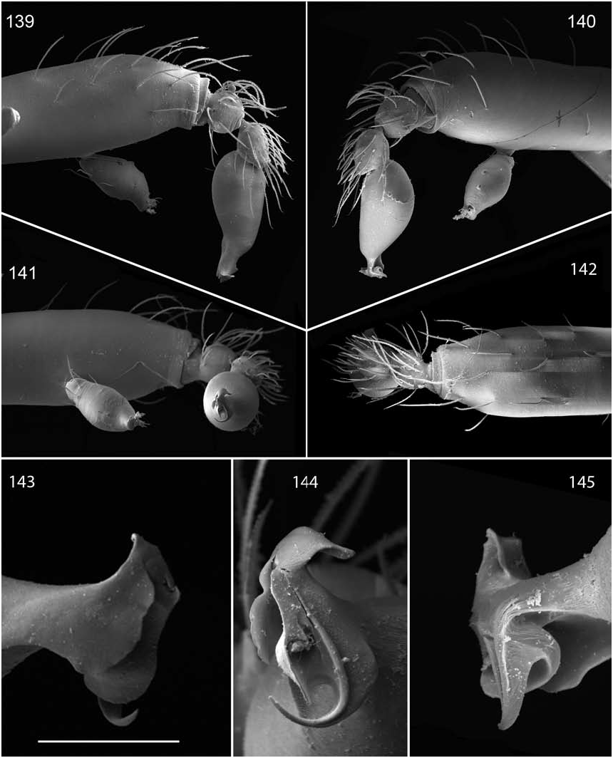
FIGURES 139–145. Camptoscaphiella paquini, new species, male (PBI_OON 02187). 139. Palp, prolateral view. 140. Same, retrolateral view. 141. Same, ventral view. 142. Same, dorsal view. 143. Embolar region, prolateral view. 144. Same, apical view. 145. Same, retrolateral view.

FIGURES 146–153. Camptoscaphiella paquini, new species, female (PBI_OON 03056: 146, 148– 150, 152; PBI_OON 03059: 147, 151; PBI_OON 03054: 153), epigynal region exterior. 146. Ventral view. 147. Sublateral view. 148. Posterior view. 149. Lateral view. 150. Posterior view showing copulatory opening (arrow). 151. Anterior view showing median row of pores. 152. Sublateral view showing apodeme orifice (O) and anterior (A) and posterior (P) spiracles. 153. Sublateral view showing edge of postepigastric scutum (arrow).

FIGURES 154–160. Camptoscaphiella paquini, new species, female (PBI_OON 03054: 154–156; PBI_OON 03059: 157–160), epigynal area interior. 154. Dorsal view of entire region. 155. Same, showing receptaculum and apodemes. 156. Receptaculum, anterior view, showing coiled duct enclosed by membrane. 157. Dorsal view of entire region, showing apparent opening of gonopore (arrow). 158. Same, receptaculum and apodemes (arrows). 159. Receptaculum, dorsal view. 160. Left apodeme, dorsal view.

FIGURES 179–181. Camptoscaphiella paquini, new species (PBI_OON 02187), male left palp. 179. Prolateral view. 180. Dorsal view. 181. Retrolateral view. Scale bars = 0.1 mm.
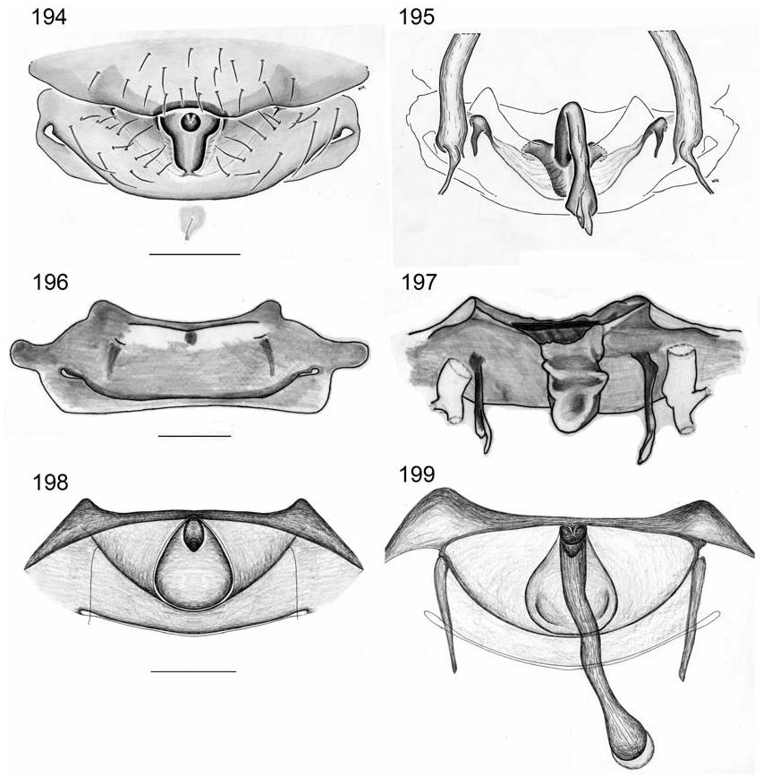
FIGURES 194–199. Camptoscaphiella species, female epigyna. 194. C. panchthar, new species (PBI_OON 15771), ventral view. 195. Same, dorsal view. 196. C. paquini, new species (PBI_OON 03059), ventral view. 197. Same, dorsal view. 198. C. nepalensis, new species (PBI_OON 23385), ventral view. 199. Same, dorsal view. Scale bars = 0.1 mm.
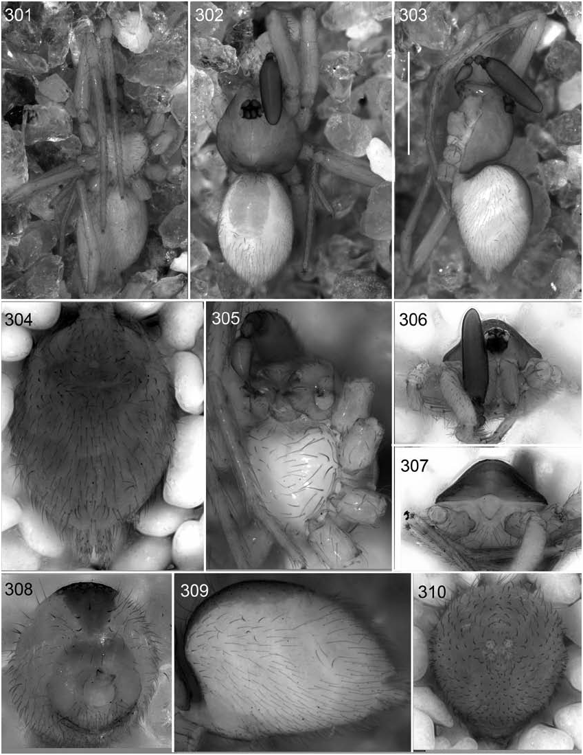
FIGURES 301–310. Camptoscaphiella paquini, new species, male (PBI_OON 02187). 301. Habitus, ventral view. 302. Same, dorsal view. 303. Same, lateral view, scale bar = 1 mm. 304. Abdomen, ventral view. 305. Sternum, ventral view. 306. Carapace, frontal view. 307. Same, posterior view. 308. Abdomen, anterior view. 309. Same, lateral view. 310. Same, posterior view.
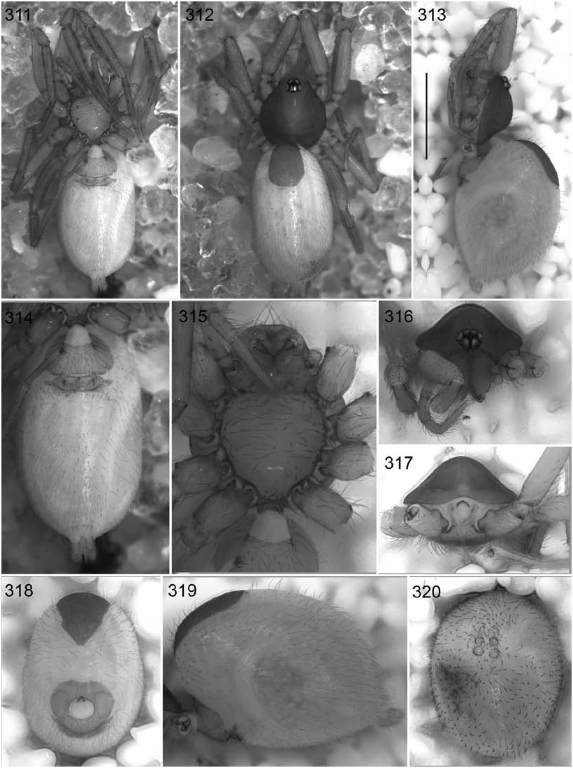
FIGURES 311–320. Camptoscaphiella paquini, new species, female (PBI_OON 03056). 311. Habitus, ventral view. 312. Same, dorsal view. 313. Same, lateral view, scale bar = 1 mm. 314. Abdomen, ventral view. 315. Sternum, ventral view. 316. Carapace, frontal view. 317. Same, posterior view. 318. Abdomen, anterior view. 319. Same, lateral view. 320. Same, posterior view.
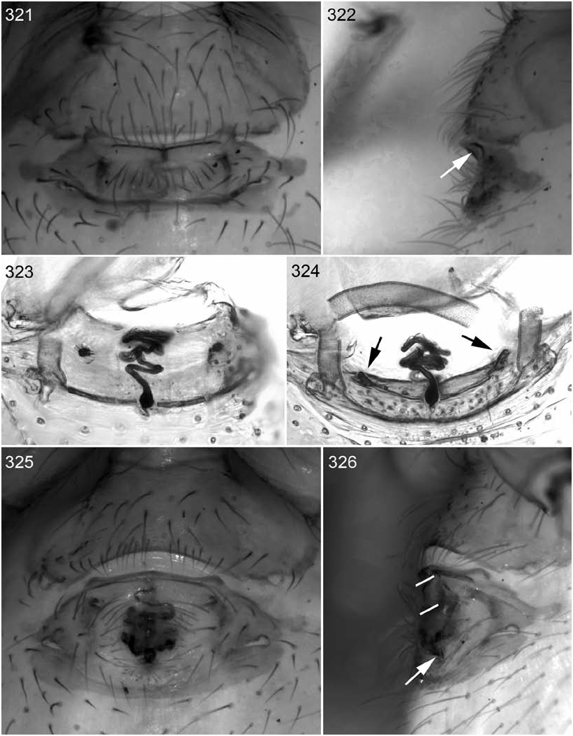
FIGURES 321–326. Camptoscaphiella and Ischnothyreus species, female epigastric regions. 321. Camptoscaphiella paquini, new species (PBI_OON 03053), ventral view. 322. Same, lateral view, arrow points to presumed copulatory opening. 323. Same (PBI_OON 03049), digested with pancreatin, dorsal view. 324. Same, posterior view, with arrows showing the bases of the (broken) apodemes. 325. Ischnothyreus sp. (PBI_OON 02188), ventral view. 326. Same, lateral view, with arrow to the presumed copulatory opening and dashes showing additional openings.
No known copyright restrictions apply. See Agosti, D., Egloff, W., 2009. Taxonomic information exchange and copyright: the Plazi approach. BMC Research Notes 2009, 2:53 for further explanation.
|
Kingdom |
|
|
Phylum |
|
|
Class |
|
|
Order |
|
|
Family |
|
|
Genus |