
Dolichopoda (Chopardina) lustriae Rampini, Di Russo, 2008
|
publication ID |
https://doi.org/10.11646/zootaxa.1923.1.1 |
|
persistent identifier |
https://treatment.plazi.org/id/9E6B87A3-FFB7-0202-FF75-81CEFBE46423 |
|
treatment provided by |
Felipe |
|
scientific name |
Dolichopoda (Chopardina) lustriae Rampini, Di Russo |
| status |
sp. nov. |
Dolichopoda (Chopardina) lustriae Rampini, Di Russo View in CoL sp. nov.
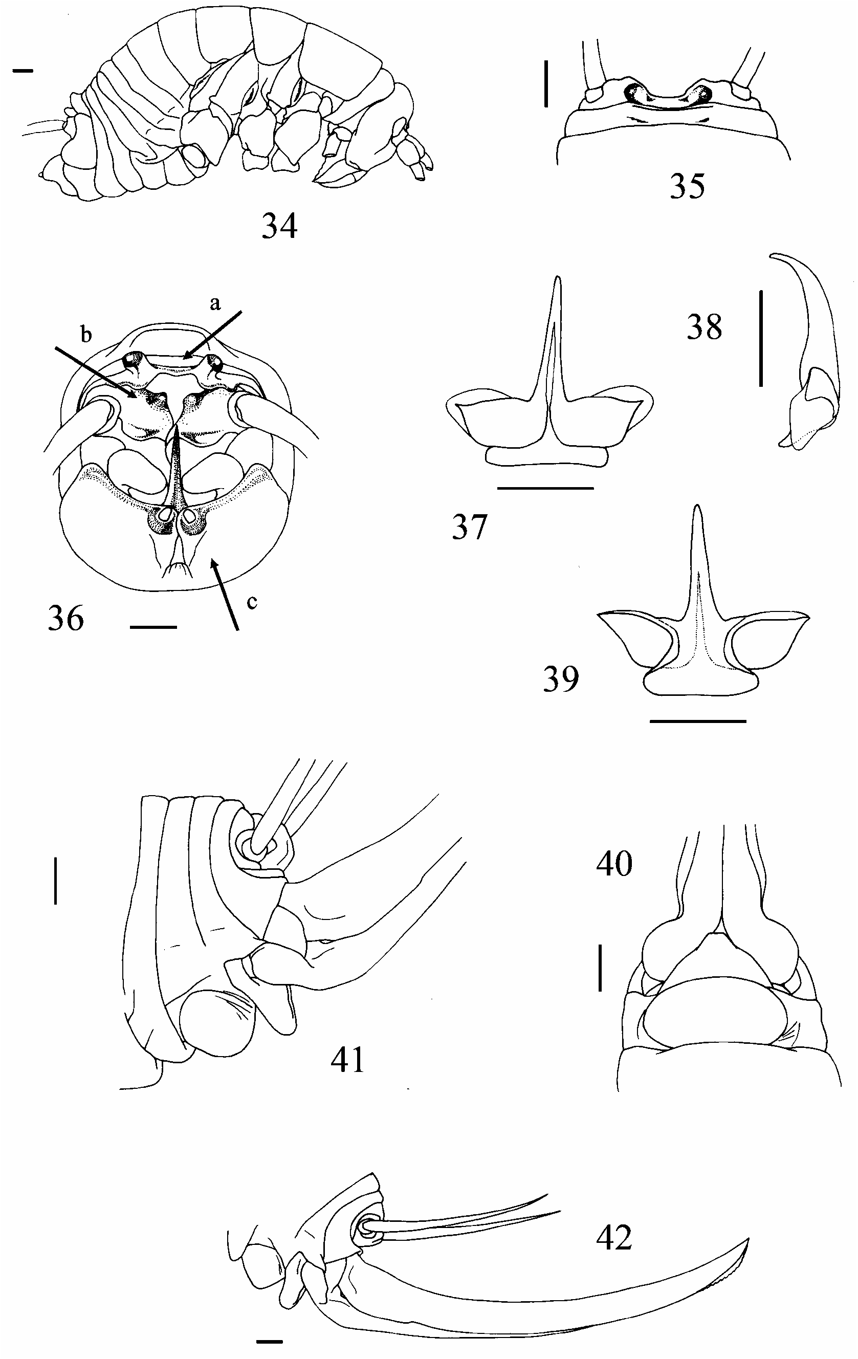
( Figs 34–42 View FIGURES 34–42 )
Diagnosis. The specimens of this species are larger than those found in the Ionic regions. The animal is yellowish-brown in colour. The edges of the pronota and the first abdominal tergite are considerably darker than the remaining tergites which are characterized by a lighter and narrower band. The head has a rounded vertex and very evident rostral tubercles. The tenth tergite has well-developed lateral expansions and tubercles similar to D. dalensi and D. matsakisi of the Peloponnesian area. The hind femora are armed with various spines on the inferior edge. Due to this characteristic, this new species must be attributed to the subgenus Chopardina present in Greece with the D. remyi species of Macedonia. However, there are other characteristics, such as the shape of the epiphallus and the tenth tergite which relate it to the species of the Peloponnesian area.
Type locality. The cave is situated in the territory of Halkiopuli (Etolia-Akarnania). It originates from an old river situated 1150 m a.s.l. on the steep western slopes of Pselovuni Mountain (Southern sector of the Valtou Mountains). The cave has a large semi-circular entrance which opens into a gallery (where the chapel dedicated to the hermit, S. Andrea, is situated). This gallery then forks. The left one is larger and better developed and has better climatic conditions. Probably for this reason, the examples of Dolichopoda were found in this gallery.
Etymology. We are happy to dedicate this new species to Lucilla Lustri for her important and assiduous speleological activity and for having participated in various missions with us (Rampini) in the caves of North- Western and Central Greece.
Material examined. Holotype male: Etolia-Akarnania , Halkiopuli , Pselovuni Mountain (1472 m a.s.l.), Aghias Andreas Cave, 1150 m a.s.l., lat. 38°59’25’’ N; long. 21°23’10’’ E, M.Rampini, G.Pintus, L.Lustri leg. GoogleMaps Paratypes: same locality, data and collectors as for holotype 1 female, 3 male nymphs and 4 female nymphs ( MZUR, PCR) GoogleMaps .
Description. Male (holotype) ( Fig. 34 View FIGURES 34–42 ). The species is big and yellowish in colour with thoracic tergites and the first abdominal segment which are decorated with large dark bands. The head is paler in colour with a rounded vertex and dark and pronounced rostral tubercles. Legs elongate and testaceous in colour. Fore and mid femora slender and have no spines. Hind femur with 1 short spine on the external and internal condyle, and 11 spines on the external edge and 23 spines on the internal part of the inferior edge. The tibia have variable numbers of spines, however, this variation is limited. Fore tibia armed with 3/3 short spines on the superior edge on both sides, whereas there are 4 on the external parts and 5 on the internal parts of the inferior edge, with two apical spurs. Mid tibiae with their upper and lower edges with 6 spines on the external side and 5 on the internal. Hind tibia with only 2 external spines on the upper edge and 21 spines on both sides of the upper edge. The tenth tergite, viewed posteriorly, appears narrow and has lateral lobes of the posterior edge short and trapezoidal, diverging, and separated by a large cavity; anterior side of the lobeswith a shallow incision in the middle ( Fig. 35 View FIGURES 34–42 ); tubercles are enlarged, cylindrical and diverging, rounded at the apex, and connected to each other by the central part of the slightly thickened upper edge ( Fig. 36a View FIGURES 34–42 ). The partially rectangular paraprocts are covered in hair at the edges, with a superior side elongated posteriorly, darker and well covered by short hair ( Fig. 36b View FIGURES 34–42 ). Subgenital plate particularly convex ventrally and well divided at the apex, while the partially rectilineal incisure of the apical part diverges at the basal portion forming a bellshape; lateral lobes triangular and terminate with large cylindrical styli which are inserted in a deep apical incisure ( Fig. 36c View FIGURES 34–42 ). Epiphallus sclerotized, the median process is elongated and very arched forwards with an acute apex, which widens at the base; basal lobes more evident and perpendicular to the median process, while the posterior ones are more developed and wing-like in shape ( Fig. 37 View FIGURES 34–42 , Fig. 38 View FIGURES 34–42 and Fig. 39 View FIGURES 34–42 ). Median process, viewed laterally, appears thickened for 2/3 of its length and considerably more slender and curved in the apical part. The accessory apparatus has a uneven part which is little sclerotized and is partially trapezoidal. The valves are very chitinous, triangular and pointed at the apex.
Length (mm): body 22,0; pronotum 4,5; fore femora 17,0; mid femora 17,5; hind femora 27,0; fore tibia 18,0; mid tibia 18,5; hind tibia 31,0 hind tarsus 12,0, 1 st article of hind tarsus 6,5.
Female. The length of the body 20 mm. The general appearance and the number of spines on the legs are very similar to the male. Subgenital plate triangular in shape, with a rounded apex with a large cylindrical protuberance which is more sclerotized and with a deep incision in the centre ( Fig. 40 View FIGURES 34–42 ). The 7 th urosternite has an evident spherical protuberance which is as large as the sternite ( Fig. 41 View FIGURES 34–42 ). Ovipositor almost as long as the body (19 mm). In this respect, it is similar to that of D. dalensi of the Peloponnisos, and is uniformly curved upwards and is slender at the apex. The inferior valves have 20 denticles ( Fig. 42 View FIGURES 34–42 ).
Dolichopoda (Dolichopoda) pavesii Galvagni, 2002
( Figs 43–45 View FIGURES 43–45 )
Thanks to the collection of new examples, it was possible to describe the morphology of the female of D. pavesii so completing the studies of Galvagni (2002) where he describes a small immature female. The new examples come from the Drogarati cave which is not far from the type locality (Drakotripa cave near the village of Aghia Nikolaos), approximately 15 Km from Sami.
Material examined. Ionian Isl. , Kephallinia (Kefalonia), near Sami, Drogarati cave, lat 38° 14’ 62’’ N, long 20° 38’ 73’’ E, 13.VIII.2003, C. Di Russo leg. 6 males, 2 females, 1 male nymph and 2 female nymphs ( MZUR, PCR); same locality, 15.VI.2004, F. Gasparo leg. 6 male nymphs and 4 female nymphs ( PCR) .
Female description. The spines on the legs are similar to those on the male. Tenth tergite similar to the male, but lacking tubercles. Lamina reticularis triangular. Subgenital plate triangular, thickened, and has a rounded apex with a large wing-like protuberance which is more sclerotized and with a deep incision in the centre ( Fig. 43 View FIGURES 43–45 ). The 7 th urosternite has a prominent coniform protuberance which is rounded at the apex; it is flattened and narrowed at the base and is not as wide as the sternite. In lateral aspect, it is very prominent compared to the preceding sternites. ( Fig. 44 View FIGURES 43–45 ). Ovipositor large basally, similar to D. kiriakii of Parga, and more curved in the first proximal portion. The superior valves have an acute apex which curves upwards. The inferior valves have a rounded apex and 19 denticles ( Fig. 45 View FIGURES 43–45 ).
Length (mm): body 17,5; pronotum 3,5; fore femora 15,0; mid femora 15,5; hind femora 26,0; fore tibia 17,5; mid tibia 17,0; hind tibia 34,5; hind tarsus 12,0; 1 st article of hind tarsus 6,0; ovipositor 12,0.
Dolichopoda (Dolichopoda) dalensi Boudou Saltet, 1972
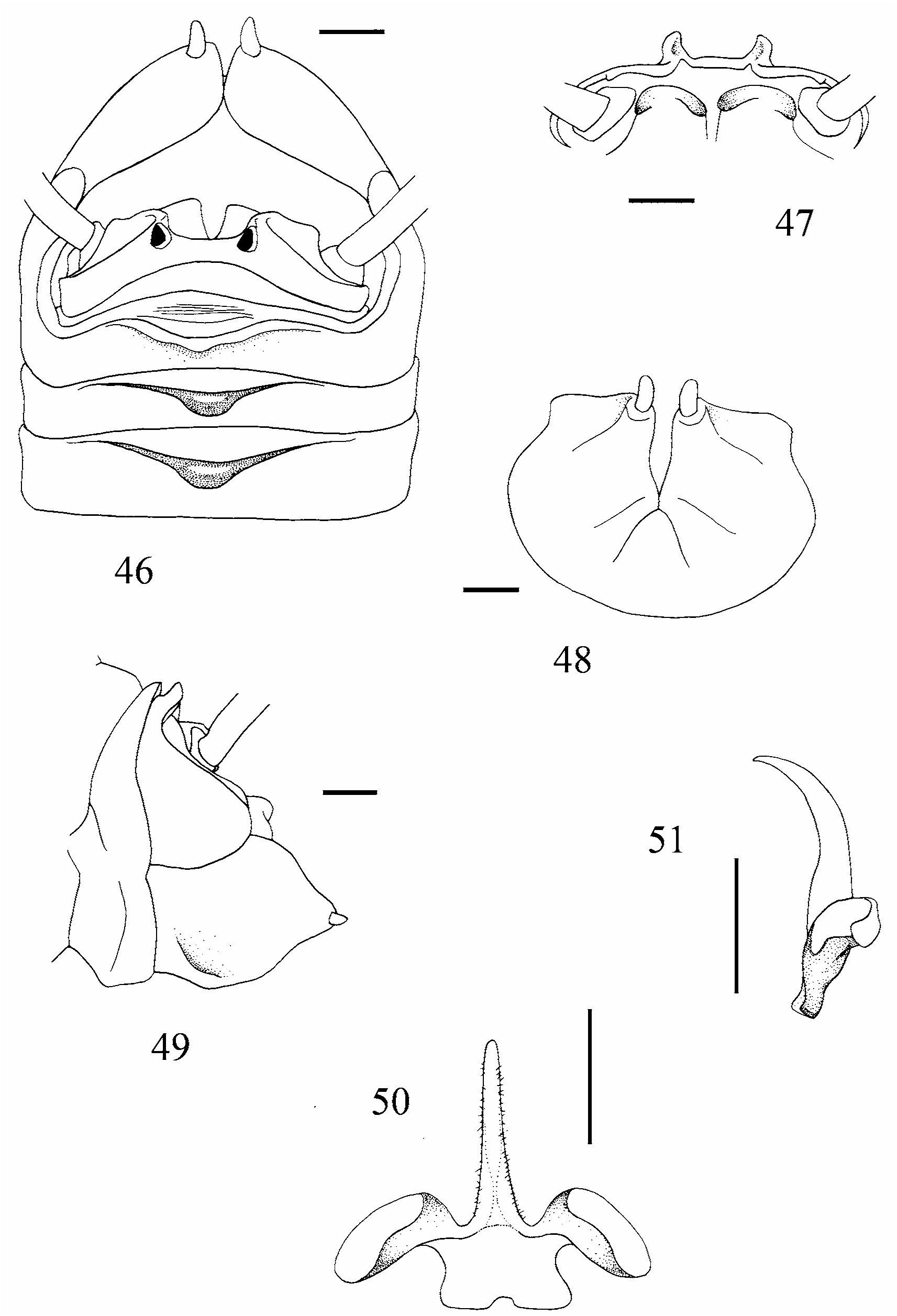
( Figs 46–51 View FIGURES 46–51 )
Also in this case, in order to complete the work of Boudou-Saltet (1972) where only one female of the new species was reported, we describe the D. dalensi male specimen on the basis of new examples we recently collected in the typical locality.
Material examined. Peloponnisos, Argolis, Kefalari, Kephalovrissi cave , 18.VIII.05, M. Rampini, C. Di Russo leg. 2 males, 2 male nymphs and 2 female nymphs ( MZUR, PCR) .
Male description. The species is large and is yellowish-brown in colour with darker posterior edges of the nota and the tergites. The ventral side uniformly lighter in colour. The head has rostral tubercles of the vertex which are dark and protruding, with clear eyespots on the sides, with deep longitudinal incisions. The legs are particularly elongated and testaceous in colour. The femora are slender. Fore tibia with 3/3 spines on both sides of the superior edge and 4/5 spines on both sides of the inferior edge. Mid tibia with 5/7 spines on the superior edge and 3/4 spines on the inferior. The hind tibia with 16/18 spines on the superior edge and 2 on the external sides of the inferior margin. In the 7 th, 8 th and particularly the 9 th abdominal tergites, the posterior edge is convex in the centre and bends backwards. The tenth tergite has two large partially-square lateral lobes with a sinuous anterior edge, which is separated by a large central depression; at the posterior corners of the incision, there are two protruding tubercles, pyramidal in shape, and which have an apex curving inwards ( Figs 46, 47 View FIGURES 46–51 ). The square-shaped paraprocts have two rounded protuberances on the sides of the superior edge which are very sclerotized. Subgenital plate spherical at the base with an incision on the posterior side which runs for half the length of the plate; the rising triangular lateral lobes have superior and ventral edges which are not particularly arched. The styli are well-developed and pubescent, and are twice as long as they are wide ( Figs 48, 49 View FIGURES 46–51 ). The epiphallus is sclerotized. In posterior view, the median process is seen to curve forwards, and is acute at the apex and very elongated, well-sclerotized and light brown in colour; the inferior edge is concave and the basal processes are lighter and perpendicular to the median process. The posterior processes more developed than the anterior processes, larger, wing-like in shape and diverge in a posterior direction ( Fig. 50 View FIGURES 46–51 ). Laterally, the median process of the epiphallus appears thickened in the third proximal and considerably more slender and curved forwards in front of the third distal ( Fig. 51 View FIGURES 46–51 ).
Length (mm): body 22,5; pronotum 4,5; fore femora 14,5; mid femora 14,0; hind femora 24,5; fore tibia 17,0; mid tibia 18,0; hind tibia 34,0; hind tarsus 13,0; 1 st article of hind tarsus 7,0.

| MZUR |
Museo di Zoologia dell'Universita "La Sapienza" |
No known copyright restrictions apply. See Agosti, D., Egloff, W., 2009. Taxonomic information exchange and copyright: the Plazi approach. BMC Research Notes 2009, 2:53 for further explanation.
|
Kingdom |
|
|
Phylum |
|
|
Class |
|
|
Order |
|
|
Family |
|
|
Genus |