
Wrightoporia subavellanea Jia J. Chen & B.K. Cui, 2014
|
publication ID |
https://doi.org/ 10.11646/phytotaxa.175.4.4 |
|
DOI |
https://doi.org/10.5281/zenodo.5149300 |
|
persistent identifier |
https://treatment.plazi.org/id/9A4EED11-DE4D-FFAB-8BF1-4AC836DBFDFC |
|
treatment provided by |
Felipe |
|
scientific name |
Wrightoporia subavellanea Jia J. Chen & B.K. Cui |
| status |
sp. nov. |
Wrightoporia subavellanea Jia J. Chen & B.K. Cui View in CoL , sp. nov. ( Figs. 1–2 View FIGURE 1 View FIGURE 2 )
MycoBank no.: MB 808216
Differs from other Wrightoporia species by annual and resupinate basidiocarps with white rhizomorphs, large pores, narrow and strongly dextrinoid skeletal hyphae, broadly ellipsoid to subglobose and strongly amyloid basidiospores, and slightly thick-walled gloeoplerous hyphae in trama.
Type.— CHINA. Guangxi Autonomous Region: Nanning, Qingxiushan Park , alt. 150 m, on rotten wood of Pinus , 9 November 2009, Dai 11484 (holotype, BJFC!) .
Etymology.— subavellanea (Lat.) referring to the species is somewhat similar to Wrightoporia avellanea (Bres.) Pouzar.
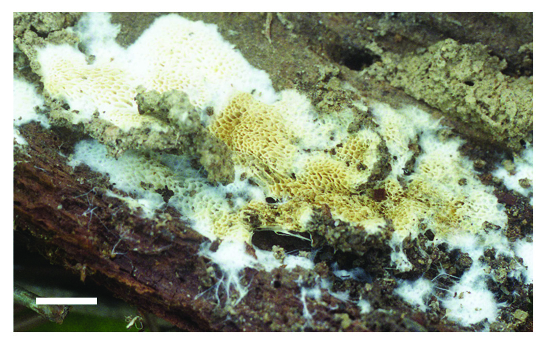
Basidiocarps. —Annual, resupinate, soft when fresh, membranous when dry; up to 13 cm long, 5.5 cm wide, and 4 mm thick at the center; pore surface cream to cream buff when fresh, cream to straw-yellow upon drying; margin with white rhizomorphs; pores circular to irregular, 2–3 per mm; dissepiments thin, entire to slightly lacerate with age; subiculum white to cream, cottony, very thin, about 0.2 mm thick; tubes concolorous with pore surface, membranous, up to 3.8 mm thick.
Hyphal structure.— Hyphal system dimitic; generative hyphae with clamp connections; skeletal hyphae strongly dextrinoid, CB +; tissues becoming brown to dark brown permanently in KOH.
Subiculum. —Generative hyphae infrequent, hyaline, thin-walled, occasionally branched, 1.5–2.8 µm in diam; skeletal hyphae dominant, hyaline to yellowish, thick-walled with a narrow lumen, rarely branched, flexuous, interwoven, 1–2.3 µm in diam.
Tubes. —Generative hyphae infrequent, hyaline, thin-walled, frequently branched, 1.3–2 µm in diam; skeletal hyphae dominant, hyaline to yellowish, thick-walled with a narrow lumen to almost solid, unbranched, often flexuous, interwoven, 0.8–1.5 µm in diam; gloeoplerous hyphae infrequent, slightly thick-walled with granular to oily contents appearing refractive in phase contrast illumination, up to 11 µm in diam, embedded in trama; cystidia absent, but cystidioles present, thin-walled, fusoid, tapering, 13–17 × 2.5–3.5 µm; basidia clavate to barrel-shaped, bearing four sterigmata and a basal clamp connection, 12–20 × 4–5 µm; basidioles in shape similar to basidia, but distinctly smaller.
Spores.— Basidiospores broadly ellipsoid to subglobose, hyaline, thick-walled, finely asperulate, strongly amyloid, CB +, (3.6–)3.8–4.2(–4.7) × (2.6–)2.8–3.2(–3.7) µm, L = 4.1 µm, W = 3.7 µm, Q = 1.31 (n = 60/2).
Additional specimens examined.— CHINA. Hainan Prov.: Changjiang County, Bawangling Nat. Res., alt. 800 m, on rotten trunk of Pinus , 10 May 2009, Dai 10826 (paratype, BJFC!) . Guangxi Autonomous Region: Nanning, Qingxiushan Park , alt. 150 m, on rotten wood of Pinus , 9 November 2009, Dai 11484 & 11492 (paratypes, BJFC!) .

| MB |
Universidade de Lisboa, Museu Bocage |
| BJFC |
Beijing Forestry University |
| CB |
The CB Rhizobium Collection |
| L |
Nationaal Herbarium Nederland, Leiden University branch |
| W |
Naturhistorisches Museum Wien |
| Q |
Universidad Central |
No known copyright restrictions apply. See Agosti, D., Egloff, W., 2009. Taxonomic information exchange and copyright: the Plazi approach. BMC Research Notes 2009, 2:53 for further explanation.
|
Kingdom |
|
|
Phylum |
|
|
Class |
|
|
Order |
|
|
Family |
|
|
Genus |