
Eidmanacris eliethae Nihei
|
publication ID |
https://doi.org/ 10.11646/zootaxa.4018.2.4 |
|
publication LSID |
lsid:zoobank.org:pub:977149FA-06A2-4C65-B7B4-3CEE96729AEF |
|
DOI |
https://doi.org/10.5281/zenodo.6095874 |
|
persistent identifier |
https://treatment.plazi.org/id/987387F1-3D1C-343A-5ADF-0E829DCE1132 |
|
treatment provided by |
Plazi |
|
scientific name |
Eidmanacris eliethae Nihei |
| status |
|
Eidmanacris eliethae Nihei & de Mello n. sp.
Figures 10–12 View FIGURE 10 View FIGURE 11 View FIGURE 12 , 13 View FIGURE 13 .
http://lsid.speciesfile.org/urn:lsid: Orthoptera .speciesfile.org:TaxonName:471411
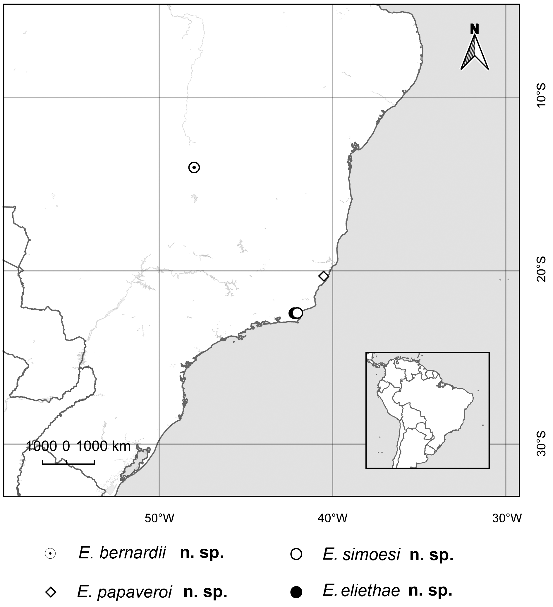
Type locality. Brazil, Rio de Janeiro State, Rio das Ostras municipality, distrito de Rocha Leão.
Type material. Holotype, allotype, 2 males paratypes ( MZSP); 1 male paratype (Departamento de Zoologia, Instituto de Biociências, Universidade Estadual Paulista, Botucatu). Brasil, Rio de Janeiro, Rio das Ostras, distrito de Rocha Leão, 22 Km from Casemiro de Abreu, i-1996, F. A. G. Mello & S. S. Nihei leg. All specimens preserved in alcohol 80%.
Material Examined. Holotype, allotype, paratypes.
Etymology. Named after Dra. Elieth F. S. Cruz, retired professor and herpetologist from the Department of Zoology at Instituto de Biociências, Universidade Estadual Paulista (UNESP, Botucatu campus).
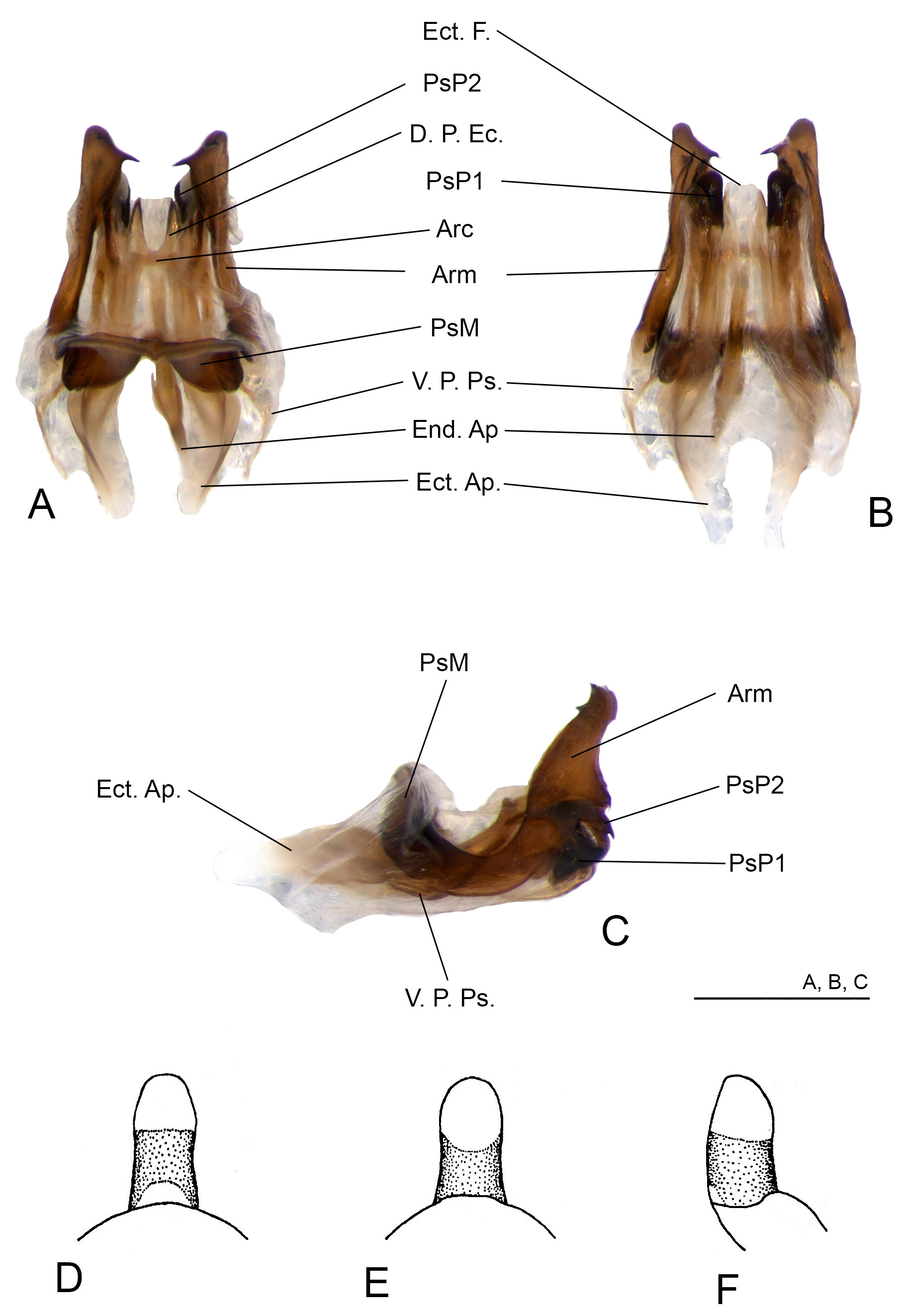
Diagnosis. Within the genus, E. eliethae n. sp. can be recognized by the following characters: male FWs dark brown, broadly rounded, margins light brown; covering metanotal gland area and surpassing the metanotum; secondary veins well visible, medium brown; internal margins touching each other only at anterior half, slightly overlapping; glandular thickening absent. Metanotal gland present, with a cluster of bristles and two short projections with rounded top. Apex of pseudepilhallic arms with two dorsal, and a ventral projection; dorsal projections of the apex pointed, one curved inwards, other up-curved; ventral projection thin, elongate, pointed, almost straight; ectophallic arc located posteriorly to the median part of pseudepiphallus, near the PsP2; dorsal projections of ectophallic invagination (D.P.Ec.) short, sclerotized, clearly separated.
Description.
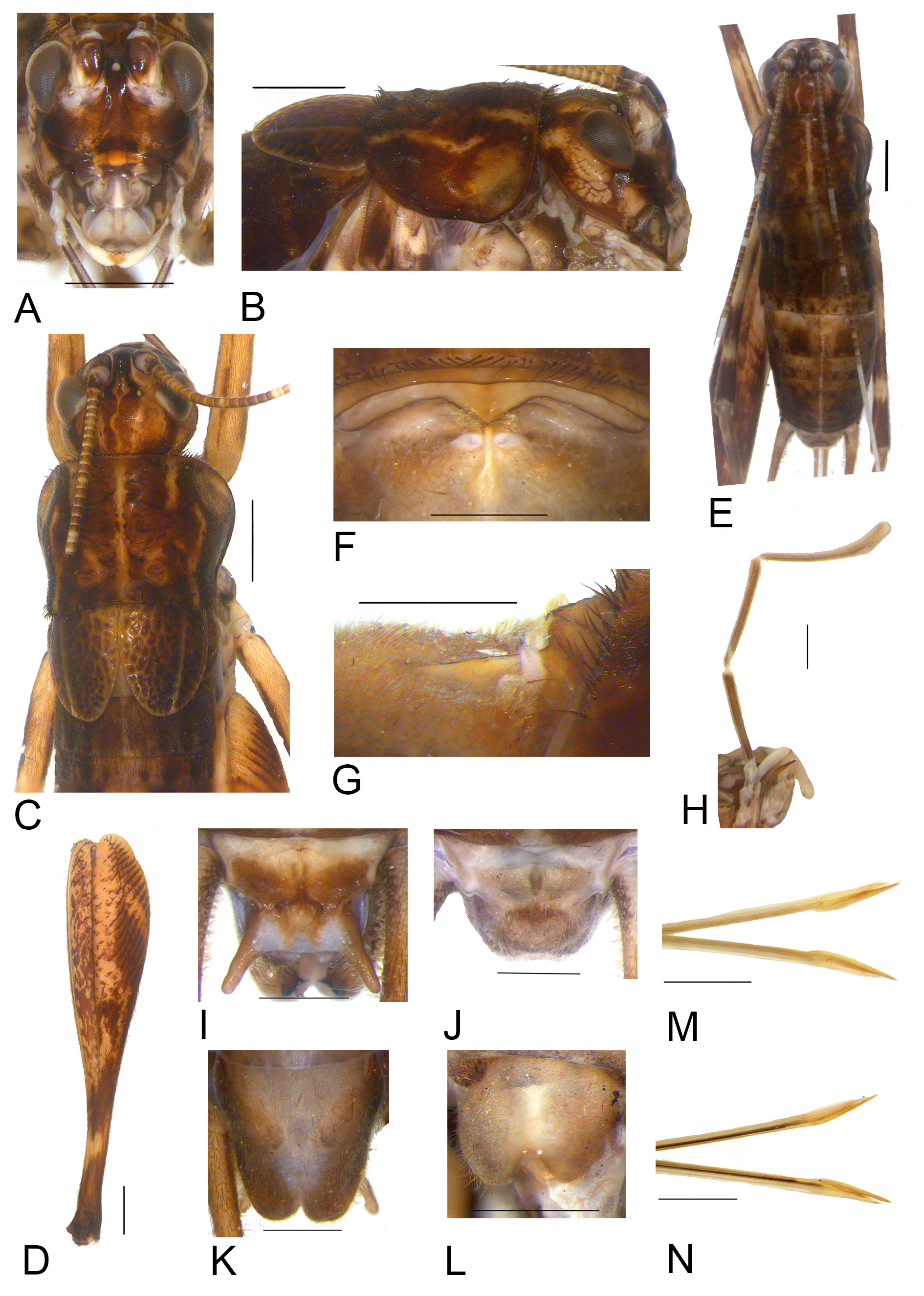
Male. General coloration dark brown, marbled with medium brown. Head. Dorsum pubescent, yellowish to medium brown, with medium and dark brown maculae and spots ( Fig. 10 View FIGURE 10 C); Occiput yellowish brown, with a medium brown band going from the occiput to the margin of each eye ( Fig. 10 View FIGURE 10 C); vertex medium to dark brown, with dark a central yellowish line descending towards the fastigium ( Figs. 10 View FIGURE 10 A, C). Fastigium dark brown, with two row of bristles ( Fig. 10 View FIGURE 10 C); longer than wide, slightly narrowed toward the apex, and narrower than scape, separated from vertex by a thick transversal line forming a “v” ( Fig. 10 View FIGURE 10 C). Frons reddish to medium brown, with a large, central yellowish to dark brown area ( Fig. 10 View FIGURE 10 A). Three large ocelli present ( Figs. 10 View FIGURE 10 A, C); eyes with an unpigmented area on supero-internal angle ( Fig. 10 View FIGURE 10 C). Maxillary palpi medium brown, long, thin, specially the joints 3 to 5( Fig. 10 View FIGURE 10 H); joints 3, 4 and 5 almost same-sized ( Fig. 10 View FIGURE 10 H); apical third of joint 5 curved, medium brown, apex whitish. In frontal view, gena dark brown, and a pair of a well visible diagonal medium brown stripe ( Fig. 10 View FIGURE 10 A); in lateral view, gena light brown, divided by a diagonal dark brown stripe that goes to the posterior part, and ascends toward the top of head, with several thick dark stripes ( Fig. 10 View FIGURE 10 B). Frontoclypeal suture yellowish brown ( Fig. 10 View FIGURE 10 A); upper portion of clypeus dark brown, with a pair of light brown maculae, lower portion greysh; labrum greyish ( Fig. 10 View FIGURE 10 A). Mandible light brown, with internal margins dark brown. Antennal scape light brown, dark brown on inner face ( Figs. 10 View FIGURE 10 A,C); antenomeres medium brown, the anterior portion of the antenna with single shite antenomeres and posterior portion with large unpigmented bands, composed by over 30 antenomeres.
Thorax. Pronotum DD wider than long, dark bworn, with sparse dark spots and maculae, slightly pubescent, divided by a distinct light brown sagittal line, and another four light brown, thick lines ( Fig. 10 View FIGURE 10 C); DD cephalic margin sub-straight, caudal margin slightly concave ( Fig. 10 View FIGURE 10 C); ventro-cephalic angle slightly rounded, ventrocaudal margin gradually ascendant ( Fig. 10 View FIGURE 10 B). Male FWs dark brown, short, but longer than in E. simoesi n. sp., rounded, margins light brown ( Fig. 10 View FIGURE 10 C); covering metanotal gland area and surpassing the metanotum ( Figs. 10 View FIGURE 10 B, C); posterior part of internal margin and apex medium brown connected to a single vein that divides the external part of FW as a lateral field; secondary veins well visible, medium brown; internal margins touching each other only at anterior half, slightly overlapping ( Fig. 10 View FIGURE 10 C); glandular thickening absent. Metanotal gland present, with a cluster of bristles and two short projections with rounded top ( Figs. 10 View FIGURE 10 F, G).
Legs. FI and FII yellowish brown, annulated with medium brown. TI and TII yellowish brown annulated with medium brown; TI with two same-sized apical spurs; TII with two inner apical spurs and one outer, smaller. FIII yellowish brown, with medium brown maculae on inferior portion, and three bands of several thin, diagonal, medium brown stripes on outer face, apical third medium brown, annulated ( Fig. 10 View FIGURE 10 D). TIII medium to dark brown, the apical third yellowish brown; subapical spurs 4/4, with serrulation between and above subapical spurs; apical spurs 3/3, more developed on inner face; inner apical spurs: dorsal one longer (iad), median shorter than dorsal (iam), ventral smallest (iav) (iad>iam>iav); outer apical spurs: median one longer (oam), dorsal (oad) and ventral (oav) almost same-sized (oam>oad>oav). Basitarsus I, II and III yellowish brown.
Abdomen. Sub-cylindrical, dark brown, pubescent, marbled, divided by a thick pale yellow thick sagittal line. Supra anal plate light brown, medium brown in the center and the distal projections, pubescent ( Fig. 10 View FIGURE 10 I); anterior margin sub-straight, lateral ones constricted on median portion, and extended distal projections; posterior margin straight ( Fig. 10 View FIGURE 10 I). Subgenital plate longer than wide, pubescent, light brown in the center and medium brown at the borders; anterior margin slightly concave; posterior margin with short, rounded distal projections ( Fig. 10 View FIGURE 10 K).
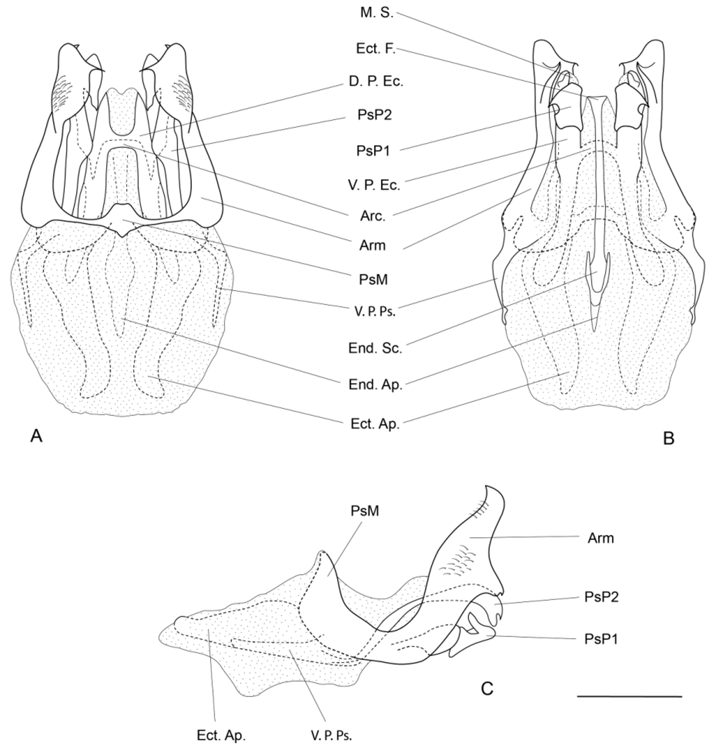
Phallic complex ( Figs. 11 View FIGURE 11 A–C; 12A–C). Pseudepiphallus: median part of pseudepiphallus sclerotized, thin; pseudepiphallic arms sclerotized, hard, with bristles near the apex ( Figs 12 View FIGURE 12 A, C); apex of pseudepilhallic arms with two dorsal, and a ventral projection; dorsal projections of the apex pointed, one curved inwards, other up-curved; ventral projection thin, elongate, pointed, almost straight ( Figs. 11 View FIGURE 11 A, B; 12A, B); pseudepiphallic arms laterally flattened, up-curved; lateral projection absent ( Figs. 11 View FIGURE 11 C, 12C); ventral projections of pseudepiphallic arms (V.P.Ps.) well visible, curved inwards ( Figs. 11 View FIGURE 11 A, B; 12A, B). PsP2 short, with small membranous sphere below apex ( Figs. 8 View FIGURE 8 A, B; 9A, B). Ectophallic invagination: ectophallic apodeme curved, shorter than in E. papaveroi n. sp. and E. simoesi n. sp. ( Figs. 11 View FIGURE 11 A, B; 12A, B); ectophallic arc located posteriorly to the median part of pseudepiphallus, near the PsP2 ( Figs. 11 View FIGURE 11 A, 12A); dorsal projections of ectophallic invagination (D.P.Ec.) short, sclerotized, clearly separated, and longer than in E. simoesi n. sp. ( Figs. 11 View FIGURE 11 A, 12A); ectophallic entirely membranous; in dorsal view, apex of ectophallic fold located between the PsP2 and pseudepiphallic arms, and connected to the D.P.Ec. ( Figs. 11 View FIGURE 11 A, B; 12A, B). Endophallus: latero-posterior projections of the endophallic sclerite similar to E. papaveroi n. sp. and E. simoesi n. sp. ( Figs. 11 View FIGURE 11 B, 12B); medio-posterior projection of the endophallic sclerite sclerotized until half of its length; endophallic apodeme well developed, not surpassing the extremity of the ectophallic apodeme ( Figs. 11 View FIGURE 11 A, B; 12A, B).
Female. Almost same size of male, general coloration similar ( Fig. 10 View FIGURE 10 E). Presence of very small FW, not reaching the metanotum border. Supra anal plate light brown, medium brown in the center, anterior margin slightly concave, posterior margin rounded with bristles ( Fig. 10 View FIGURE 10 J). Subgenital plate medium brown, short, pubescent, wider than long, anterior margin almost straight, posterior margin rounded with a central concavity ( Fig. 10 View FIGURE 10 L). Ovipositor as in figs. 10M, N. Female genitalia. Copulatory papilla as in Figs. 11 View FIGURE 11 D–F.
Obs. Copulatory papilla missed.
Measurements (mm).
Males (n=3): Hw, 3.24±0.09 (3.16–3.34); iod, 1.65±0.14 (1.48–1.73); Lpron, 3.41±0.21 (3.16–3.53); awpron, 3.43±0.09 (3.34–3.53); pwpron, 3.7±0.2 (3.47–3.84); wpron, 4.52±0.25 (4.27–4.77); LFW, 2.34±0.32 (1.98–2.6); wFW, 1.94±0.09 (1.86–2.04); LFIII, 17.9±0.98 (16.95–18.9); wFIII, 3.45±0.15 (3.3–3.6); LTIII, 18.8±0.23 (18.6– 19.05); Ltars 1-III, 5.05±0.38 (4.65–5.4).
Female (n=1): Hw, 3.53; iod, 1.79; Lpron, 3.53; awpron, 3.41; pwpron, 4.09; wpron, 4.71; LFIII, 17.55; wFIII, 3.75; LTIII, 18.45; Ltars 1-III, 4.5; OL, 18.15.

| MZSP |
Sao Paulo, Museu de Zoologia da Universidade de Sao Paulo |
No known copyright restrictions apply. See Agosti, D., Egloff, W., 2009. Taxonomic information exchange and copyright: the Plazi approach. BMC Research Notes 2009, 2:53 for further explanation.
|
Kingdom |
|
|
Phylum |
|
|
Class |
|
|
Order |
|
|
Family |
|
|
Genus |
|
Kingdom |
|
|
Phylum |
|
|
Class |
|
|
Order |