
Hypatopa phoebe Adamski
|
publication ID |
https://doi.org/ 10.11646/zootaxa.3618.1.1 |
|
publication LSID |
lsid:zoobank.org:pub:B548B139-E8D9-4F10-956E-E0001E6C7586 |
|
DOI |
https://doi.org/10.5281/zenodo.6147530 |
|
persistent identifier |
https://treatment.plazi.org/id/985F879D-DF08-7248-C2DD-FE55FB347007 |
|
treatment provided by |
Plazi |
|
scientific name |
Hypatopa phoebe Adamski |
| status |
sp. nov. |
Hypatopa phoebe Adamski View in CoL , new species
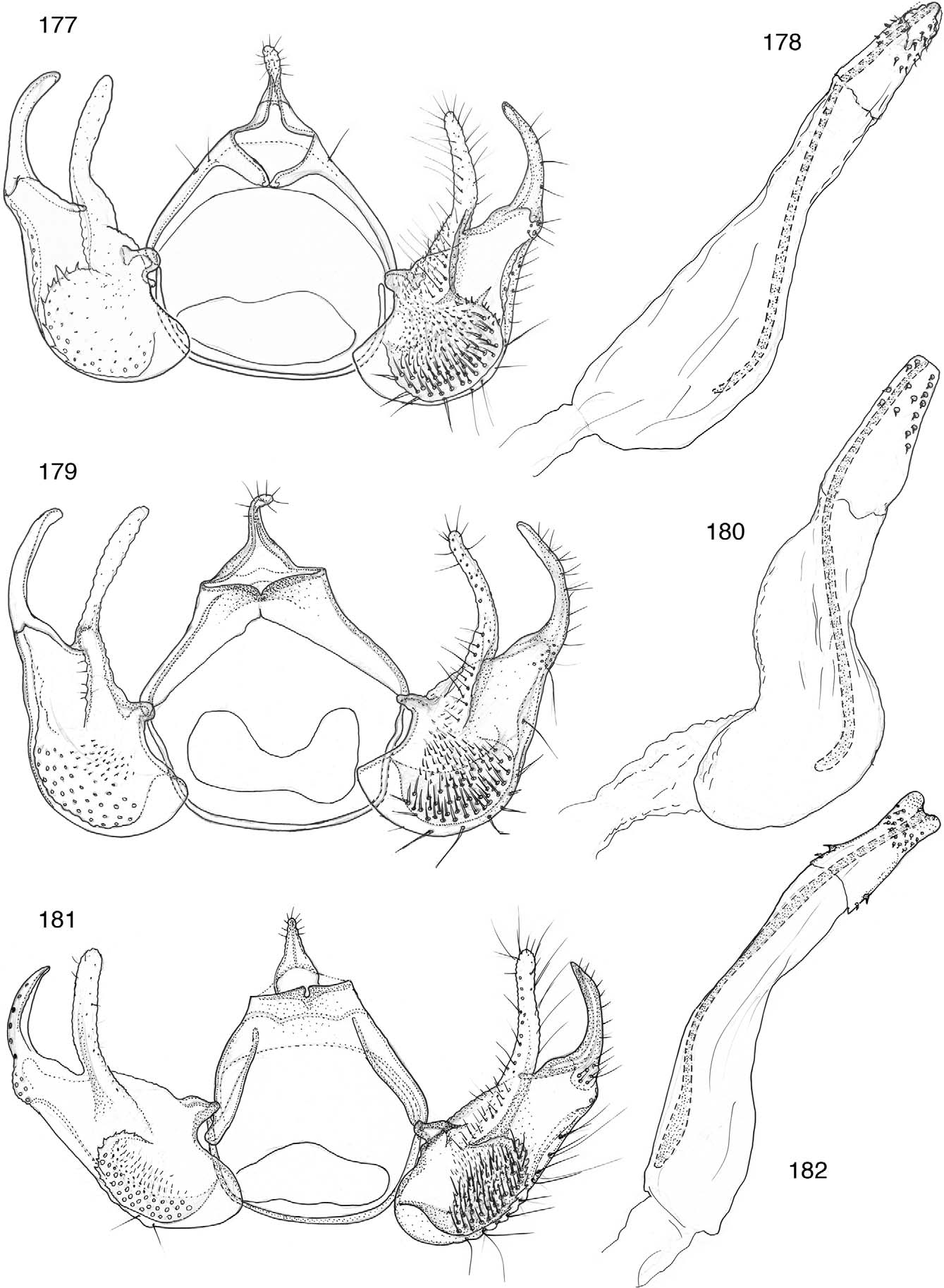
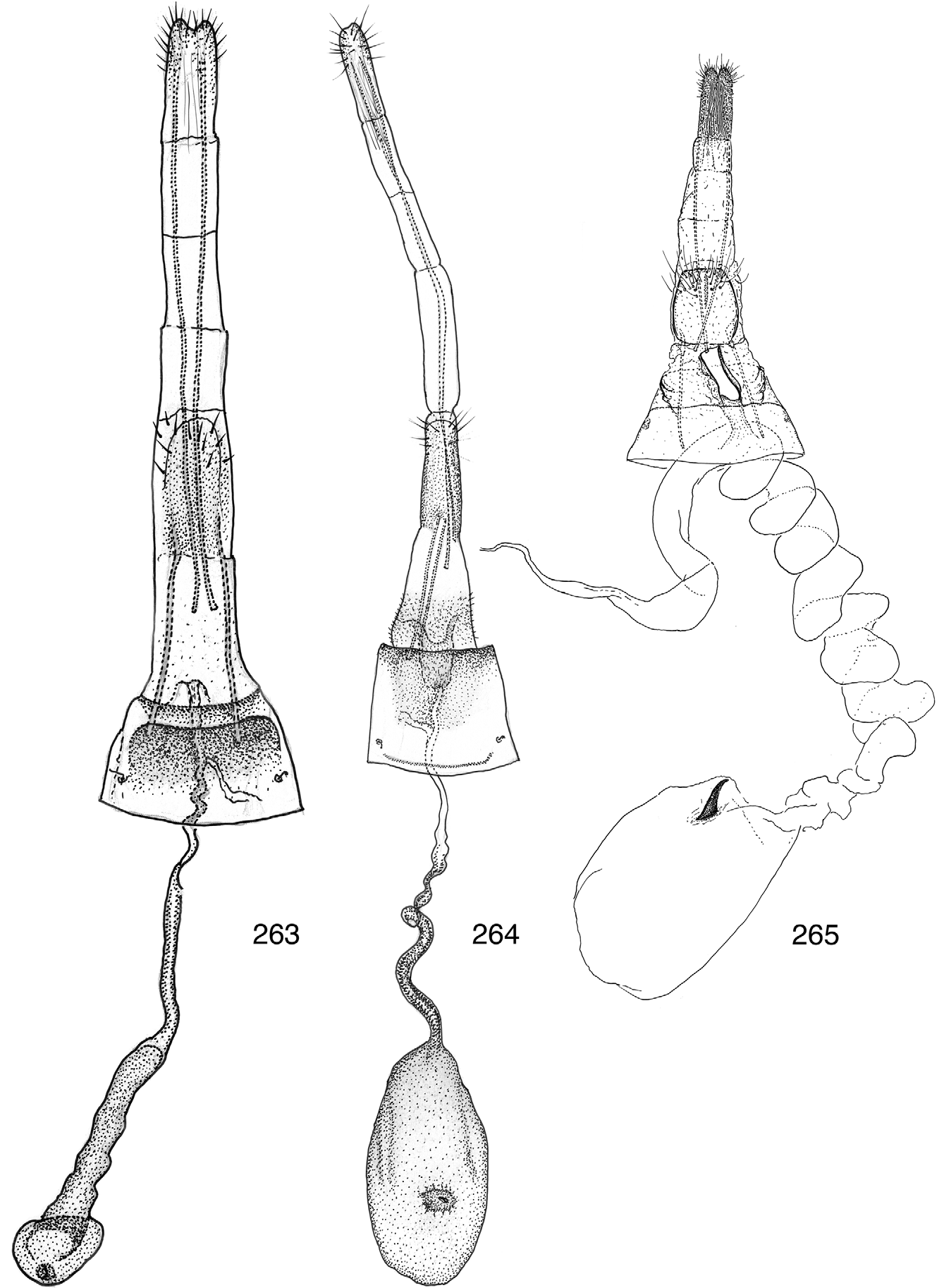
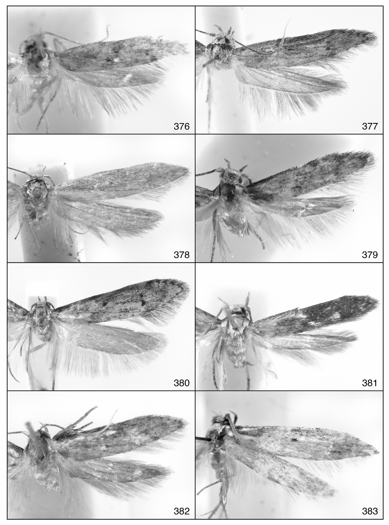
( Figs. 36 View FIGURES 31 – 40 , 181–182 View FIGURES 177 – 182 , 264 View FIGURES 263 – 265 , 377 View FIGURES 376 – 383 , Map 36)
Diagnosis.— Hypatopa phoebe is similar to H. semela in facies but differs from the latter by having a more acutely curved apical process of the ventral part of the valva; and a longer phallus. H. phoebe also has a shallowly crenulate margin of proximal flange of the dorsal part of the valva; a basally broadened anellus with two lateral lobes, and a widely notched apex that is setose on the apical 1/3 and bisetose along the margin near base that are lacking in H. semela .
Description.—Head: Scales on vertex and frontoclypeus grayish brown tipped with pale grayish brown. Outer surface of labial palpus grayish brown intermixed with pale grayish-brown scales along apical margin of segment 2, inner surface paler. Antennal scape pale brown, pecten grayish brown, flagellum grayish brown. Proboscis pale grayish brown.
Thorax: Tegula with basal 1/2 brown, apical 1/2 pale brown; mesonotum with basal 1/5 brown, apical 4/5 pale brown. Legs brown intermixed with pale-brown scales near midsegments and on apical margins of all segments and tarsomeres. Forewing ( Fig. 377 View FIGURES 376 – 383 ): Length 7.5–8.4 mm (n = 12), pale brown intermixed with brown scales, paler basally gradually darkening to apex; cell with three spots, one near middle, two on apical end along crossvein; a short apical portion of radial and medial veins darkly streaked. Undersurface brown. Venation ( Fig. 36 View FIGURES 31 – 40 ) with M3 and CuA1 arising from a common point on distoposterior part of cell; cubital veins divergent from bases with CuA1 straight and CuA2 acutely curved basally. Hindwing: Translucent pale brown. Venation ( Fig. 36 View FIGURES 31 – 40 ) with cubitus 3- branched; M2+M3 branched with CuA1 slightly beyond distoposterior part of cell.
Abdomen: Male genitalia ( Figs. 181–182 View FIGURES 177 – 182 ): Uncus gradually narrowed from widened base to narrowly rounded apex, nearly straight, sparsely setose, shorter than width of anal opening. Gnathos wide, confluent with tegumen, ventroposterior margin emarginate mesially. Sockets of tergal setae not extending beyond midlength of tegumen. Valva divided; ventral part slightly projecting inwardly, near parallelsided, narrowing abruptly, forming large, inwardly curved, apical process; process setose on outer surface, slightly concave on inner margin; ventral margin slightly upturned beyond middle, forming narrow fold to near small setose lobe at base of apical process; dorsal part with apical portion of costa extending dorsally, forming setose digitate process; process broadly curved inwardly; basal ridge of digitate process extending ventrally fusing with dorsolateral ridge of proximal flange; flange subelliptical, sparsely microtrichiate on dorsal 1/4, densely setose on ventral 3/4; margin shallowly crenulate. Juxta bandlike. Vinculum semicircular. Phallus and sclerite of phallus longer than valva; phallus broadly curved from middle; sclerite of phallus acutely curved from 1/3; anellus broad basally, constricted near middle, slightly widening, forming two lateral lobes, apex widely notched, setose on apical 1/3, bisetose along lateral margins near base. Female Genitalia ( Fig. 264 View FIGURES 263 – 265 ): Apophyses posteriores slightly more than 3X longer than apophyses anteriores. Ostium bursae within sparsely microtrichiate membrane, slightly posterior to seventh segment; antrum elongate, deeply emarginate. Posterior margin of seventh sternum straight. Ductus bursae about 3X longer than apophyses posteriores; with two rows of imbricate platelets within anterior 1/2, gradually becoming sparser posteriorly. Inception of ductus seminalis arising from ductus bursae slightly posterior to anterior margin of seventh sternum. Corpus bursae ovoid, more spinulate posteriorly than anteriorly; signum, short laterally flattened, subtriangilar process arising from ovoid base.
Holotype, 3, “Est[ación] La Casona, R[eserva] B[iológica] Monteverde, Prov[incia] Punta[renas], COSTA RICA, 1520 m, Mar[zo] 1994, N. Obando, L-N-253250, 449700, # 2819, “INBio: COSTA RICA: CRI001, 764788 [barcode label], “INBio, 3 Genitalia Slide by D. Adamski, No. 2544 [yellow label].
Paratypes (6 3, 5 ƤƤ): 5 3, same data as for holotype except, “CRI001, 764719, “Slide No. 2545, “Wing Slide No. 7004; “CRI001, 764764, “Slide No. 2560, “Wing Slide No. 7007; “CRI001, 764755, Slide No. 2547; “CRI001, 764761, “Slide No. 2549; “CRI001, 764791, “Slide No. 2557, “USNM 83979; 2 ƤƤ, “ Feb. 1992, “CRI000, 801525, “Slide No. 4525, “USNM 83980; “CRI000, 801587, “Slide No. 4526, “USNM 83981; 1 3, 1 Ƥ, “ Dic. 1992, “CRI001, 358307, “Slide No. 2574; “CRI001, 358294, “Slide No. 4528, “USNM 83982; 1 Ƥ, “ Mar. 1991, “CRI001, 320122, “Slide No. 4527, “USNM 83983; 1 Ƥ, “ Ene. 1994, “CRI001, 867221, “Slide No. 4556, “USNM 83984 [5 in INBio, 6 in USNM].
MAP 36. Distribution of Hypatopa phoebe (●) and H. semela (˔).
Distribution (Map 36). Hypatopa phoebe is known from one collecting site on the Cordillera de Tilarán in west-central Costa Rica.
Etymology. The specific epithet phoebe , is chosen in honor of Phoebe , goddess of the moon.

No known copyright restrictions apply. See Agosti, D., Egloff, W., 2009. Taxonomic information exchange and copyright: the Plazi approach. BMC Research Notes 2009, 2:53 for further explanation.
|
Kingdom |
|
|
Phylum |
|
|
Class |
|
|
Order |
|
|
SuperFamily |
Gelechioidea |
|
Family |
|
|
Genus |