
Qarunavus meyeri Simons & Gingerich, 1974
|
publication ID |
https://doi.org/ 10.1093/zoolinnean/zlad065 |
|
DOI |
https://doi.org/10.5281/zenodo.10474122 |
|
persistent identifier |
https://treatment.plazi.org/id/9803BE57-FF8B-FFAC-FF7C-790E420FFBD4 |
|
treatment provided by |
Plazi |
|
scientific name |
Qarunavus meyeri Simons & Gingerich, 1974 |
| status |
|
Qarunavus meyeri Simons & Gingerich, 1974
Amended diagnosis: Differs from the other ptolemaiids by having the following unique combination of characters: a single small incisor next to a large canine, a submolariform p4 with four cuspids in its talonid, and moderately hypsodont molars with precingulids. Qarunavus meyeri differs further from both Ptolemaia species by having three to four mental foramina, no diastema between the canine and the premolars, and almost equal-sized molars, bearing well-formed and distinct trigonids, with separated protoconid, paraconid, and metaconid, and talonids, with four cuspids. It also differs further from both Cleopatrodon species by having narrower and more hypsodont molars with a well-developed lingual talonid notch (modified asser Gunnell et al. 2010).
Holotype: NHMUK PV M 10189, less hemimandible.
Stratigraphical distribution: ‘Lower fossil wood horizon’, which corresponds to the lower sequence of the Late Eocene to Early Oligocene Gebel ºatrani Formation, Fayum Depression ( Egypt).
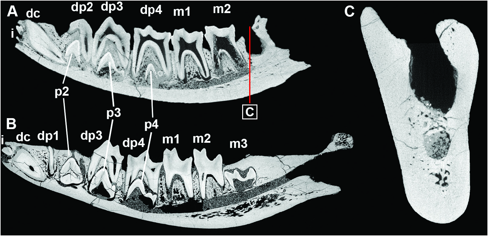
Referred material: NHMUK M 10189, less hemimandible with erupted dc, dp2, dp3, dp4, m1, and m2; SMNS-P-44287, right hemimandible with erupted dc, dp1, dp3, dp4, m1, and m2, and a portion of the less hemimandible with dc, dp1, and dp2, and isolated less p2, part of p3 or p4, m1, and m2, belonging to the same individual.
Remarks: Originally, Schlosser (1911) assigned SMNS-P-44287, including all elements from the SMNS studied herein along with an upper molar and a radius, to P. lyonsi . A few years later, Maưhew (1918) suggested that the mandible represented a distinct taxon. Van Valen (1966, 1967) followed this notion and referred to it as a yet unnamed genus of ptolemaiidan. Simons and Gingerich (1974) erected the new genus and species Q. meyeri , based on another hemimandible (NHMUK M 10189), and included the right mandible of SMNS-P- 44287 in this species. They also mentioned that the two hemimandibles are identical, morphologically and ontogenetically (preserving the same deciduous and permanent teeth), and that they probably belong to the same individual, a notion that was also sustained in later works ( Bown and Simons 1987, Gunnell et al. 2010). However, Schlosser (1911) described, along with the right hemimandible SMNS-P-44287 itself, also an anterior portion of a less hemimandible and some isolated teeth, which he referred to the same individual. This mandibular portion exhibits the dp1 and dp2, and the isolated teeth represent the erupted less m1 and m2, and the unerupted less p2 and the tip of either the unerupted less p3 or p4. Based on the fact that the two mandible portions exhibit the same ontogenetic stage and that their symphyses fit very well together, this less mandibular portion and the right hemimandible housed in the SMNS, both belong to the same individual. Thus, the view of Schlosser (1911) is herein confirmed. Furthermore, the fact that NHMUK M 10189 represents a different individual is also confirmed by slight differences in the morphology and the different developmental stage of the permanent teeth, which infers a different age for this individual, as discussed below. Therefore, the less hemimandible of NHMUK M 10189 represents another distinct individual, which exhibits a very similar morphology and was at a similar ontogenetic age to SMNS-P-44287, though slightly younger.
DESCRIPTION
Mandible
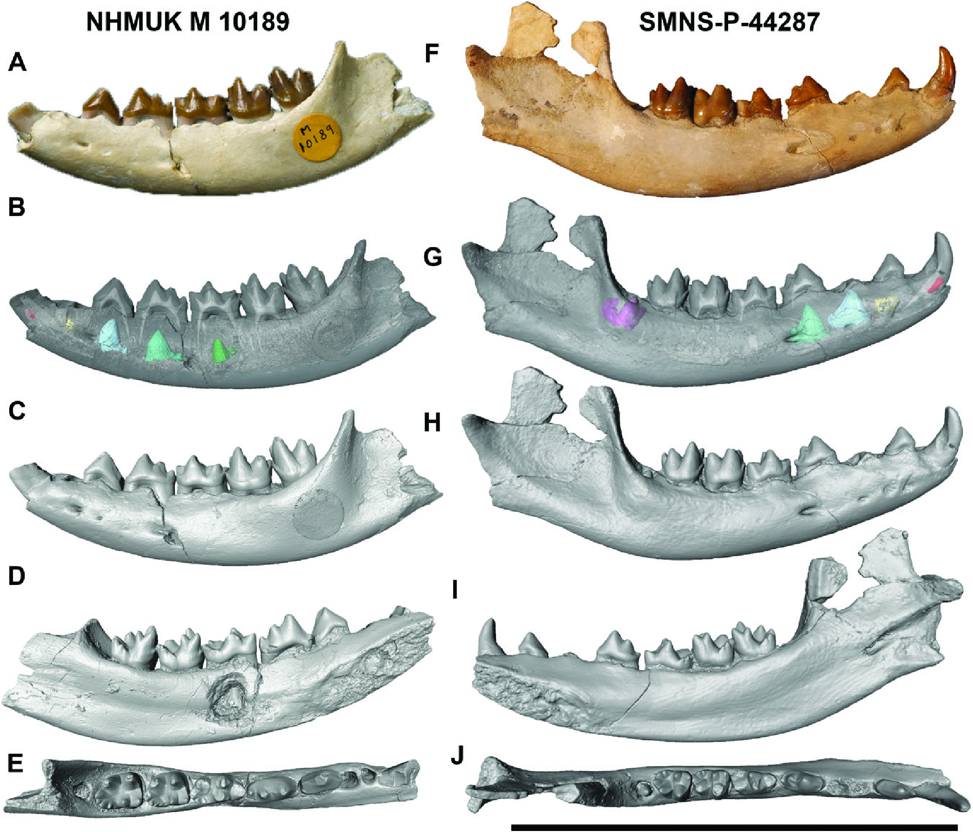
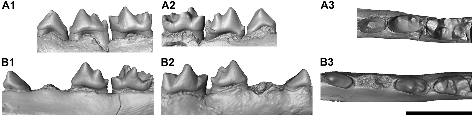
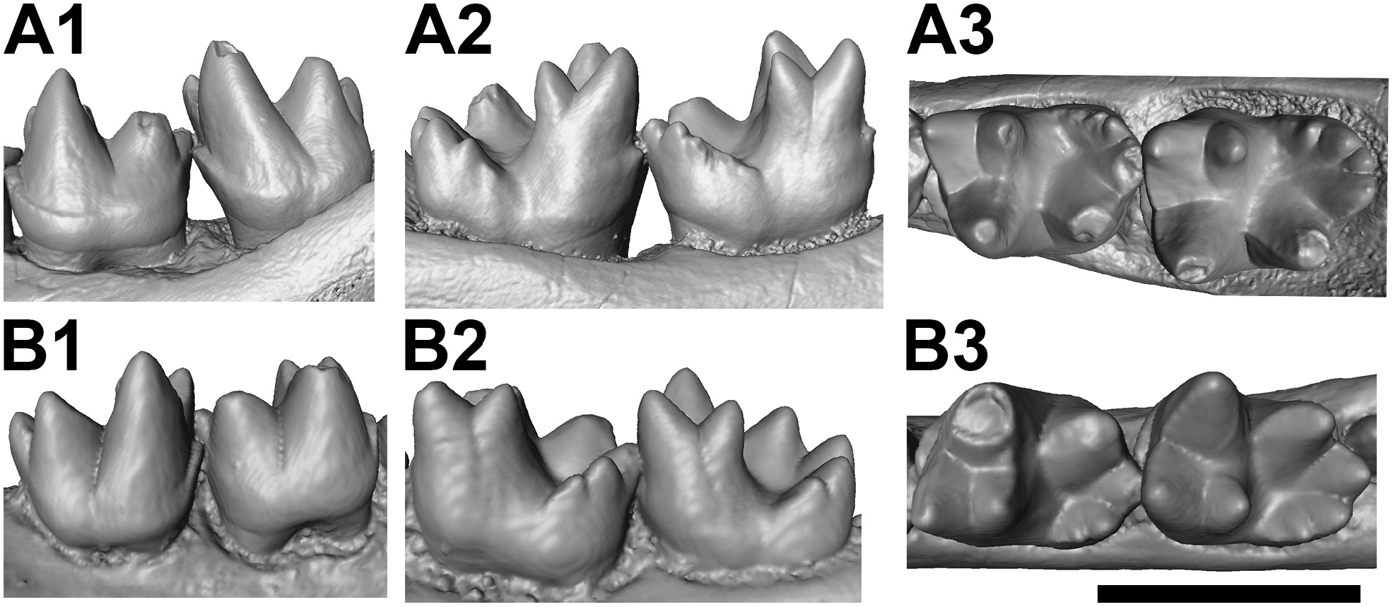
Only two specimens are known from this species, the holotype NHMUK M 10189 ( Fig. 2A View Figure 2 ) and the referred mandible SMNS-P-44287 ( Fig. 2F View Figure 2 ). Both mandibles belonged to juvenile or subadult individuals, as demonstrated by the retained deciduous dentition and the yet unerupted m3 ( Fig. 3 View Figure 3 ). They are morphologically very similar to each other, and have the same dental formula, both have a long and ventrally slightly convex horizontal mandibular ramus and similar dimensions. The horizontal ramus is 14 mm high at the level of the m 1 in both specimens. The tooth row, measured from the mesial end of the dp1 alveolus to the distal end of the m2 alveolus, is 54 mm long in NHMUK M 10189 and 56 mm long in SMNS-P-44287. In both specimens, the symphyseal region is elongated and ends distally at the level of the mesial portion of the dp3. Both mandibles exhibit several mental foramina. In NHMUK M 10189, three are present: two below the dp2 and one slightly larger one below the distal root of the dp3. In SMNS-P-44287, four mental foramina are present, one below the centre of the dp1, two below the dp2 and one somewhat larger one below the contact between the dp3 and the dp4.
The vertical mandibular ramus is only preserved in SMNS-P-44287. However, even in this specimen, the dorsal portion of the coronoid process is broken off, the angular process is broken, and the condylar process is slightly damaged. The angular process is positioned approximately at the same level as the tooth crown bases, but its size cannot be assessed. The condylar process is positioned fairly high, above the level of the teeth. The coronoid process is relatively long and forms a slightly obtuse angle to the horizontal ramus.
Incisor
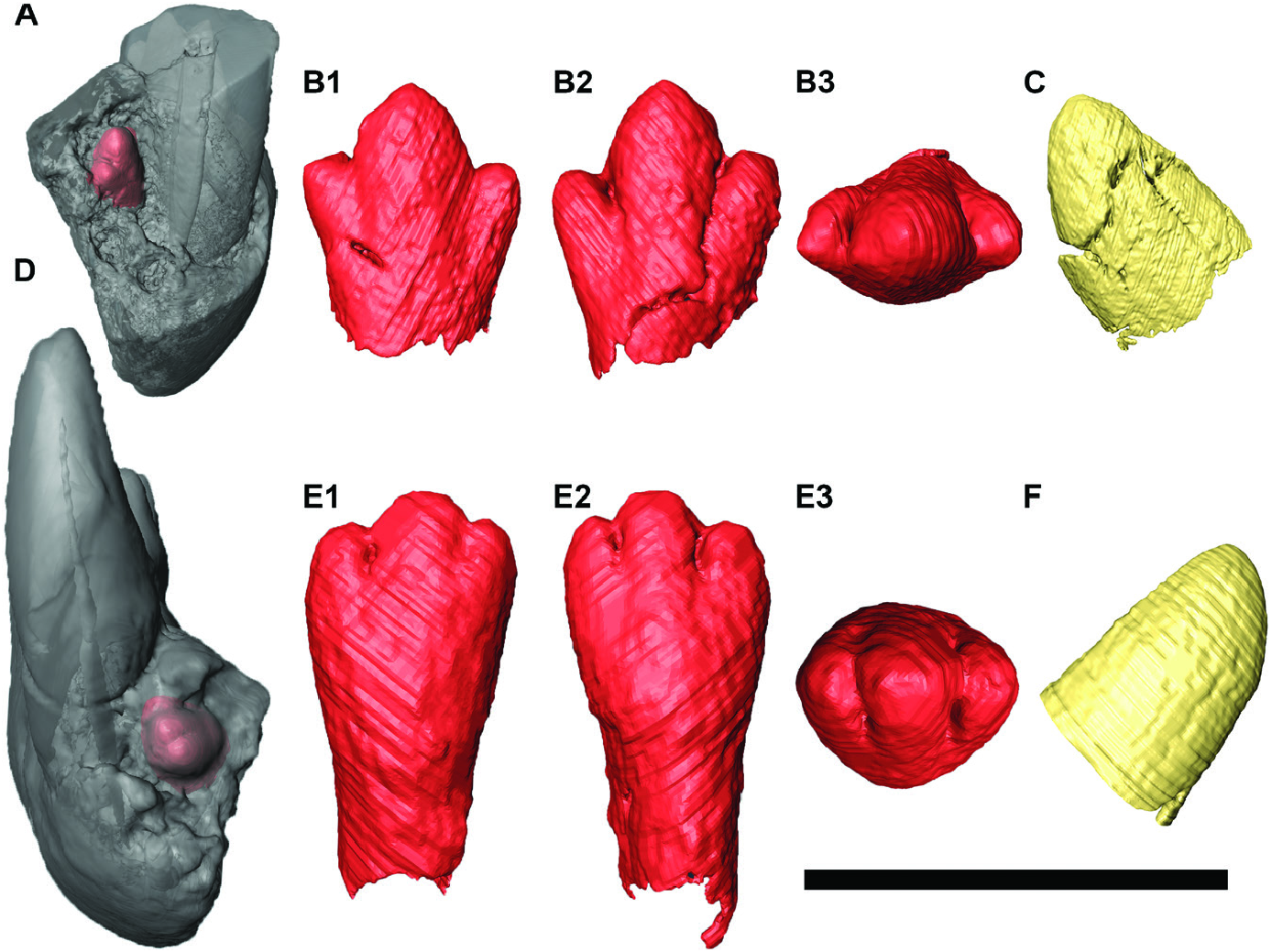
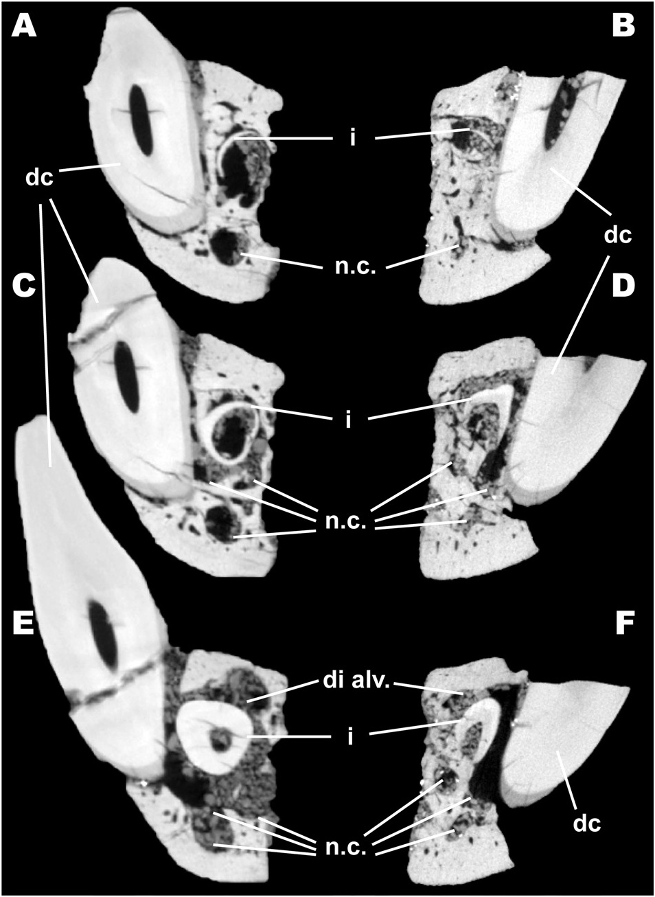
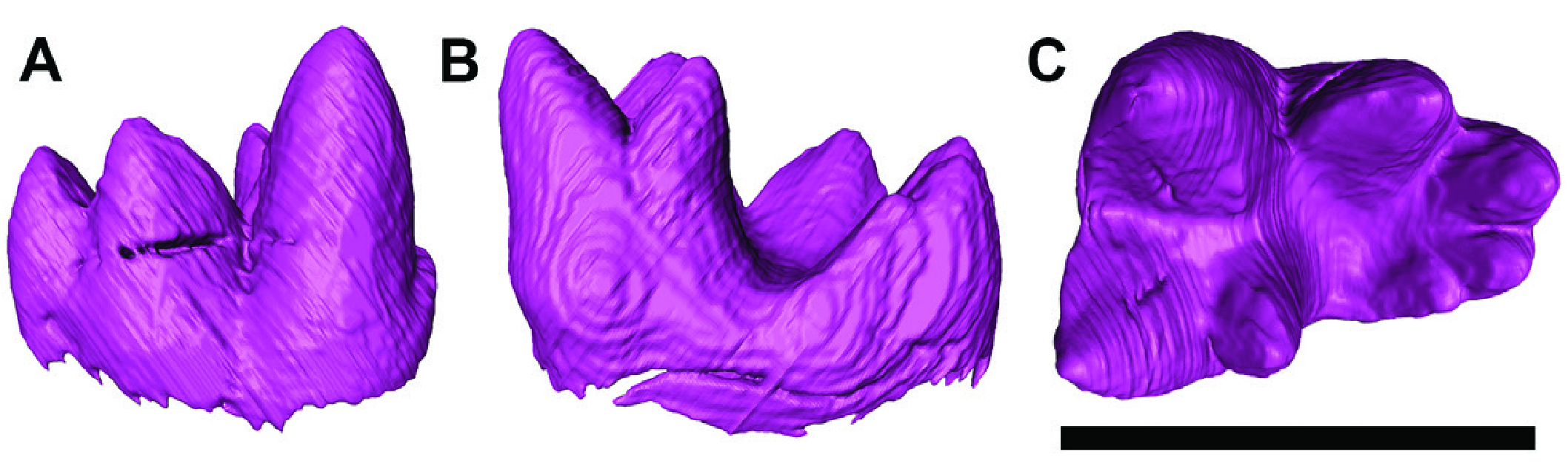
Both specimens ( NHMUK M 10189 and SMNS-P-44287) exhibit a single permanent incisor ( Fig. 4 View Figure 4 ); the tip of the crown is visible due to minor damage to the anterior part of the mandible in both specimens. The anterior portions and the symphyseal areas of both specimens are almost completely preserved, confirming that the lack of any other incisor is not related to damage to the mandible ( Figs 4 View Figure 4 , 5 View Figure 5 ). Consecutive transverse cross-sections at different levels of the symphyseal portion ( Figs 4 View Figure 4 , 5A–F View Figure 5 ) reveal a large cavity and a smaller one ventral to it. The large one is the alveolus for the permanent incisor, which is still preserved inside it. The smaller one is positioned right below the incisor and is even more ventrally placed than the ventral edge of the canine. This thin but long cavity stretches anteroposteriorly beyond the alveolus of the permanent incisor, and we thus identify it as a nutrient canal. In the more anterior cross-sections ( Fig. 5C–F View Figure 5 ), two additional cavities are visible, partially or fully connected with the already mentioned nutrient canal (seen in Fig. 5A, B View Figure 5 ) and therefore, represent branches of the same canal. In the last cross-section ( Fig. 5E, F View Figure 5 ), another cavity is visible that is situated dorsomedially to the incisor. This cavity appears only in the anterior portion of the unerupted permanent incisor and does not connect to any other cavity, beside the alveolus of this tooth. Therefore, it seems most plausible that this cavity represents the posterior portion of the alveolus of the deciduous incisor, which is not preserved. The possibilities of another already formed incisor not being preserved or another developing later in ontogeny seem also very unlikely, as all other permanent teeth are already at least partially formed. Therefore, Q. meyeri bears only a single permanent lower incisor.
In both specimens the incisor has a moderately high, polycuspid crown, bearing three distinct cuspids, a large central one and two smaller ones placed symmetrically laterally to the large one and are separated by two notches. In NHMUK M 10189, the crown is 1.8 mm long and 2.2 mm wide, though the length would have been higher when fully developed, whereas in SMNS-P-44287 the crown of the incisor is 2.4 mm long and 2.4 mm wide. The cuspids are much more distinct and further diverging from each other, with the lateral cuspids forming a slightly greater angle to each other. The cuspids are also taller and much more acute in NHMUK M 10189, whereas in SMNS-P-44287, they are placed much closer to each other, with the two lateral cuspids being almost parallel to, and only slightly smaller than, the central cuspid. The cuspids of SMNS-P-44287 are also completely rounded, without any distinct peak, and are shorter than in NHMUK M 10189. Lastly, in NHMUK M 10189 the tooth is not fully formed, completely lacking its root, whereas in SMNS-P-44287, it is almost fully formed, retaining a single open root.
Canine
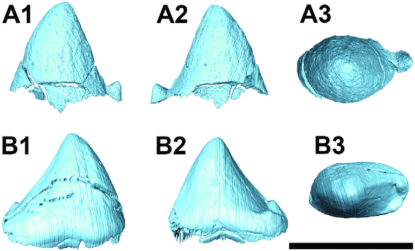
Deciduous: In both specimens, the deciduous canine is at least partially retained within the mandible, with a single root. The end of the root is partially resorbed, through the formation of the permanent canine, making it mesiodistally flaưened. However, its crown is only preserved in SMNS-P-44287, where it is high (10.3 mm), acute, and curved upwards, with its tip turning slightly backwards. The cross-section of the crown at the base is 5.7 mm long and 4.1 mm wide. It bears a long wear facet along the distal side of the crown, probably from wear with the upper deciduous canine; this wear facet is bigger in the right deciduous canine than in the less one in SMNS-P-44287. No other wear facet is visible, which indicates that there was probably no contact between the deciduous canine and the deciduous incisor.
Permanent: In both specimens, the germ of the permanent canine is present, but it has only just started to form, and it is positioned below the dp1. At this stage, the canine comprises a single, simple cone. In SMNS-P-44287, much more of the tooth has already been formed compared with the canine in NHMUK M 10189 and it is 3.8 mm long and 3.1 mm wide.
Premolars
Deciduous: The dp1 is preserved in both mandibular portions of SMNS-P-44287 but missing in NHMUK M 10189 ( Figs 2 View Figure 2 , 6 View Figure 6 ). It has a simple crown, which is 6.9 mm long and 3.6 mm wide, with a single tip that is placed slightly mesially. A faintly visible crest runs from the occlusal tip of the dp1 to its distal crown base and an extremely small distolingual cingulid is present. The dp1 is the only tooth without a permanent replacement, as confirmed in both specimens. Although it is very likely, it is not certain whether the dp1 would have been retained into adulthood as in Cleopatrodon ( Bown & Simons, 1987) , or would have been shed during the course of the animal’s life.
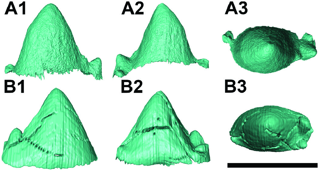
The dp2 is preserved in the less mandibular portion of SMNS-P-44287 and in NHMUK M 10189 (7.9 mm long and 4 mm wide). It is bigger than the dp1, but has a similar morphology, bearing a simple crown with a single tip. The tip is placed slightly mesially to the centre of the tooth in SMNS-P-44287 with a very slight curve dorsally, whereas in NHMUK M 10189, the tip is placed in the centre of the tooth, giving it a more symmetrical profile. A very weakly developed crest runs from the occlusal tip of the dp2 to its distal crown base in both specimens. In SMNS-P-44287, at the crown base, where the small crest ends, a minuscule distal cuspule is present. In NHMUK M 10189, this part is not preserved, probably due to lateral wear with the neighbouring dp3.
The dp3 is present in NHMUK M 10189 (10.1 mm long and 4.6 mm wide) and in the right mandibular portion of SMNS-P-44287 (9.8 mm long and 4.5 mm wide). It is larger than the dp2 and bears a large main cuspid almost in the centre of the tooth. A smaller, but still prominent cuspid is placed behind it, in the middle of the posterior half of the tooth. Moreover, a weak anterior cingulid and a somewhat stronger distal one, bearing a small cuspule. The pre- and postcingulids are stronger and more extensive in NHMUK M 10189 and the cuspid on the postcingulid is much stronger in the NHMUK specimen, being developed into a dorsally projecting cuspid, in comparison to the smaller and more distally projecting cuspule in SMNS-P-44287.
The dp4 is present in both NHMUK M 10189 (10.7 mm long and 5.6 mm wide) and SMNS-P-44287 (10.9 mm long and 5.6 mm wide). It is the largest tooth in the mandible, being longer than the molars, in both specimens. It is molariform, with a well-formed trigonid and talonid, morphologically very similar to the molars, but with a shorter crown. It is heavily worn, representing the most worn tooth in the mandible. The metaconid is the only cuspid that is almost completely preserved and is therefore the tallest one, in its current state. However, when unworn the protoconid was the largest cuspid and the paraconid might have been similarly large as the metaconid, since the paraconid is almost completely worn off. The paraconid is placed mesially, much more so than in the molars, and is distinct from the other two cuspids of the trigonid, in contrast to the molars. In contrast, the protoconid and the metaconid are placed much closer to each other and are connected for most of their height. The talonid comprises four cuspids, similar to the molars. These cuspids decrease in size from the buccal-most one to the lingual-most one and probably represent in that order the hypoconid, hypoconulid, entoconid, and entoconulid.
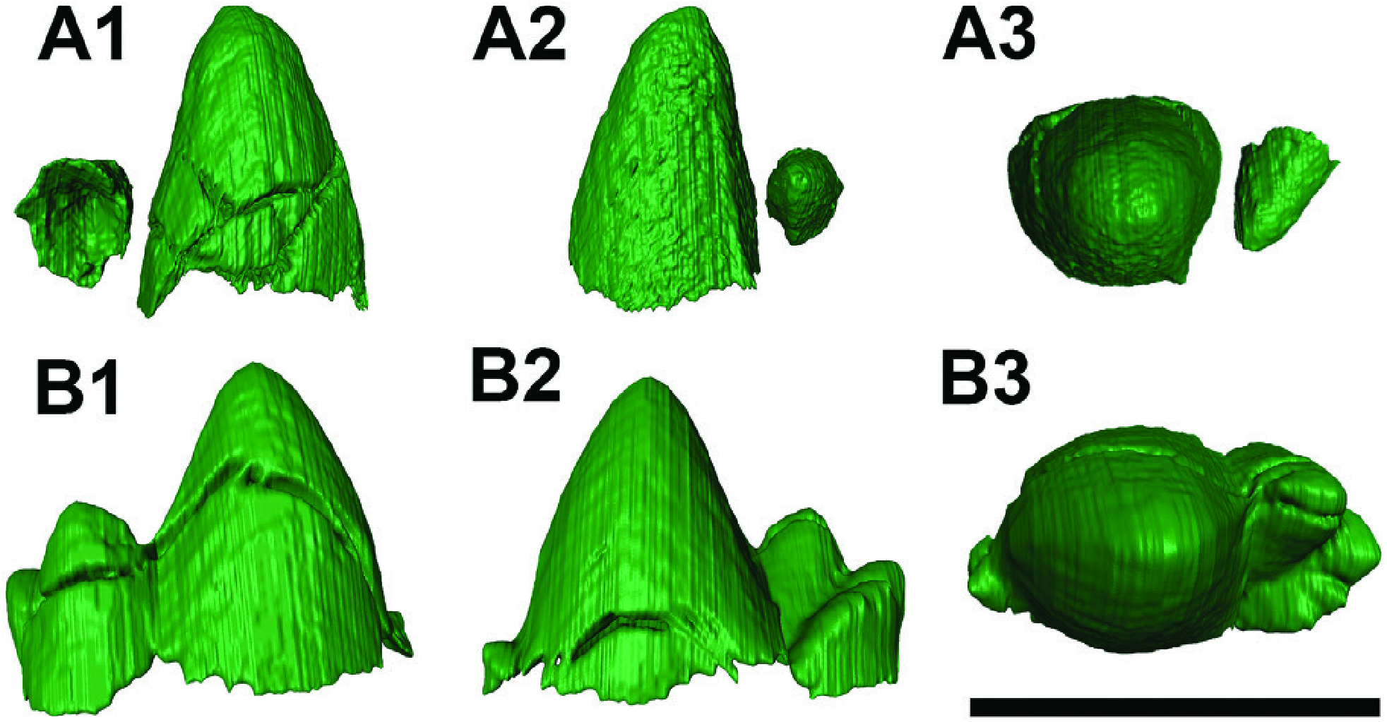
Permanent: The p2 is preserved in both specimens ( Fig. 2B, G View Figure 2 ). In fact, in SMNS-P-44287, the less p2 is isolated from the mandible, due to damage to the mandible, whereas the less p2 of NHMUK M 10189 and the right p2 of SMNS-P-44287 are still contained inside the mandible and their morphology can only be studied through the μCT-scans of the specimens ( Fig. 7 View Figure 7 ). In NHMUK M 10189, the not fully formed p2 has a length of 7.4 mm and a width of 3.9 mm. In SMNS-P-44287, the crown of the p2 has a length of 9.3 mm and a width of 5.3 mm. Both exhibit a large central cuspid, representing the protoconid. The distal side of the protoconid is slightly concave. A weakly developed crest runs from its tip to its distal base, connecting the protoconid with a small cuspid that probably represents the hypoconid. No mesial cingulid is visible, although a distal cingulid is present in SMNS-P-44287. The distal cingulid bears the hypoconid and a smaller cuspid, situated more lingually, which may represent the entoconid. In NHMUK M 10189, the ventral part of the tooth is not fully developed and lacks the basal part of the enamel, therefore the existence of cingulids cannot be assessed. However, the hypoconid is preserved and is much larger than in SMNS-P-44287.
The p3 is preserved within the mandibles of both specimens ( Fig. 8 View Figure 8 ). It is similar to the p2, but slightly larger and bears much stronger distal cuspids and a mesial cingulid. In NHMUK M 10189, the not fully formed p3 is 8.7 mm long and 4.5 mm wide. In SMNS-P-44287, the p3 is 9.9 mm long and 5.4 mm wide. In NHMUK M 10189, the mesial cingulid bears a prominent cuspule, which is absent in SMNS-P-44287. Additionally, in NHMUK M 10189, the hypoconid is much larger and a faintly visible cuspule is present in front of the hypoconid.
The p4 is also preserved in both mandibles, NHMUK M 10189 and SMNS-P-44287 ( Fig. 9 View Figure 9 ). However, in NHMUK M 10189 most of the tooth had not yet developed by the time of the animal’s death ( Fig. 9A View Figure 9 ). More specifically, only two cuspids, which were not yet connected by mineralised tissue, are visible; one is much larger than the other. The larger one corresponds to the protoconid, whereas the smaller one most probably represents the hypoconid. The p4 of SMNS-P-44287 is 10 mm long, about 5.5 mm wide and submolariform. It exhibits a large central cuspid, which corresponds to the protoconid, and a distinct talonid with a well-developed hypoconid and three additional, smaller cuspids decreasing in size from the most buccal to the most lingual one, similarly to the molars. Mesially, a cingulid is present that bears a prominent cuspid. The protoconid and the hypoconid seem to be connected by a faintly visible crest. The preserved portion of the hypoconid in NHMUK M 10189 indicates that it would have been bigger, with a sharper tip than in SMNS-P-44287 when complete.
Molars
Three molars would be present in each specimen. However, in NHMUK M 10189, the m3 is not preserved, as demonstrated by the empty alveolus behind the erupted m2 ( Fig. 3C View Figure 3 ). In SMNS-P-44287, all three molars are preserved but the m3 is just starting to erupt ( Figs 2B View Figure 2 , 11 View Figure 11 ), with only the protoconid protruding, and its roots have not formed yet. In both specimens, the m1 and m2 are fully erupted and have already begun being worn ( Fig. 10 View Figure 10 ). In NHMUK M 10189, the m1 is 9.5 mm long and 7.1 mm wide and the m2 is 9.1 mm long and 7.2 mm wide. In SMNS-P-44287, the m1 is 9.7 mm long and 7.2 mm wide, the m2 is 9.7 mm long and 7.3 mm wide, and the m3 is 9.1 mm long and 6.7 mm wide.
All three lower molars are almost identical and have a distinct trigonid and talonid ( Figs 9 View Figure 9 , 10 View Figure 10 ). The trigonid has relatively high crowns. The protoconid is the largest cuspid, being significantly larger than the paraconid and the metaconid. The paraconid is placed rather distally, being fused to the protoconid and the metaconid along most of their height and only separating approximately 2 mm below the crown tip of the paraconid. Well-formed valleys separate the cusps from each other, while a low paracristid connects the paraconid with the protoconid. In the m 1 in both specimens, a very small preprotocristid is fused to a similarly well-developed postmetacristid, thereby connecting the protoconid with the metaconid. The metaconid is slightly smaller than the paraconid in all teeth. A precingulid is present in all lower molars. In the m1 of NHMUK M 10189, the precingulid extends towards the buccal side of the tooth, covering the mesial half of the tooth, below the trigonid.
The talonid is well separated from the trigonid, with well-formed lingual talonid notches, and comprises several smaller cuspids that are connected to each other. These cuspids probably correspond to the hypoconid, hypocinulid, entoconid, and entoconulid, from the buccal-most one to the lingual-most one and reduce in size in this order. A slight cristid obliqua is present in all molars. The m2 of NHMUK M 10189 is the only tooth that has an additional cuspid between the hypoconulid and the entoconid ( Fig. 9A View Figure 9 3 View Figure 3 ).
| NHMUK |
Natural History Museum, London |
No known copyright restrictions apply. See Agosti, D., Egloff, W., 2009. Taxonomic information exchange and copyright: the Plazi approach. BMC Research Notes 2009, 2:53 for further explanation.