
Phyllodicola Delamare Deboutteville & Laubier, 1961
|
publication ID |
https://doi.org/ 10.11646/zootaxa.4579.1.1 |
|
publication LSID |
lsid:zoobank.org:pub:A4015309-D9B3-4BB7-ABCB-B88A1F8CE5FC |
|
persistent identifier |
https://treatment.plazi.org/id/97720E2D-FFF0-D607-CBF7-BBB904B3F7BF |
|
treatment provided by |
Plazi |
|
scientific name |
Phyllodicola Delamare Deboutteville & Laubier, 1961 |
| status |
|
Genus Phyllodicola Delamare Deboutteville & Laubier, 1961 View in CoL
Syn: Phyllocola Delamare Deboutteville & Laubier, 1960 (preoccupied)
Non Phyllocola Gistl, 1848 (Coleoptera)
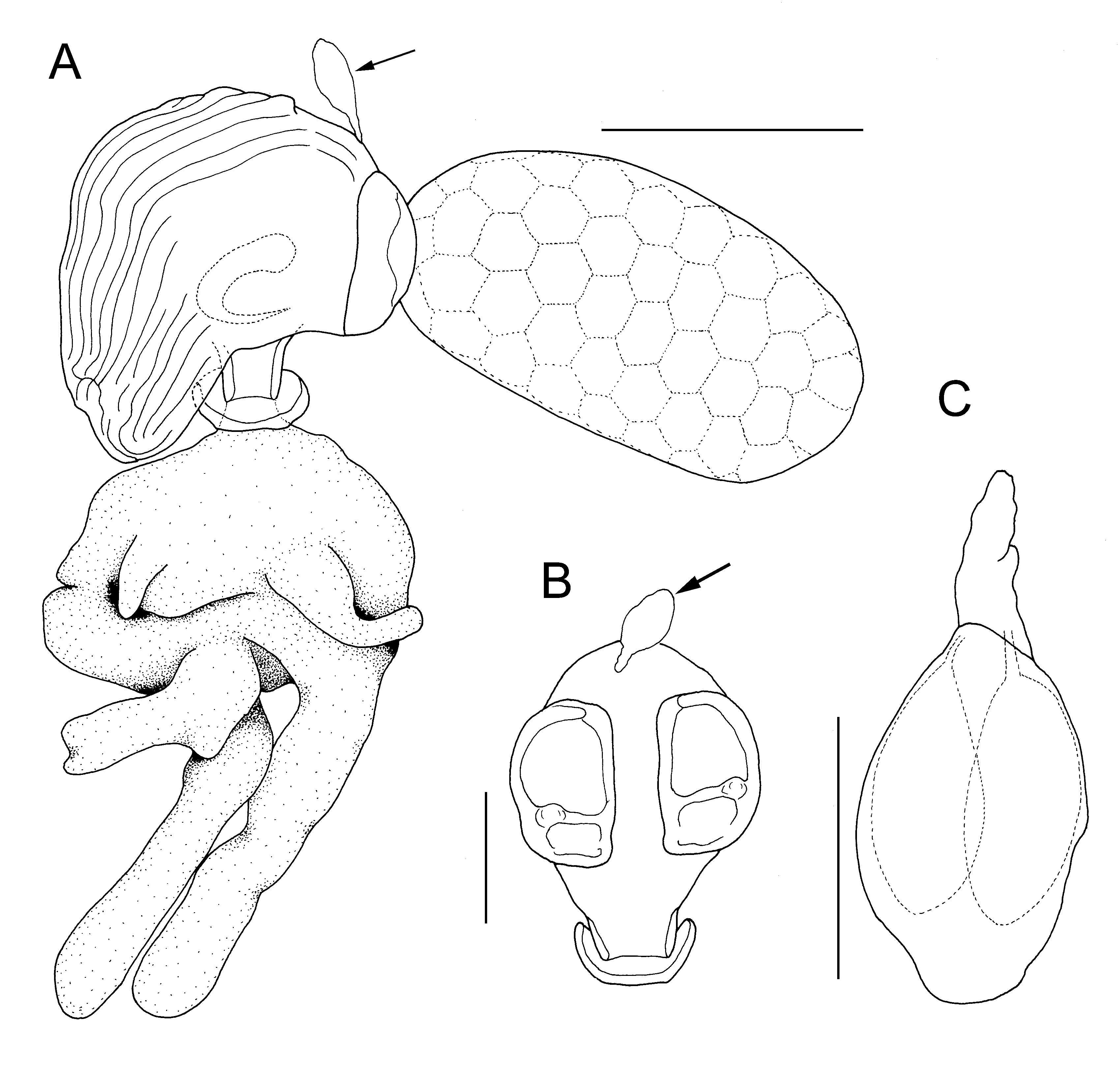
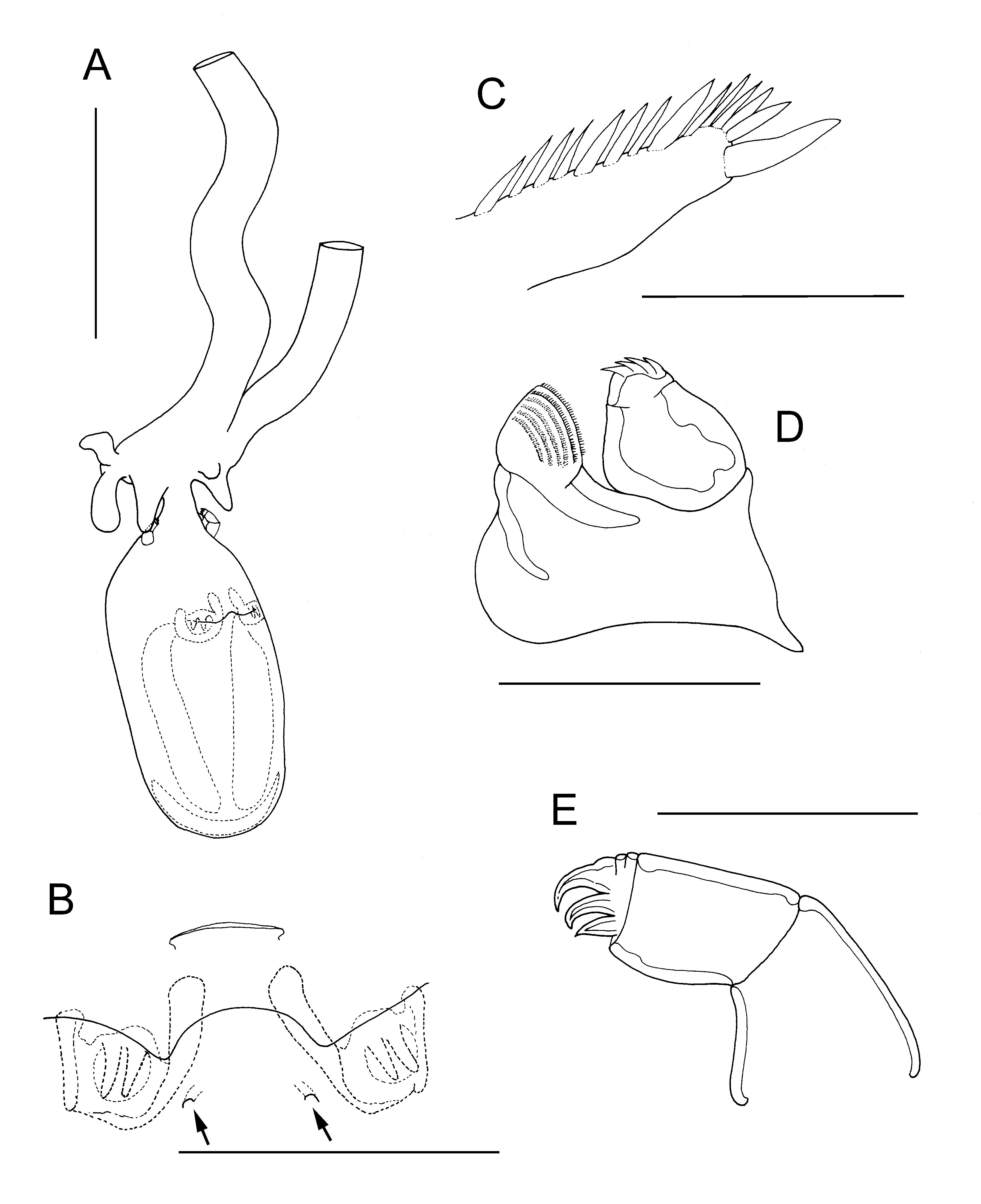
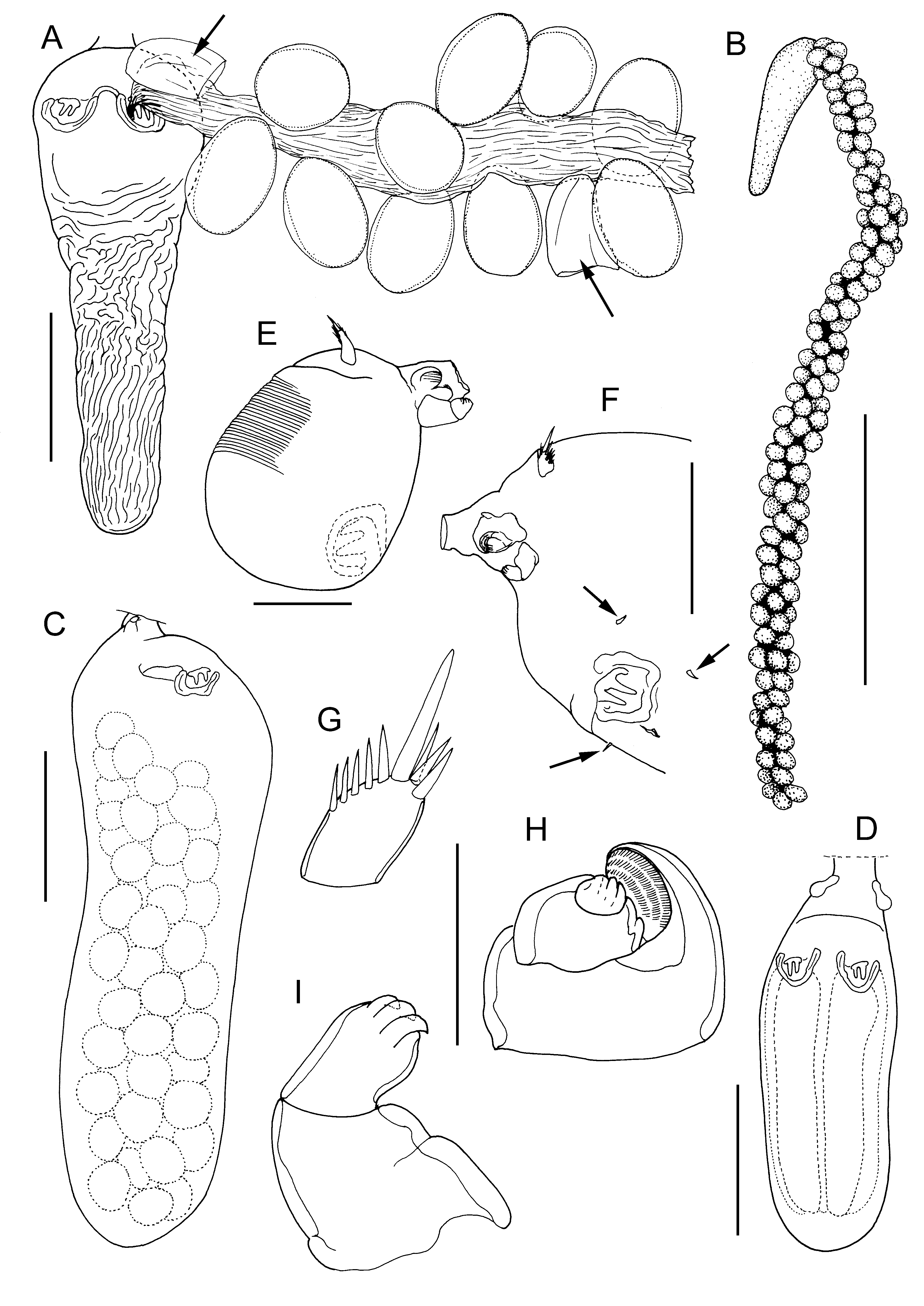
Diagnosis. Adult female body (ectosoma) highly transformed, unsegmented, attached via short broad stalk to endosoma embedded within host. Ectosoma ovoid, about 400 to 900 µm in length ( Fig. 9A View FIGURE 9 ), varying with reproductive state (cf. Fig. 13 View FIGURE 13 A–C). Anus lacking. Paired genital apertures present, located on ventral surface anterior to mid-level; each aperture with two strong tooth-like elements ( Fig. 11B View FIGURE 11 ). Paired copulatory pores (arrowed in Fig. 11B View FIGURE 11 ) located on ventral surface adjacent to genital apertures. Egg strings elongate, with large eggs attached to central axial filament (cf. Fig. 13A View FIGURE 13 ). Antennule ( Fig. 9C View FIGURE 9 ) located dorsal to stalk, unsegmented, tapering towards tip, armed with array of stout setal elements along anterior and apical margins. Antenna ( Fig. 11D View FIGURE 11 ) with broad basal segment bearing, posteriorly, robust, indistinctly 2-segmented endopod armed with 4 apical claws; basal segment produced anteriorly into lobe ornamented with minutely spinulose lamellae. Maxilliped ( Fig. 11E View FIGURE 11 ) subchelate, comprising robust basal segment plus subchela consisting of long proximal segment and compound apical segment armed with 3 strong claws on distal margin. Stalk originating on underside of ectosoma in oral region. Endosoma taking form of two elongate rootlets penetrating body cavity of host, with several short digitiform processes originating around base of primary rootlets. Male unknown. Nauplius lecithotrophic, with nauplius eye.
Type species: Phyllodicola petiti (Delamare Deboutteville & Laubier, 1960)
Remarks. The development of the ectosoma can be reconstructed by reference to Laubier (1961): in the very early stage a vestigial abdominal process is still visible ( Laubier 1961: Fig. 3b View FIGURE 3 ) but this is absorbed during subsequent development so the mature adult female has an ovoid ectosoma lacking any trace of a defined abdomen.
We interpret the uniramous antenna slightly differently from Laubier (1961). We interpret the ramus as ornamented with claws that are flattened and very broad at the base so they are rather lamellate in form. The compound apical segment of the maxilliped shows bands of thickening ( Fig. 11E View FIGURE 11 ) which appear to correspond to at least two incompletely expressed segments. It is this segmentation pattern that provides the evidence supporting the interpretation of these limbs as maxillipeds rather than maxillae, since the maxilla exhibits a maximum of two expressed segments in all members of the poecilostome lineage within the order Cyclopoida ( Huys & Boxshall 1991) .

No known copyright restrictions apply. See Agosti, D., Egloff, W., 2009. Taxonomic information exchange and copyright: the Plazi approach. BMC Research Notes 2009, 2:53 for further explanation.
|
Kingdom |
|
|
Phylum |
|
|
Class |
|
|
Order |
|
|
Family |
|
Kingdom |
|
|
Phylum |
|
|
Class |
|
|
Order |
|
|
Family |