
Lanassicola arcticus, Boxshall & O’Reilly & Sikorski & Summerfield, 2019
|
publication ID |
https://doi.org/ 10.11646/zootaxa.4579.1.1 |
|
publication LSID |
lsid:zoobank.org:pub:A4015309-D9B3-4BB7-ABCB-B88A1F8CE5FC |
|
DOI |
https://doi.org/10.5281/zenodo.5927058 |
|
persistent identifier |
https://treatment.plazi.org/id/97720E2D-FFC7-D633-CBF7-BC6D0105F2AA |
|
treatment provided by |
Plazi |
|
scientific name |
Lanassicola arcticus |
| status |
gen. et sp. nov. |
Lanassicola arcticus View in CoL gen. et sp. nov.
Type material: Holotype ovigerous ♀ from Lanassa venusta (Malm, 1874) , Stjernsund, Stn 12-1 (70.27983°N, 22.4015°E), depth 402 m, 15 August 2012; collected by A. Sikorski; NHMUK Reg. No. 2015.465. 3 paratype ♂♂ attached to holotype female; same locality and habitat data. GoogleMaps
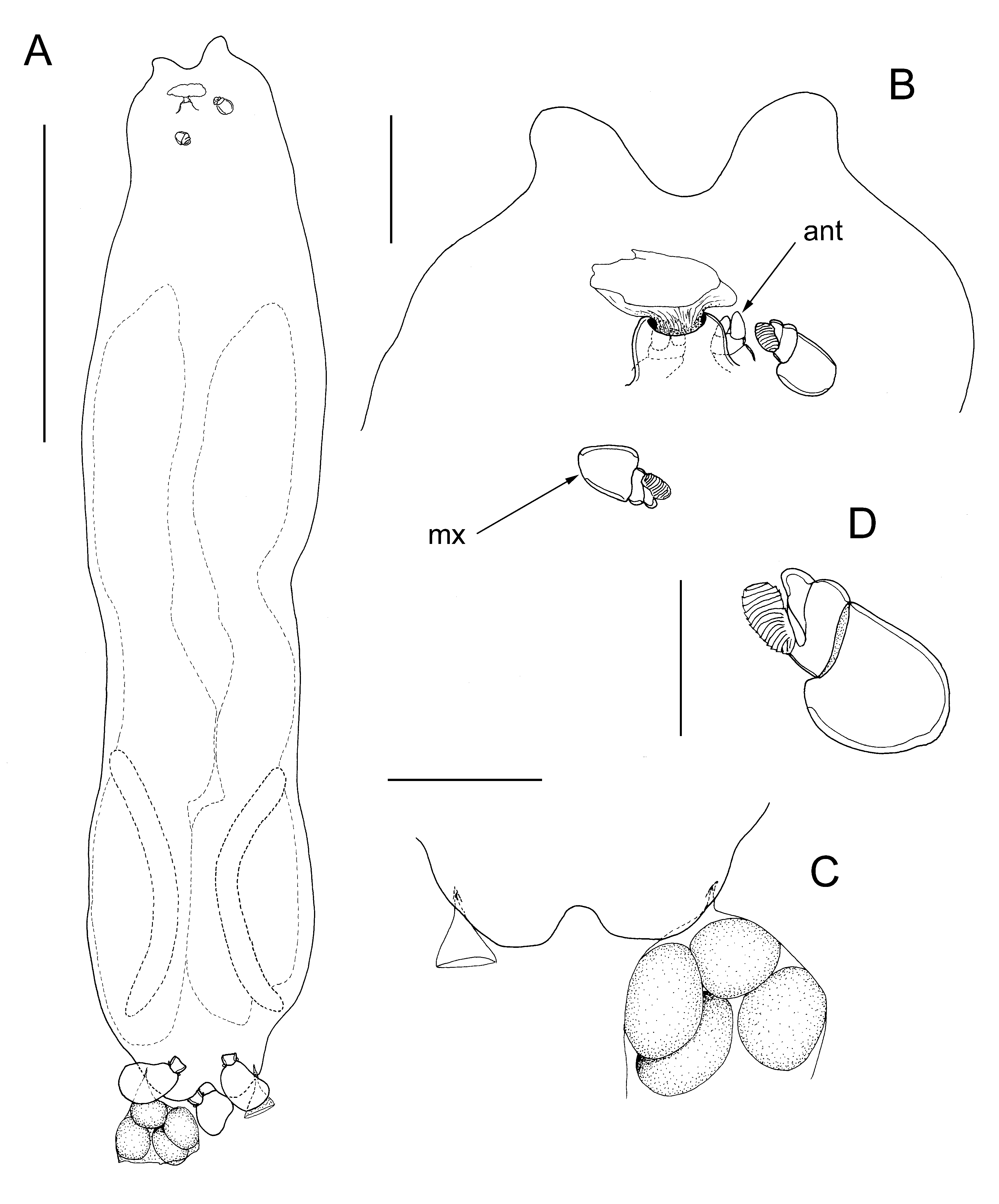
Differential diagnosis. Female body ( Fig. 17A View FIGURE 17 ) with elongate ectosoma, about 5.8 times longer than wide, connected via short stalk to small bulla within host. Ectosoma dorsoventrally flattened, about 2.19 mm in length and with maximum width of 0.38 mm in anterior half. Frontal margin produced into paired, slightly asymmetrical lobes, probably representing antennules ( Fig. 17B View FIGURE 17 ). Lateral margins weakly sinuous, widest in anterior half, narrowing slightly in mid-region, becoming wider posteriorly. Posterior margin terminating in paired posterolateral lobes bearing genital apertures ( Fig. 17C View FIGURE 17 ). Stalk located on mid-ventral surface close to frontal margin ( Fig. 17B View FIGURE 17 ), connecting to bulla (endosoma). Bulla small, expanding within host, incomplete. Paired antennae located close to ventral line just anterior to origin of stalk ( Fig. 17B View FIGURE 17 , ant). Each antenna 2-segmented; first segment unarmed, second segment bearing paired distal adhesion pads. Maxillae located posterior to stalk ( Fig. 17B View FIGURE 17 , mx); each maxilla 2-segmented, with robust first segment and broad second segment bearing 2 distal pads, one strongly corrugated, one smooth ( Fig. 17D View FIGURE 17 ). Cement glands slender, curved, about 0.4 mm in length, located in posterior quarter of ectosoma.
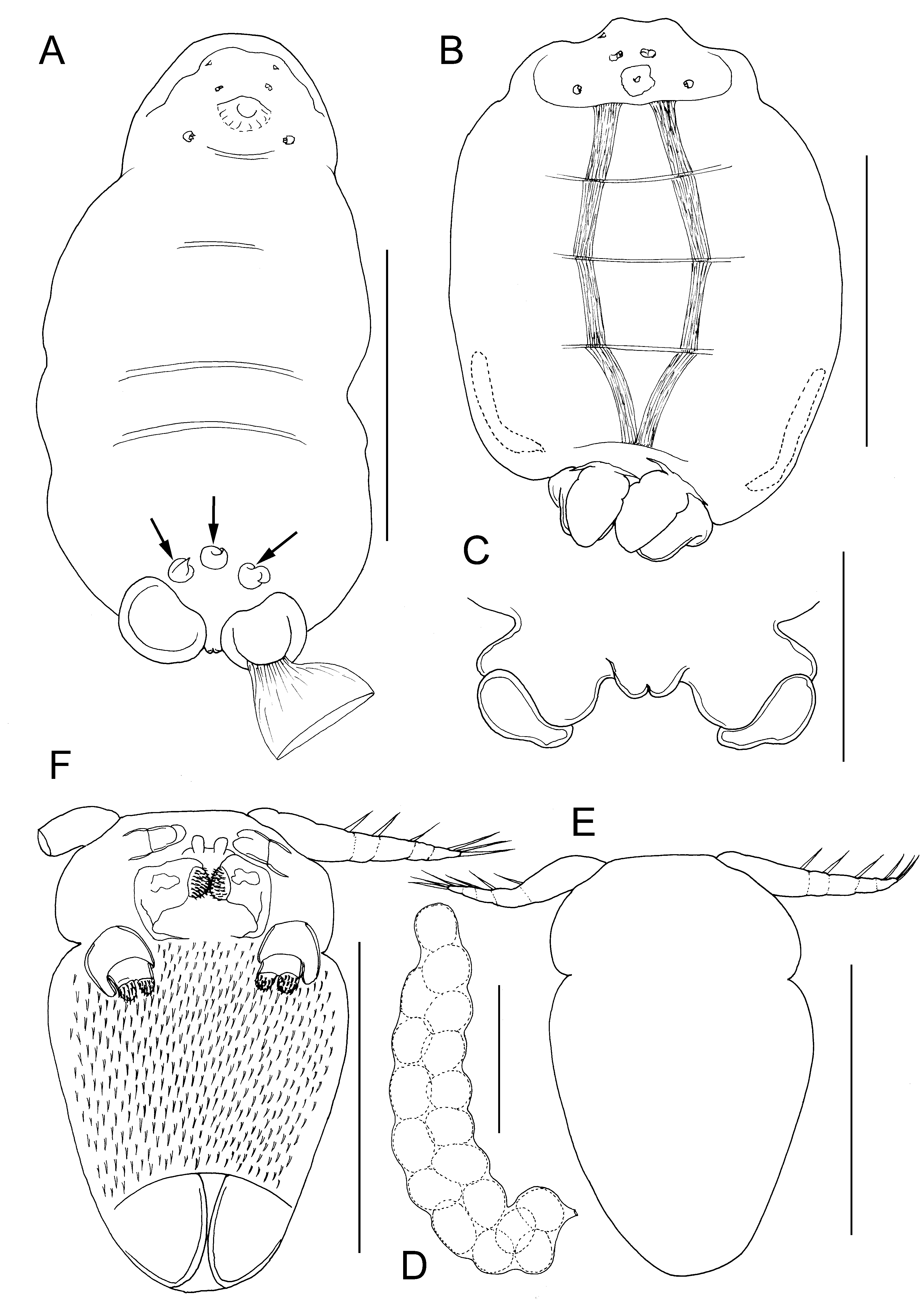
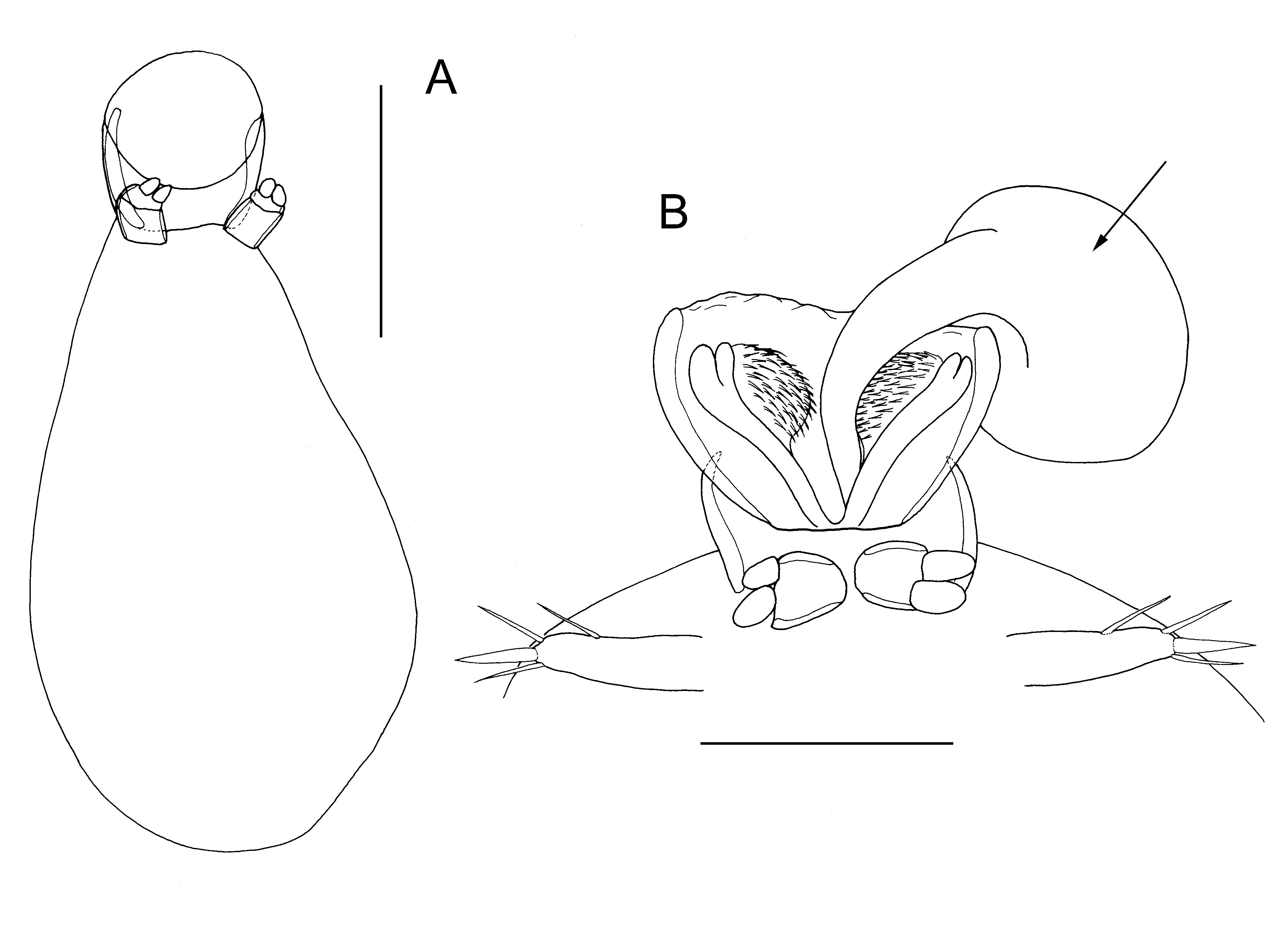
Male sac-like ( Fig. 18A View FIGURE 18 ), comprising anteriorly-directed oral funnel plus lobate trunk about 126 µm in length; trunk pear-shaped with narrow anterior end, broadening posteriorly, maximum width of about 77 µm at about two thirds of distance along trunk. Paired antennules located anterodorsally; unsegmented ( Fig. 18B View FIGURE 18 ), tapering, armed with 4 setae along anterior margin and on apex. Paired antennae located on frontal margin either side of midline ( Fig. 18B View FIGURE 18 ). Oral region modified into funnel-like structure located anteriorly. Oral funnel enclosing paired spinulate pads. Maxillae similar in structure to those of female, located posterior to oral funnel on ventral surface ( Fig. 18A View FIGURE 18 ). Male discharging twisted, conical spermatophores from oral funnel; each spermatophore with globular basal part (arrowed in Fig. 18B View FIGURE 18 ) attached to surface of female, and tapering distal tubule.
Etymology. The name of the new species, arcticus , alludes to the type locality within the Arctic Circle.
Remarks. The male in figure 18B is in the process of discharging a tapering conical spermatophore. This spermatophore is being extruded via a funnel-like structure in the oral region of the male, and the circular base that adheres to the female emerges first. The spermatophore is attached in the genital region of the female. The spinulate pads in the oral region of the male form part of the extrusion mechanism in Lanassicola gen. nov., and we infer this mechanism is also exhibited by Melinnacheres which has the same oral morphology in the male ( Fig. 14F View FIGURE 14 ) and the same shape of spermatophore ( Fig. 14A View FIGURE 14 ). This mode of spermatophore transfer, where spermatophores are extruded anteriorly through an opening in the cephalothorax of the male is similar to that described for the members of the family Herpyllobiidae , although the bottle-shaped male herpyllobiids are considerably more reduced and lack specialised structures such as the spinulate pads present in these saccopsine males.
The adult female is positioned in a longitudinal indentation in the host’s body, near its anterior end, lying between the worm and its tube, with its head end directed down the tube towards the posterior end of the host.
| NHMUK |
Natural History Museum, London |
No known copyright restrictions apply. See Agosti, D., Egloff, W., 2009. Taxonomic information exchange and copyright: the Plazi approach. BMC Research Notes 2009, 2:53 for further explanation.
|
Kingdom |
|
|
Phylum |
|
|
Class |
|
|
Order |
|
|
Family |
|
|
Genus |