
Toxoplasma gondii, (Nicolle & Manceaux, 1908)
|
publication ID |
https://doi.org/ 10.1016/j.ijppaw.2023.03.005 |
|
DOI |
https://doi.org/10.5281/zenodo.10914355 |
|
persistent identifier |
https://treatment.plazi.org/id/930287D6-FFAA-FF8D-FC92-CA1DEEC15AA7 |
|
treatment provided by |
Felipe |
|
scientific name |
Toxoplasma gondii |
| status |
|
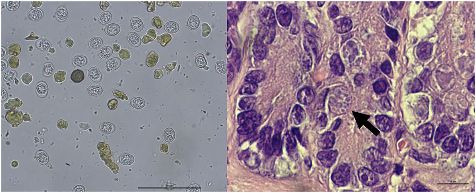
3.2. Detection of T. gondii oocysts and enteroepithelial stages
T. gondii -like oocysts were found in 3/176 (1.7%, 95% CI: 0–3.6) faecal samples ( Fig. 2 View Fig ). All three samples tested positive for T. gondii - DNA and negative for H. hammondi . All the three lynx with oocysts in faecal samples were juvenile animals, with two of them being antibodypositive. T. gondii enteroepithelial stages were detected in small intestine sections from two of the animals (W20_8385 and W21_4446) ( Fig. 2 View Fig ). In the third animal (W14_3480) such evaluation was compromised by autolysis.
3.3. PCR on fresh tissue samples
A total of 11 out of 150 analysed fresh tissue samples from 10 out of 92 (10.9%, 95% CI: 4.5–17.2) lynx, from which fresh tissue samples were available, were positive for T. gondii -DNA by qPCR. Positive samples were 7/88 skeletal muscle, 2/26 heart muscle and 2/36 brain tissue. One lynx tested positive in two tissues (skeletal and heart muscle).
No known copyright restrictions apply. See Agosti, D., Egloff, W., 2009. Taxonomic information exchange and copyright: the Plazi approach. BMC Research Notes 2009, 2:53 for further explanation.
|
Kingdom |
|
|
Phylum |
|
|
Order |
|
|
Family |
|
|
Genus |