
Lurymare utarum Marcus 1952
|
publication ID |
https://doi.org/ 10.11646/zootaxa.3873.5.3 |
|
publication LSID |
lsid:zoobank.org:pub:687DC4E0-9B78-4AF0-9DD2-8B868E3B8EB5 |
|
DOI |
https://doi.org/10.5281/zenodo.6143928 |
|
persistent identifier |
https://treatment.plazi.org/id/927D87F1-FFD8-320C-FF78-7E726B61FC68 |
|
treatment provided by |
Plazi |
|
scientific name |
Lurymare utarum Marcus 1952 |
| status |
|
Lurymare utarum Marcus 1952 View in CoL
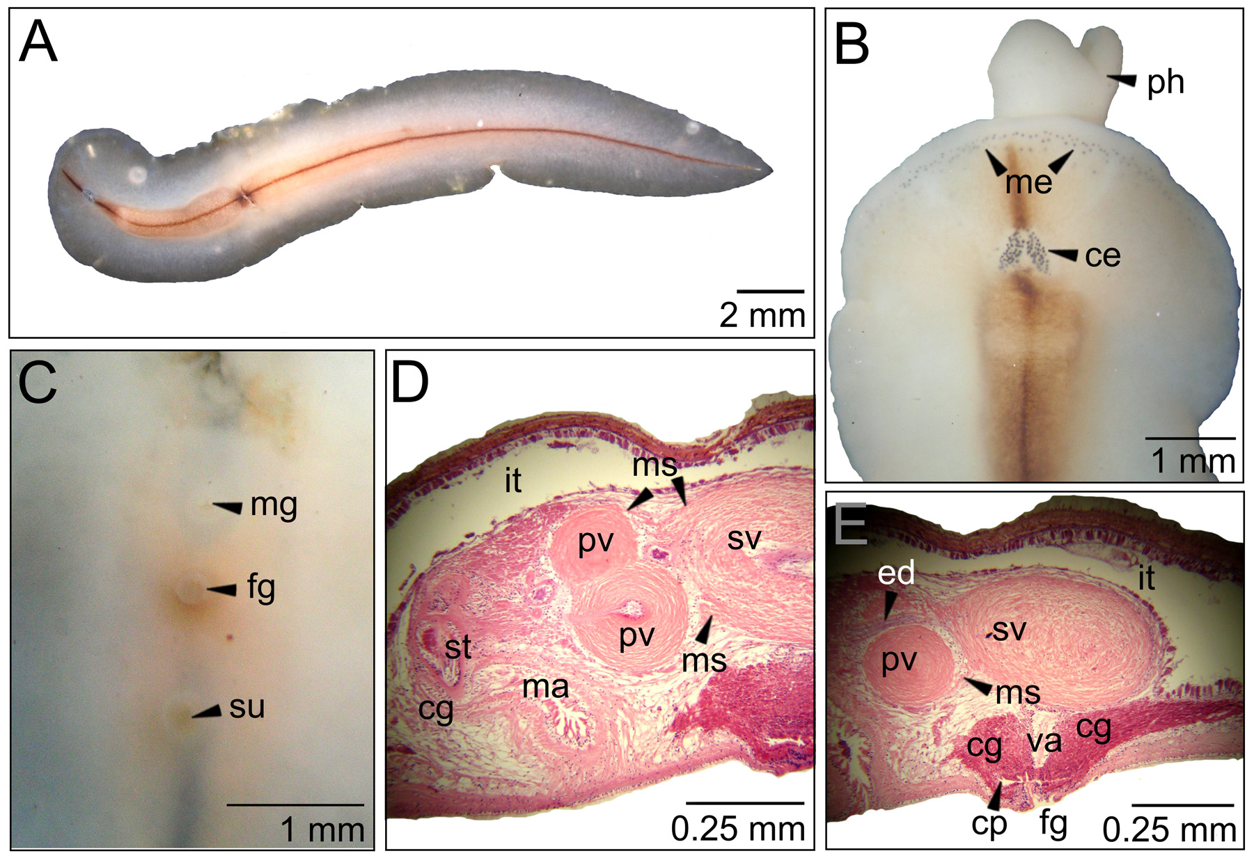
( Fig. 15 View FIGURE 15 )
Material examined. One specimen (MNRJ-PLAT 62, 22 x 5 mm) as sagittal sections of reproductive structures (13 slides), remainings preserved in ethanol 70%. Collected 0 8.12.2007 at Praia das Conchas, Cabo Frio, Brazil, under rocks (22°52'33.05"S, 41°58'39.27"W).
Distribution. This very rare species was described from Ilha de São Sebastião, São Paulo (type locality; Marcus 1952) and also reported from Florida and Colombia ( Quiroga et al. 2004a). It is the first time that this species is recorded from Rio de Janeiro State.
Diagnosis. Translucid body; brown median line. Cerebral eyespots in two oval groups; marginal eyespots reach the brain level. Two prostatic vesicles and one seminal vesicle included in the same muscle sheath.
Description. Color: Translucent body cream to ivory, with brown median line ( Fig. 15 View FIGURE 15 A).
Form: Body characteristic of the family Prosthiostomidae , elongated, with anterior end rounded and posterior end pointed.
Tentacles: Absent.
Eyespots: Cerebral eyespot groups oval at 1.5 mm from the anterior margin ( Fig. 15 View FIGURE 15 B). Contain 54–59 eyespots. Marginal eyespots disposed in three approximately parallel lines ( Fig. 15 View FIGURE 15 B) that reach the brain level. About 70 eyespots present at each side of the median line.
Digestive system: Pharynx tubular and voluminous measures 4 mm, in our specimen it is protracted ( Fig. 15 View FIGURE 15 B). Main intestine extends over ¾ of the body length.
Epidermis and body wall: Dorsal epidermis (40 µm) taller than ventral epidermis (13 µm). Well-developed muscular layer composed by external longitudinal layer, intermediary circular and internal diagonal layer. Dorsal muscular layer measures 42 µm and ventral layer 39 µm. Sucker with 0.5 mm diameter.
Gonopores: One male gonopore and one female gonopore present ( Fig. 15 View FIGURE 15 C). Measure 0.25 mm. Male gonopore at 5 mm from the anterior margin, followed by the female gonopore at 1 mm distance and by the sucker at 1.5 mm.
Male reproductive system: Male atrium, with 0.47 mm length, curves backwards and opens perpendicularly to the body ( Fig. 15 View FIGURE 15 D) forming a half “s”. Penis papilla measures 130 µm. Two prostatic vesicles present ( Fig. 15 View FIGURE 15 D), rounded, and located one over the other, measuring 230 x 220 µm. Seminal vesicle oval, located over the female gonopore, measuring 470 x 320 µm. The three vesicles are found in a same muscle sheath ( Fig. 15 View FIGURE 15 D), a diagnostic character of the genus. Seminal duct passes through the prostatic vesicles to join the prostatic ducts at 0.16 mm from the penis papilla.
Female reproductive system: Vagina 145 µm, surrounded by cement glands densely disposed ( Fig. 15 View FIGURE 15 E).
Disposition of cement glands and vagina limited by the presence and size of the voluminous seminal vesicle, located right above. Cement pouch elongated, measure 79 µm. Female atrium 61 µm deep ( Fig. 15 View FIGURE 15 E).
Taxonomic remarks. This species was originally described in the genus Prosthiostomum ( Marcus 1950) , then Marcus & Marcus (1968) transferred it to Lurymare , based on the presence of a muscle sheath containing prostatic and seminal vesicles, an exclusive characteristic of this genus. This was supported by Prudhoe (1985), but Poulter (1975) considered that Lurymare should be a subgenus of Prosthiostomum .
The drawings made by Marcus (1950) show the prostatic vesicles and the seminal vesicle together in a muscular sheath. Our specimen is in accordance with the original description ( Marcus, 1950) and the amendment ( Marcus & Marcus, 1968). Therefore this species should be included in Lurymare as this is the only prosthiostomid genus that possesses this character. This is something both Faubel (1984) and Prudhoe (1985) agreed on in their polyclad taxonomical revisions. Of the species recently found in Brazil and analyzed by us, both Lurymare matarazzoi and Lurymare utarum should be included in this genus, as both have prostatic and seminal vesicles included in a muscular sheath. However, Prudhoe (1989) later said that the validity of Lurymare as a genus seems unsure, due to variation in the presence of the muscle sheath around the accessory vesicles according to specimen size. Thus, it would be possible that Lurymare represents a late stage of Prosthiotomum, but this should be checked through a specific morphological and molecular study. Our specimen has approximately the same measure (22 x 5 mm) as found in the specimen of the original description (22.5 x 6 mm).
Other Lurymare View in CoL species, such as L. katoi View in CoL , have different eyespot arrangements, with the cerebral group shorter and more close together. Also, this species has a different coloration pattern with gold to orange-red pigment granules ( Poulter 1975). L. monosorum View in CoL and L. drygalski View in CoL present different coloration patterns, eyespot arrangements, and disposition of prostatic vesicles ( Bock 1913). The other Brazilian Lurymare View in CoL species, L. matarazzoi View in CoL , does not have the brown median line ( Marcus 1950). L. utarum View in CoL is the only one in the genus with this coloration pattern.
No known copyright restrictions apply. See Agosti, D., Egloff, W., 2009. Taxonomic information exchange and copyright: the Plazi approach. BMC Research Notes 2009, 2:53 for further explanation.
|
Kingdom |
|
|
Phylum |
|
|
Class |
|
|
Order |
|
|
Family |
|
|
Genus |