
Pseudoceros juani, Bahia, Juliana, Padula, Vinicius, Lavrado, Helena Passeri & Quiroga, Sigmer, 2014
|
publication ID |
https://doi.org/ 10.11646/zootaxa.3873.5.3 |
|
publication LSID |
lsid:zoobank.org:pub:687DC4E0-9B78-4AF0-9DD2-8B868E3B8EB5 |
|
DOI |
https://doi.org/10.5281/zenodo.6143916 |
|
persistent identifier |
https://treatment.plazi.org/id/927D87F1-FFC2-3213-FF78-7F6E68DBFE27 |
|
treatment provided by |
Plazi |
|
scientific name |
Pseudoceros juani |
| status |
sp. nov. |
Pseudoceros juani sp. nov.
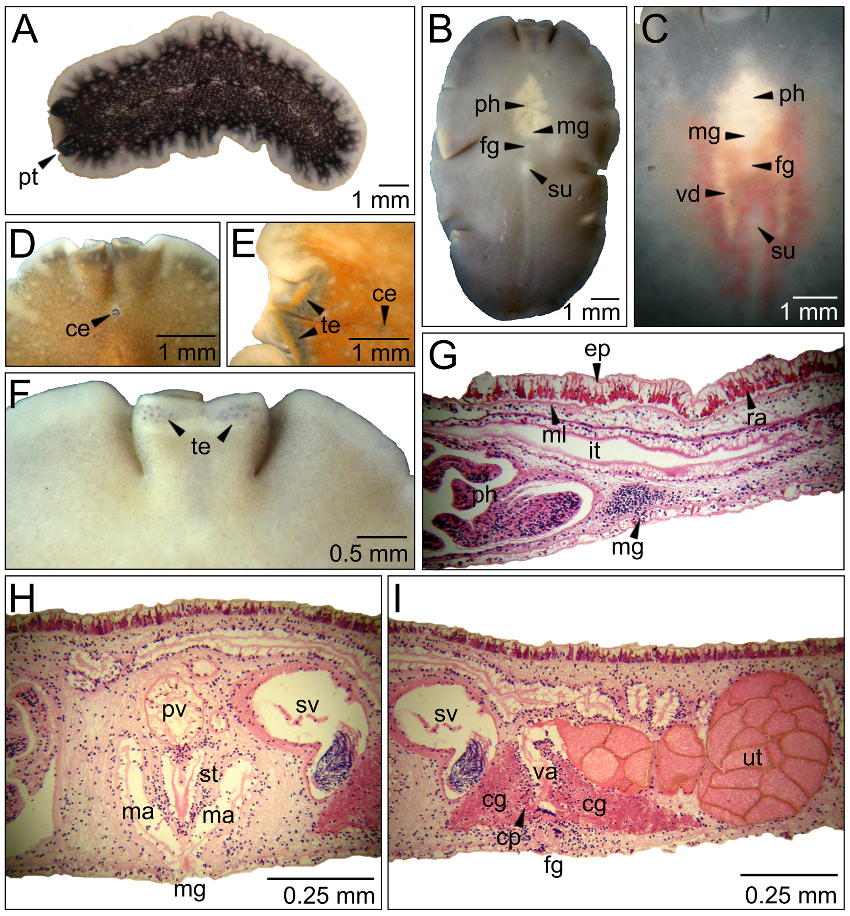
( Fig. 8 View FIGURE 8 )
Etymology. This species is named after Juan Piñeiro-Maceira, father of the first author, in acknowledgment of his support throughout her research on polyclads.
Type material. Holotype: One specimen (MNRJ-PLAT 40, 36 x 18 mm) as sagittal sections of reproductive structures (35 slides). Collected 31.12.2008 at Enseada da Hípica, Ilha do Papagaio, Cabo Frio, Brazil (22°53'53.95"S, 41°58'42.11"W).
Distribution. Species only known so far from the type locality.
Diagnosis. Background color brick orange with scattered white and dark spots; whitish translucent marginal band with a thin light yellow outermost line.
Description. Color: Background color brick orange. ( Fig. 8 View FIGURE 8 A and G). Translucent white marginal band 0.5 mm wide. Externally to it there is a thin light yellow line that runs all over the body margin, including pseudotentacle tips; it disappears after fixation. White and dark spots scattered in the dorsal surface except on the margin, giving the appearance of a granulated background. Tentacles are darker than the rest of the body ( Fig. 8 View FIGURE 8 A). Ventral surface milky but with the same coloration pattern as presented dorsally.
Form: Body oval and elongated. ( Fig. 8 View FIGURE 8 A and B). Length is three times the width. Margin slightly ruffled.
Pseudotentacles: Broad, tubular and simple rounded marginal folds ( Fig. 8 View FIGURE 8 A and C), 2.5 mm long.
Eyespots: Cerebral eyespots at 1.5 mm from the anterior margin ( Fig. 8 View FIGURE 8 C). Cluster is about 0.2 mm large and rounded horse shoe shaped. There are about 37 cerebral eyespots. A band of tentacular eyespots ( Fig. 8 View FIGURE 8 C) is located in the border of the tentacles (approximately 68 eyespots in each fold), some other ocelli are scattered in between the tentacle folds (around 36 eyespots) and ventrally near the margin (30 eyespots on each side).
Digestive system: Pharynx tipical of the genus, radially ramified, almost as long as wide with lots of intricate folds ( Fig. 8 View FIGURE 8 B). It is 5 x 4.5 mm large and located 2.5 mm from the anterior margin. Mouth opens slightly anteriorly to the middle of the pharynx, 6 mm from the anterior margin. Main intestine extends about 80% of the body length and reaches to 4.8 mm from the posterior margin ( Fig. 8 View FIGURE 8 B).
Epidermis and body wall: Dorsally, epidermis and muscular layer are more developed. Body wall thickness is 81 µm dorsally and 27 µm ventrally. Rhabdites present on both surfaces ( Fig. 8 View FIGURE 8 E) but more abundant dorsally. Pigment granules present in the epidermis. Muscular fibers disposed as longitudinal, circular and diagonal layers, respectively. Sucker at 14 mm from the anterior margin and 4.9 mm from the female gonopore ( Fig. 8 View FIGURE 8 B).
Gonopores: Male and female gonopores are 2 mm from one another ( Fig. 8 View FIGURE 8 B). Male gonopore 9 mm distant from the anterior margin and measures 0.5 mm. Female gonopore mesures 0.4 mm.
Male reproductive system: Prostatic vesicle round and located slightly left from the body median line ( Fig. 8 View FIGURE 8 E). It measures 0.2 x 0.22 mm and is located near the base of the penis papillae, anteriorly to the seminal vesicle. Seminal vesicle large (840 x 510 µm), elongated parallel to the longitudinal axis of the body ( Fig. 8 View FIGURE 8 F). Its wall is densely muscularized (110 µm thick). Ejaculatory duct stretches perpendicularly to the vesicle and joins the prostatic duct at the penis papillae. Penis papillae 260 µm long. Male atrium 270 µm deep and with folds ( Fig. 6 View FIGURE 6 F). Stylet 180 µm long.
Female reproductive system: Vagina 180 µm long, connecting to oviducts perpendicularly ( Fig. 8 View FIGURE 8 D). Oviducts directed backwards. Numerous cement glands present ( Fig. 8 View FIGURE 8 D) and cement pouch (90 µm) relatively long. Female atrium 110 µm long ( Fig. 8 View FIGURE 8 D).
Taxonomic remarks. There are three Pseudoceros species described or reported from Brazil ( Marcus 1949; Bahia & Padula 2009). Pseudoceros chloreus Marcus 1949 has a different color pattern and according to Marcus’ illustrations ( Marcus 1949, pg: 153) has a pharynx more elongated and with fewer folds than we find in our specimen. Some P. b i c o l o r and P. rawlinsonae specimens have a similar pattern of white dorsal spots, but not the dark spots, and the general coloration pattern (especially the marginal band that is striated in P. b i c o l o r and P. rawlinsonae but is not in our specimen) is different ( Verrill 1901; Bolaños et al. 2007; Bahia & Padula 2009; Litvaitis et al. 2010).
The general coloration pattern of P. j u an i sp. nov. resembles that of the Mediterranean and Canarian ( Vera et al. 2008) species Yungia aurantiaca (Delle Chiaje 1822) . However, this genus is distinguished from Pseudoceros by the presence of intestinal pores and “ Pseudobiceros -like” pseudotentacles ( Faubel 1984; Prudhoe 1985; Newman & Cannon 1994). The Australian species Pseudoceros devisii Woodworth, 1898 possesses yellowish orange coloration, but its pattern is dark orange marginal band and median line ( Woodworth 1898). The picture of P. prudhoei (Newman & Cannon 1994; pg: 261) resembles our specimen. However, its marginal band is blue to mauve. Internally this species differs from P. j u an i sp. nov. in the position of the prostatic vesicle not being in front of the seminal vesicle but slightly under it (Newman & Cannon 1994; pg: 235). The arrangement of tentacular eyespots is similar to the one illustrated in Newman & Cannon (1994) figures 3A and E.
The internal features of P. juani sp. nov. resemble those of Pseudoceros lactolimbus Newman & Cannon, 1998 and P. uniarborensis Newman & Cannon, 1994 , such as a long and large seminal vesicle and prostatic vesicle located in front of the seminal vesicle. However, these species coloration patterns (Newman & Cannon 1994 pg: 254, 263; 1998 pg: 309, 319) differ from that observed in P. juani sp. nov. ( Fig. 8 View FIGURE 8 A). The specimen found at Cabo Frio and studied herein does not fit any of the known Pseudoceros species ( Yeki & Kaburaki 1918; Kaburaki 1923; Hyman 1953; Hyman 1954; Prudhoe 1989; Newman & Cannon 1994, 1995, 1998; Newman & Schupp 2002), therefore the species is herein described and named.
No known copyright restrictions apply. See Agosti, D., Egloff, W., 2009. Taxonomic information exchange and copyright: the Plazi approach. BMC Research Notes 2009, 2:53 for further explanation.
|
Kingdom |
|
|
Phylum |
|
|
Class |
|
|
Order |
|
|
Family |
|
|
Genus |