
Diphtherophora eldarica, Ghaderi & Hesar & Karegar & Pereira, 2020
|
publication ID |
https://doi.org/ 10.11646/zootaxa.4851.2.10 |
|
publication LSID |
lsid:zoobank.org:pub:43C5F7B3-1C03-4738-84CC-FF17B4FE0889 |
|
DOI |
https://doi.org/10.5281/zenodo.4477047 |
|
persistent identifier |
https://treatment.plazi.org/id/924E87E3-964C-425D-FF4C-3FCD19D6F7E3 |
|
treatment provided by |
Plazi |
|
scientific name |
Diphtherophora eldarica |
| status |
sp. nov. |
Diphtherophora eldarica n. sp.
( Figs. 1 View FIGURE 1 , 2 View FIGURE 2 , Table 1 View TABLE 1 )
Description. Female: Body habitus straight to slightly curved ventrally after fixation. Cuticle regularly striated from the anterior end to the level of secretory-excretory pore, but appearing smooth posteriorly; relatively thick, about one-fifth to two-fifths of the body width. Subcuticle layer finely striated along the entire body and about 2 µm thick. Head region offset from the body contour. Outer labial papillae not readily observed. Amphid aperture oval in shape and as wide as about one-third of the head width at its base. Spear typical of the genus, with a sclerotized acute tip. Spear guiding apparatus arched, moderately sclerotized. Pharynx typical of the genus and composed of two parts, the anterior region (corpus) slender and expanding gradually to end as a rounded or sometimes slightly pyriform terminal bulb. Nerve ring located at mid-pharynx region or posterior to it. Secretory-excretory pore opposite the posterior end of the slender part of pharynx or at the beginning of the pharyngeal bulb. Cardia not observed under LM.
Reproductive system didelphic, amphidelphic, with branches equally developed, ovaries reflexed with germinal zones directed toward vagina, spermatheca not present at the junction of oviduct and uterus. Sperm cells with rodshaped nuclei throughout the uteri. Vulva a transverse slit as seen in a ventral view. Cuticle invaginated just anterior and posterior to the vulva. Vagina orthogonal to the body axis, with moderately to well-developed musculature. Rectum 14–16 µm or 54–73 % of the anal body width. Cuticle overhanging the anterior lip of the anus. Tail conoid, bent dorsally near tail tip, with a rounded terminus. The tail terminus lacks lateral caudal pores.
Male: In general, morphologically similar to female. Body habitus almost straight. Cervical ventromedian papillae absent. No lateral body pores observed in the anterior region of the body. Reproductive system monorchic, extending to near mid-body, and containing numerous rod-shaped sperm. Two ventromedian precloacal supplements were observed, the anteriormost near the proximal end of spicules and the posterior one level with the distal ends of spicules. Spicules with marked capitulum and narrow shaft, expanding again at beginning of the blade part and then gradually tapering to the distal end. Spicular capsule weakly developed. Gubernaculum ventrally arcuate at anterior tip. Tail similar to female, with a rounded terminus. Overhanging cuticle on cloacal lips was not observed, as the cuticle did not separate from the body over a short distance anterior or posterior to cloaca.
Diagnosis. The new species is characterized by females with striated cuticle at the anterior end of the body, head offset from the body contour, spear 15–17 µm in length, rod-shaped sperm, overhanging cuticle on the anterior lip of the anus and a conical tail bent dorsally near the terminus. Additionally, males lack ventromedian neck papillae but have two ventromedian precloacal supplements at the level of the spicules.
Type host and locality. The specimens were collected from the rhizosphere of pine trees ( Pinus eldarica Medw. ) in Dalampar valley in Margavar, Urmia city, West Azerbaijan province, Northwestern Iran by A. Mokaram Hesar in September 2015 (GPS coordinates: 37º10’65’’N, 44º52’73’’E).
Type material. Holotype, three female paratypes and one male paratype, mounted in pure glycerin on a slide, were deposited in the collection of the Department of Plant Protection , School of Agriculture , Shiraz University, Shiraz, Iran.
Etymology. The specific epithet refers to the associated plant species, Pinus eldarica , from the rhizosphere of which the new Diphtherophora species was recovered.
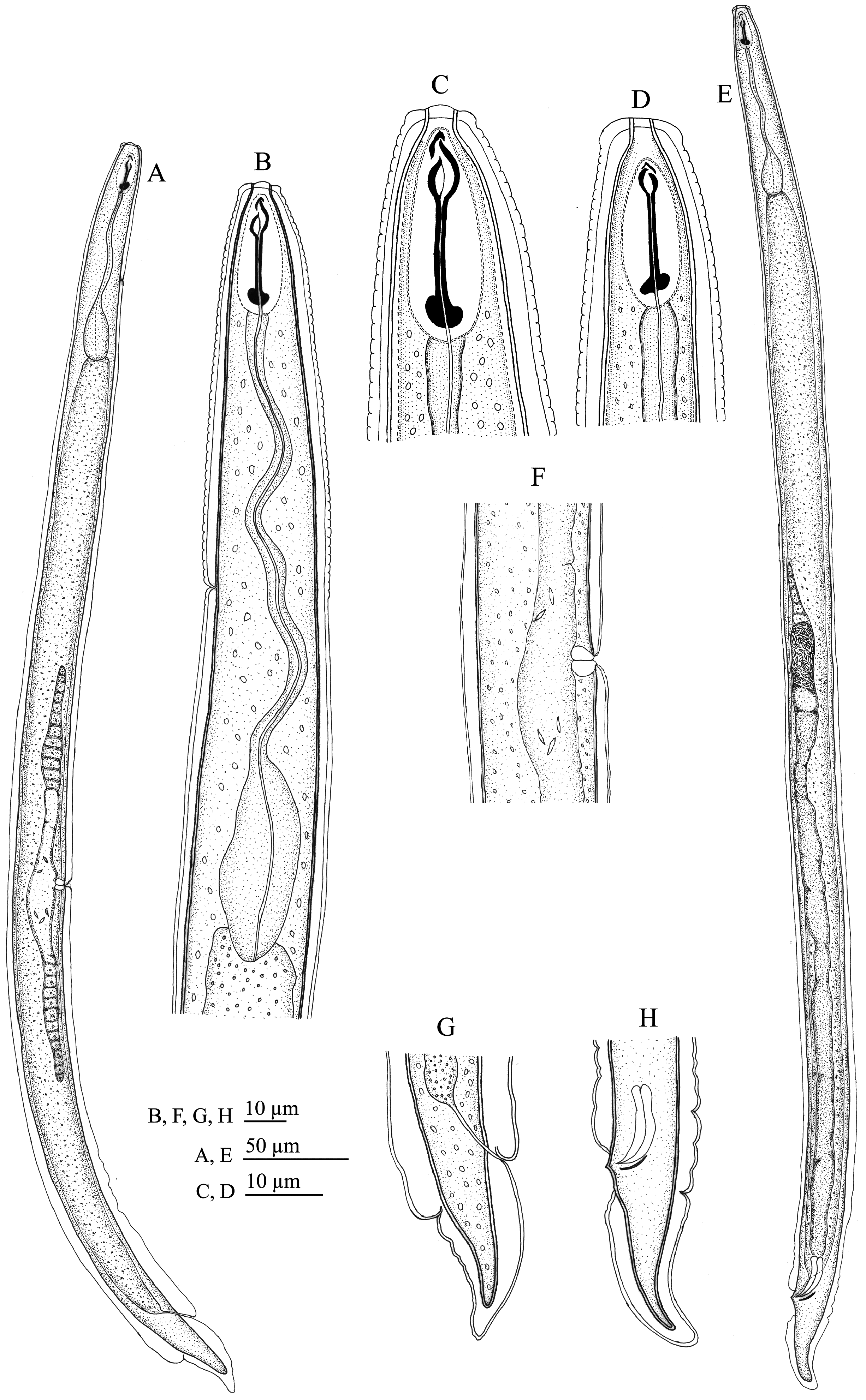
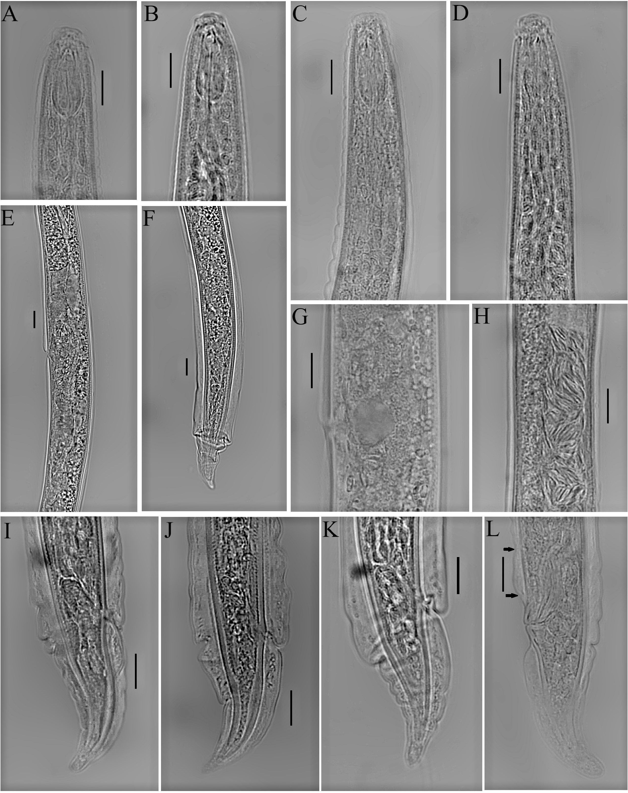
TABLE 1. Morphometrics of the new species Diphtherophora eldarica n. sp. and an Iranian population of D. caudata (measurements are in µm).
| Characters | D. eldarica n. sp. | D. caudata | |||
|---|---|---|---|---|---|
| Holotype female | 3 paratype, females | 1 paratype male | 6 females | 1 male | |
| L | 623 | 645 ± 33.1 | 679 | 604 ± 26.8 | 643 |
| (616–681) | (558–633) | ||||
| a | 23.1 | 20.2 ± 3.6 | 27.2 | 19.4 ± 1.8 | 21.4 |
| (17.7–24.3) | (16.4–21.8) | ||||
| b | 4.2 | 4.6 ± 0.2 | 4.6 | 3.8 ± 0.4 | 4.1 |
| (4.4–4.8) | (3.3–4.1) | ||||
| c | 16.4 | 16.7 ± 1.5 | 16.6 | 15.9 ± 2.0 | 11.3 |
| (15.0–17.7) | (13.3–19.0) | ||||
| c’ | 1.7 | 1.6 ± 0.2 | 2 | 1.8 ± 0.3 | 2.2 |
| (1.3–1.8) | (1.4–2.1) | ||||
| V | 56.2 | 55.9 ± 1.2 | – | 56.0 ± 2.0 | – |
| (54.9–57.3) | (52.3–57.9) | ||||
| Spear | 17 | 16.2 ± 0.8 | 16 | 15.9 ± 0.6 | 15 |
| (15.5–17.0) | (15.0–17.0) | ||||
| Pharynx | 147 | 141 ± 0.6 | 148 | 159 ± 18.6 | 156 |
| (140–141) | (136–188) | ||||
| S.E. pore | 72 | 74.3 ± 1.5 (73-76) | 66.5 | 82.1 ± 4.7 (77-87) | 83.5 |
| Head–vulva | 350 | 360 ± 11.8 | – | 338 ± 14.6 | – |
| (353–374) | (319–359) | ||||
| Tail length | 38 | 38.7 ± 2.5 | 41 | 38.5 ± 5.8 | 57 |
| (36.0–41.0) | (32–47) | ||||
| Body width | 27 | 32.3 ± 4.0 (28.0–36.0) | 25 | 31.3 ± 2.0 (29.0–34.0) | 30 |
| Anal body width | 23 | 25.0 ± 2.6 (22.0–27.0) | 21 | 21.7 ± 2.1 (19.0–25.0) | 26 |
| Spicules | – | – | 20 | – | 17 |
No known copyright restrictions apply. See Agosti, D., Egloff, W., 2009. Taxonomic information exchange and copyright: the Plazi approach. BMC Research Notes 2009, 2:53 for further explanation.
|
Kingdom |
|
|
Phylum |
|
|
Class |
|
|
Order |
|
|
Family |
|
|
Genus |