
Katianna maryae, Bernard, Ernest C., 2014
|
publication ID |
https://doi.org/10.11646/zootaxa.3754.2.4 |
|
publication LSID |
lsid:zoobank.org:pub:9C5CC352-B8FA-43D5-B3A8-1993DF1441C9 |
|
DOI |
https://doi.org/10.5281/zenodo.6135343 |
|
persistent identifier |
https://treatment.plazi.org/id/903A2131-FFCF-FFF7-92FD-FC1150CCF837 |
|
treatment provided by |
Plazi |
|
scientific name |
Katianna maryae |
| status |
sp. nov. |
Katianna maryae n. sp.
Figs. 1–6 View FIGURE 1 View FIGURE 2 View FIGURE 3 View FIGURE 4 View FIGURE 5 View FIGURE 6
Specimens examined. Holotype female and numerous paratypes, USA, Tennessee, Knox County, Knoxville, University of Tennessee, west (agricultural) campus, between Morgan Hall and Ellington Plant Sciences, pan sweeps of grassy lawns with scattered herbaceous dicots, May 1995, C. L. Williver, M. M. Gibbs, E.C. Bernard, collectors; many additional specimens collected from same locality in following years in all seasons, E.C. Bernard, collector; 25 specimens, Tennessee, Anderson County, Oak Ridge, leaf litter, 19 February 1995, M.M. Gibbs, collector; 7 specimens, Tennessee, Blount County, Great Smoky Mountains National Park, Abrams Falls ranger station, grass lawn, 10 August 2007, E.C. Bernard, collector.
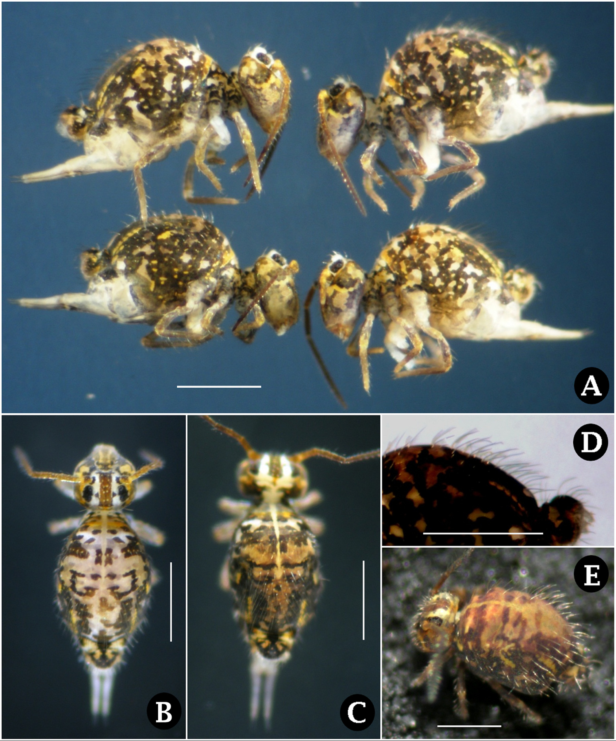
Description of female. Length 1.1–1.3 mm. Large abdomen rounded ( Figs. 1 View FIGURE 1 A, 2G); Abd. V and VI separate ( Figs. 2 View FIGURE 2 G, 3G). In ethanol, head posterior largely gray-violet with violet-black network of lines ( Fig. 4 View FIGURE 4 B); grayviolet color extending anteriorly and surrounding orange spot on gena ( Figs. 1 View FIGURE 1 A, B); fronto-clypeal region with median violet stripe flanked by orange stripes; copper-brown median stripe between black eye patches, flanked by white stripes sometimes ornamented with small black spots ( Fig. 1 View FIGURE 1 B); postocular tubercles white. Antennal segments I–III orange-brown, Ant. IV violet or orange-brown basally then violet. Body with intricate mosaic of violet-black, orange and white in varying proportions ( Figs. 1 View FIGURE 1 A–C), with narrow median stripe extending to midbody, this stripe usually crossed by transverse white stripe in region of Th. III-Abd. I; midbody generally with G or J-shaped black figures ( Figs. 1 View FIGURE 1 B, C); small abdomen black dorsally with two rectangular orange spots. Leg color variable, usually whitish proximally and light violet distally, sometimes with orange pigment. Ventral tube and furcula white or lightly washed with violet. Each macrosetal socket set in small white dot.
Color of live females similar but body with mosaic of yellow, orange and dark violet ( Fig. 1 View FIGURE 1 E). Median abdominal stripe yellow, transverse stripe yellow-orange.
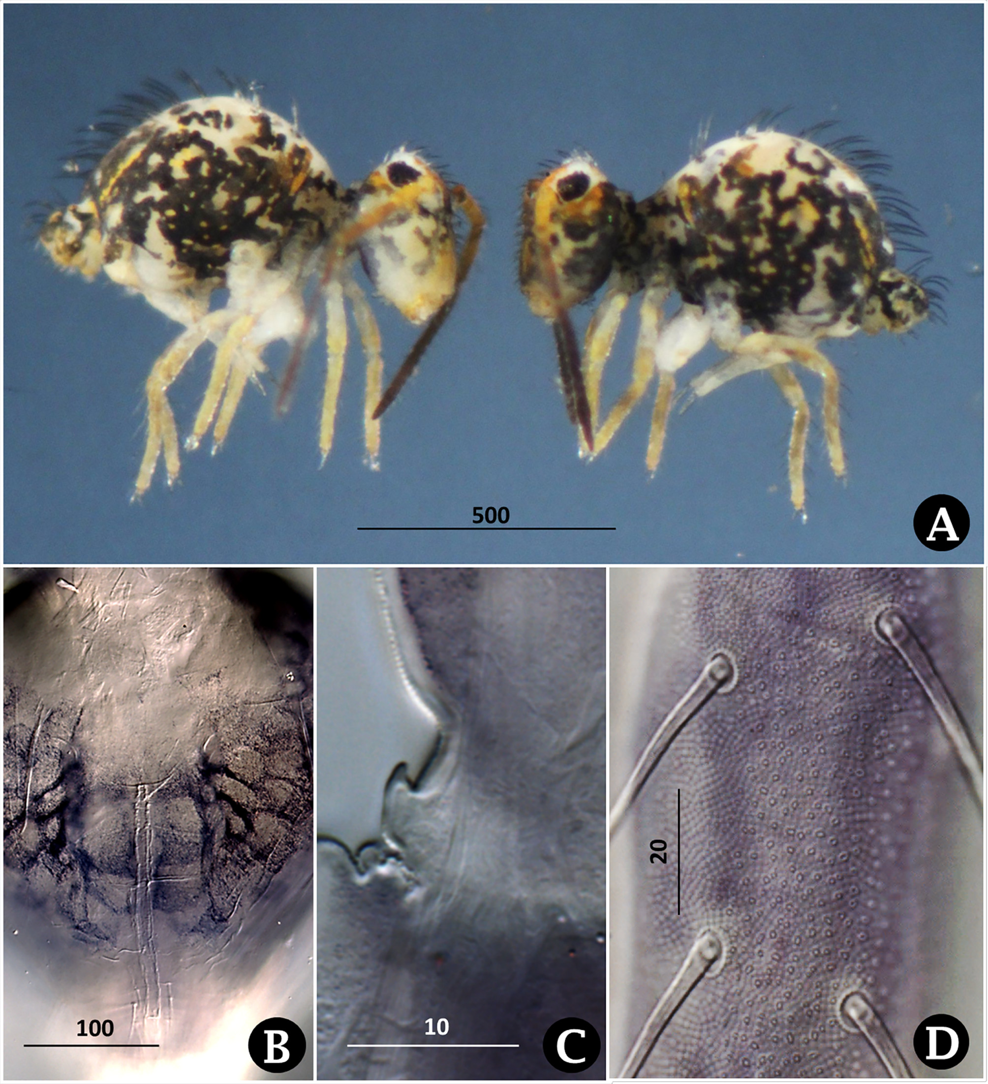
Cuticle finely granulate over most of its surface ( Figs. 5 View FIGURE 5 A, B); antennal segments and leg segments distal to coxae with numerous and more prominent circular or oval granules extending above surface ( Fig. 4 View FIGURE 4 D); raised base of bothriothrix D with granules in partial spiral arrangement ( Fig. 5 View FIGURE 5 C); more posteriorly, small abdomen with pattern of raised clusters of granules ( Fig. 5 View FIGURE 5 C, E).
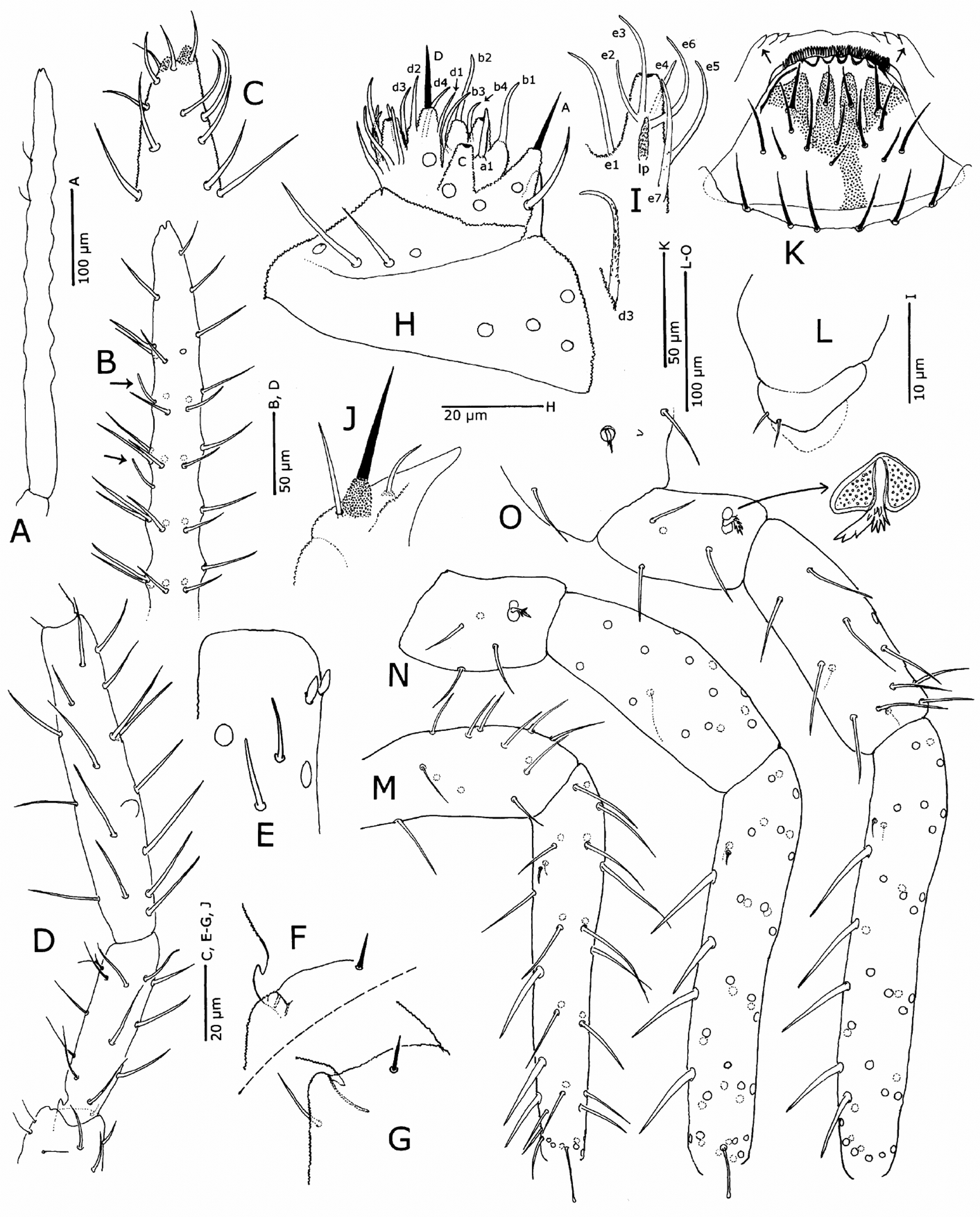
Antennal segment IV about twice the length of Ant. III ( Figs. 6 View FIGURE 6 A, D), annule-like but not subsegmented; basal fourth with parallel sides, more anteriorly with nine equally spaced enlargements each bearing a whorl of seven pointed setae; subapically, two slender distal sensilliform setae on exterior side ( Figs. 6 View FIGURE 6 A, B), segment apparently devoid of other specialized sensillum-like setae; apex of Ant. IV with two conical processes, apical bulb absent ( Fig. 6 View FIGURE 6 C). Papilla of Ant. III simple, unlobed ( Fig. 6 View FIGURE 6 D); Ant. III sense organ composed of two exposed oval sense clubs and two sensillum-like setae slightly posterior to clubs ( Fig. 6 View FIGURE 6 E). Base of Ant. II with apparent sense organ consisting of pit and conical projection ( Figs. 4 View FIGURE 4 C, 6D–F); this projection exposed when antenna is outstretched ( Fig. 4 View FIGURE 4 C, 6F), covered by Ant. I and partially in pit when antennae angled at the Ant. I–II joint ( Fig. 6 View FIGURE 6 G). First antennal segment with seven setae, dorsally and Laterally with three thicker and three thinner setae, ventrally with one small seta at apex ( Fig. 6 View FIGURE 6 D). Three proprioreceptors (oval organs) near antenna base ( Fig. 2 View FIGURE 2 A).
Labial palpus with all papillae and guard setae ( Fig. 6 View FIGURE 6 H); proximal region with five stout, slightly curved setae, basolateral field with four setae. Guard seta a1 straight, spike-like; b1 long, on papillate base; d3 microserrated on most of its length; lateral process granulate, reaching base of e3 ( Fig. 6 View FIGURE 6 I). Maxillary outer lobe with simple palpus and two sublobal hairs, the more basal hair straight ( Fig. 6 View FIGURE 6 J). Maxillary lamellae not extending past teeth of maxillary head. Usually six prelabral setae present ( Fig. 6 View FIGURE 6 K), occasionally median seta present to give seven. Labral setae in three rows, proximal to distal 5-5-4 setae; middle three setae of basal row and median seta of middle row shorter than other setae; outer setae of distal row thickened, spike-like, on small papillae. Dorsal edge of labrum with five acute lobes and apical fringe; ventral edge sinuate, with three teeth at each corner ( Fig. 6 View FIGURE 6 K). Eight ocelli in each eyepatch, ocelli E and G slightly smaller than adjacent ocelli, C and D strongly reduced, D smaller than C ( Fig. 2 View FIGURE 2 E). Linea ventralis without spine-like process ( Fig. 4 View FIGURE 4 B).
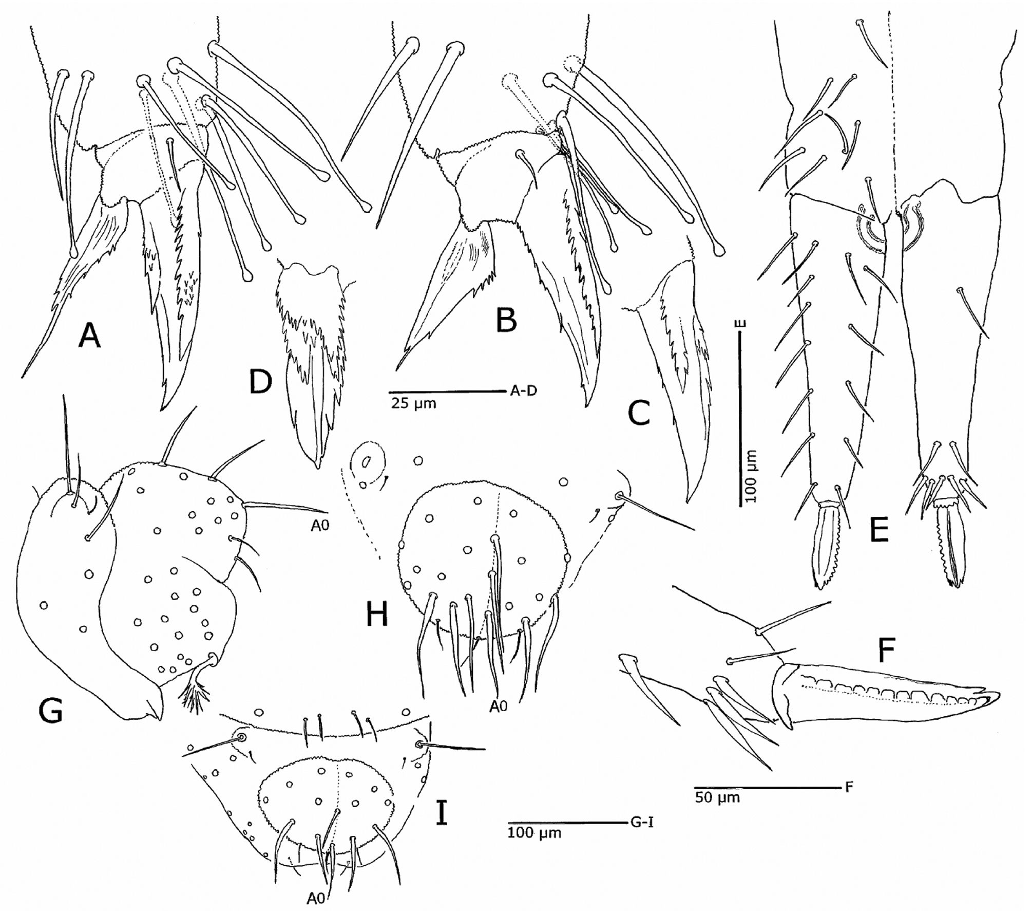
Middle coxa with two setae and conical papilla ( Fig. 5 View FIGURE 5 A), hind coxa with two setae, forked sensillum in circular pit and conical papilla ( Figs. 5 View FIGURE 5 B, 6 O). Middle and hind trochanters with similar trochanteral organs composed of oval or heart-shaped pit surrounding thick serrated seta ( Fig. 6 View FIGURE 6 O); middle trochanter with three setae, hind trochanter with four setae ( Figs. 6 View FIGURE 6 N, O). Fore, middle and hind femora with 13, 15, and 13 setae, respectively; interior middle seta of middle femur twice the length of its hind femur counterpart ( Figs. 6 View FIGURE 6 N, O). Tibiotarsi each with six whorls of setae and with a pair of minute subventral, sub-basal setae. Ventral surface of fore, middle and hind tibiotarsi with 3, 4, 5 spine-like setae ( Figs. 6 View FIGURE 6 M– O) and distally with 8, 8, 6 clavate tenent hairs, respectively ( Figs. 3 View FIGURE 3 A, B). Foot structure similar on all legs, except filament long and reaching tip of unguis on fore and hind unguiculi, very short on hind unguiculus ( Figs. 3 View FIGURE 3 A, B). Unguis with two teeth on inner side, multiple proximal denticles in line or clustered along inner edge, and prominent multidentate pseudonychia on both lateral sides ( Figs. 3 View FIGURE 3 A–D), terminating in large lateral teeth ( Fig. 3 View FIGURE 3 D); viewed from exterior side pseudonychia often fused into several multiserrated layers. Pseudonychial region often with clusters of minute denticles ( Fig. 3 View FIGURE 3 A). Unguiculus with rows of denticles along one or both edges.
Ventral tube with 2+2 laterodistal setae ( Fig. 6 View FIGURE 6 L). Tenaculum with 3+3 teeth and two setae. Manubrium with 8+8 dorsal setae; dens ventrally with two subapical setae and one proximal seta, dorsally with six exterior, two inner dorsal, one dorsal, and five lateral setae ( Fig. 3 View FIGURE 3 E). Mucro notched at apex, with smooth exterior edge, coarsely toothed inner edge; mucronal seta absent ( Fig. 3 View FIGURE 3 F). Subanal appendage palmate with three multifurcate major branches ( Figs. 5 View FIGURE 5 D, E).
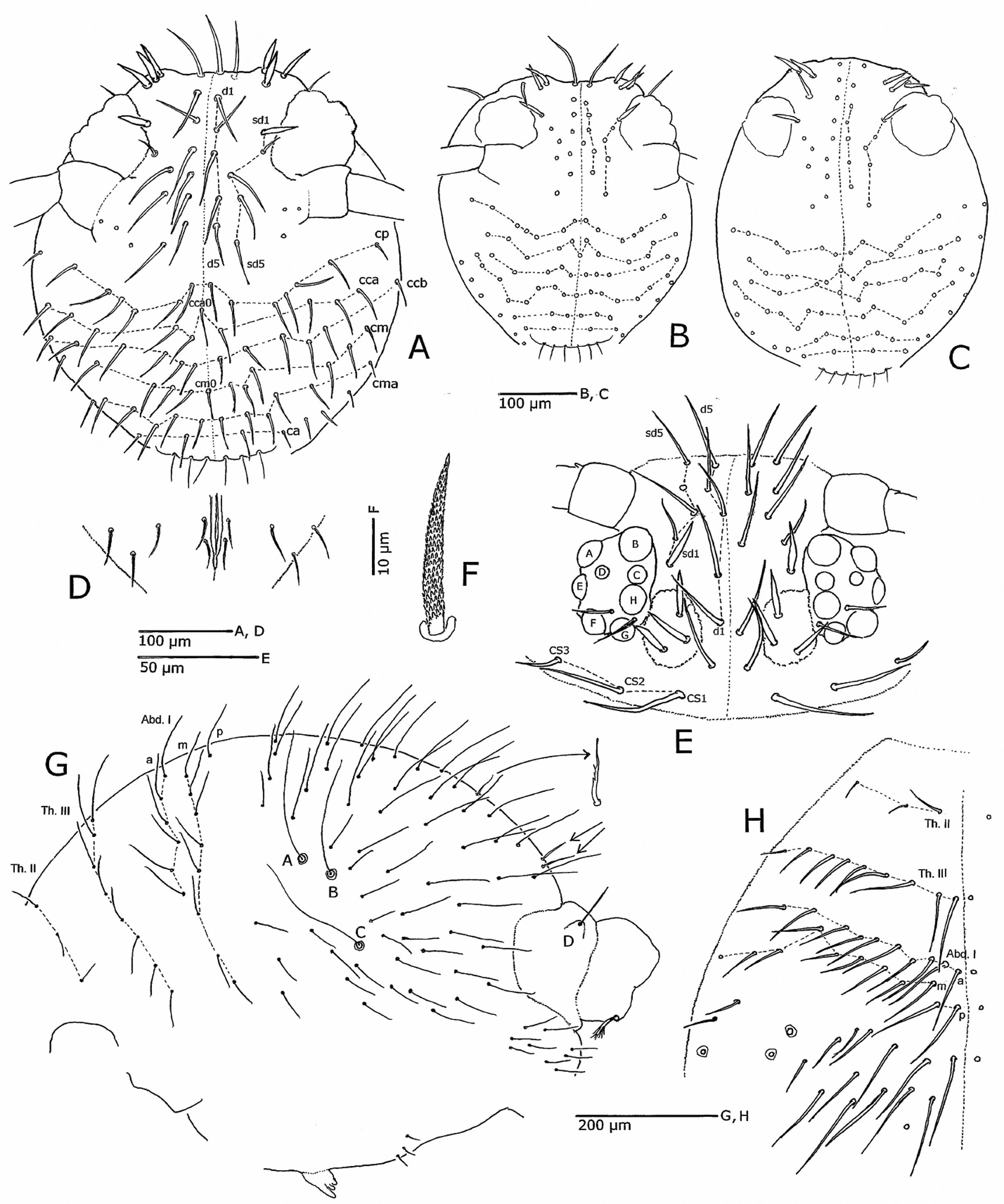
Chaetotaxy. Setae appearing smooth at low magnification; slender setae roughened at high magnification, cephalic spines minutely scaled in spiraling rows ( Fig. 2 View FIGURE 2 F); setae normally simple, antennae or legs rarely with a random forked seta ( Fig. 5 View FIGURE 5 A). Setae of head and body usually displaying minor asymmetry. Clypeal region with six transverse rows of setae posterior to pre-labral setae ( Figs. 2 View FIGURE 2 A–C): anterior clypeal (ca), anterior clypeal-medial (cma), clypeal-medial (cm), anterior clypeal-central 1 (ccb), posterior clypeal-central 2 (cca) and clypeal-posterior (cp); genal row absent; medial setae cm0 and cca0 usually present ( Figs. 2 View FIGURE 2 A, B), occasionally absent or displaced ( Fig. 2 View FIGURE 2 C). Interantennal-interocular area with d and sd setal rows, seta sd1 spine-like ( Figs. 2 View FIGURE 2 A–C); medial seta occasionally present ( Fig. 2 View FIGURE 2 B); sd’ row and seta a0 absent. Postocular tubercles each with 3 stout spine-like setae; 2+2 setae between tubercles ( Fig. 2 View FIGURE 2 E). Cervical setae 3+3, divergent, innermost pair (CS1, CS2) longer than CS3 ( Fig. 2 View FIGURE 2 E). Posterior of head with 2+2 setae near the linea ventralis and 3 postgenal setae arranged in a triangle on each side ( Fig. 2 View FIGURE 2 D).
Mesothorax with 3+3 setae, metathorax with 8+8 or 9+9 setae in a single row; Abd. I with three rows of setae, each side with 14–18 setae in a+m rows and 1 or 2 p-setae ( Figs. 2 View FIGURE 2 G, H). Bothriothrix B slightly posterior to line of bothriotricha A and C; B-C distance twice that of AB. Region of Abd. II–IV with numerous long setae not arranged in discernible pattern; posterior margin with 2+2 short, smooth setae ( Figs. 2 View FIGURE 2 G, 3I). One specimen with small, serrate, clavate medial seta ( Fig. 2 View FIGURE 2 G). Venter of abdomen usually with 3+3, occasionally 2+2 short setae.
On Abd. V bothriothrix D straight, tip tapering, slightly bent ( Fig. 5 View FIGURE 5 C), arising from small rounded papilla bearing one long seta and one short basal seta ( Fig. 3 View FIGURE 3 G). Four medial setae on Abd. VI, A-row setae slightly swollen near base; A0 simple, not bifurcate ( Figs. 3 View FIGURE 3 G, H).
Description of male. Males similar to females in most respects. Length 0.7–0.9 mm. Head with variable violet patches on gena; large abdomen with large white patches; legs pale orange ( Fig. 4 View FIGURE 4 A). Three medial setae on Abd. VI ( Fig. 3 View FIGURE 3 I).
Etymology. This species is named in loving memory of the late Mary Fitzpatrick, spouse of the noted nature photographer Kevin Fitzpatrick and a skilled photographer in her own right.
Relationships. Because of the relatively little attention given to Katianna for many years, the relationships of K. maryae n. sp. to other species of the genus are difficult to determine. In the key to South American species ( Heckman 2001) K. maryae n. sp. traces to K. willincki ( Delamare Deboutteville & Massoud, 1963) . Based on length (~700 µm) and less complex head chaetotaxy the single specimen probably is a juvenile, and thus may not be comparable to adults of K. maryae n. sp. The two species have a somewhat similar color patterns, both have G or J-shaped markings on the dorsum of the large abdomen, and they have the same ventral setal arrangement on the dens. However, they differ in the following ways, which may be due to the juvenile status of K. willincki : nine annule-like enlargements on Ant. IV in K. maryae n. sp. (six in K. willincki ); hind tibiotarsus with six clavate tenent hairs (seven in K. willincki ); two internal teeth and complex pseudonychia on the unguis (one tooth, no pseudonychia in K. willincki ), hind unguiculus triangular, much shorter than unguis, several denticles on inner edge (in K. willincki , hind unguiculus tapering, same length as unguis, without denticles). Morphologically K. maryae n. sp. is similar to K. jeanneli Delamare Deboutteville & Massoud, 1963 in the presence of pseudonychia and numerous denticles and minute teeth on the unguis and unguiculus, but K. maryae n. sp. has only six clavate tenent hairs on the hind tibiotarsus (seven in K. jeanneli ). Additionally, the mucronal tip of K. maryae n. sp. is straight, not hooked as in K. jeanneli . The color patterns are also different, with K. jeanneli having a stripe of black pigment on each side of the large abdomen as well as a black blotch toward the posterior end. Delamare Deboutteville & Massoud (1963) described but did not illustrate the habitus of K. jeanneli ; however, they stated that the pattern much resembled K. wygodzinskyi Delamare Deboutteville & Massoud (1963) , which they did illustrate as having dark lateral bands and a large medial spot on the large abdomen.
Katianna maryae n. sp. differs from the Australian and New Zealand species in color pattern and in number of clavate tenent hairs (eight on foreleg in K. maryae n. sp., six or less in the Australian and New Zealand species).

No known copyright restrictions apply. See Agosti, D., Egloff, W., 2009. Taxonomic information exchange and copyright: the Plazi approach. BMC Research Notes 2009, 2:53 for further explanation.
|
Kingdom |
|
|
Phylum |
|
|
Class |
|
|
Order |
|
|
Family |
|
|
Genus |