
Stigmacoccus asper Hempel
|
publication ID |
https://doi.org/ 10.5281/zenodo.177176 |
|
DOI |
https://doi.org/10.5281/zenodo.6237295 |
|
persistent identifier |
https://treatment.plazi.org/id/900A87FC-FFFC-FFDB-FF37-F92B3EA7ADB6 |
|
treatment provided by |
Plazi |
|
scientific name |
Stigmacoccus asper Hempel |
| status |
|
Stigmacoccus asper Hempel View in CoL
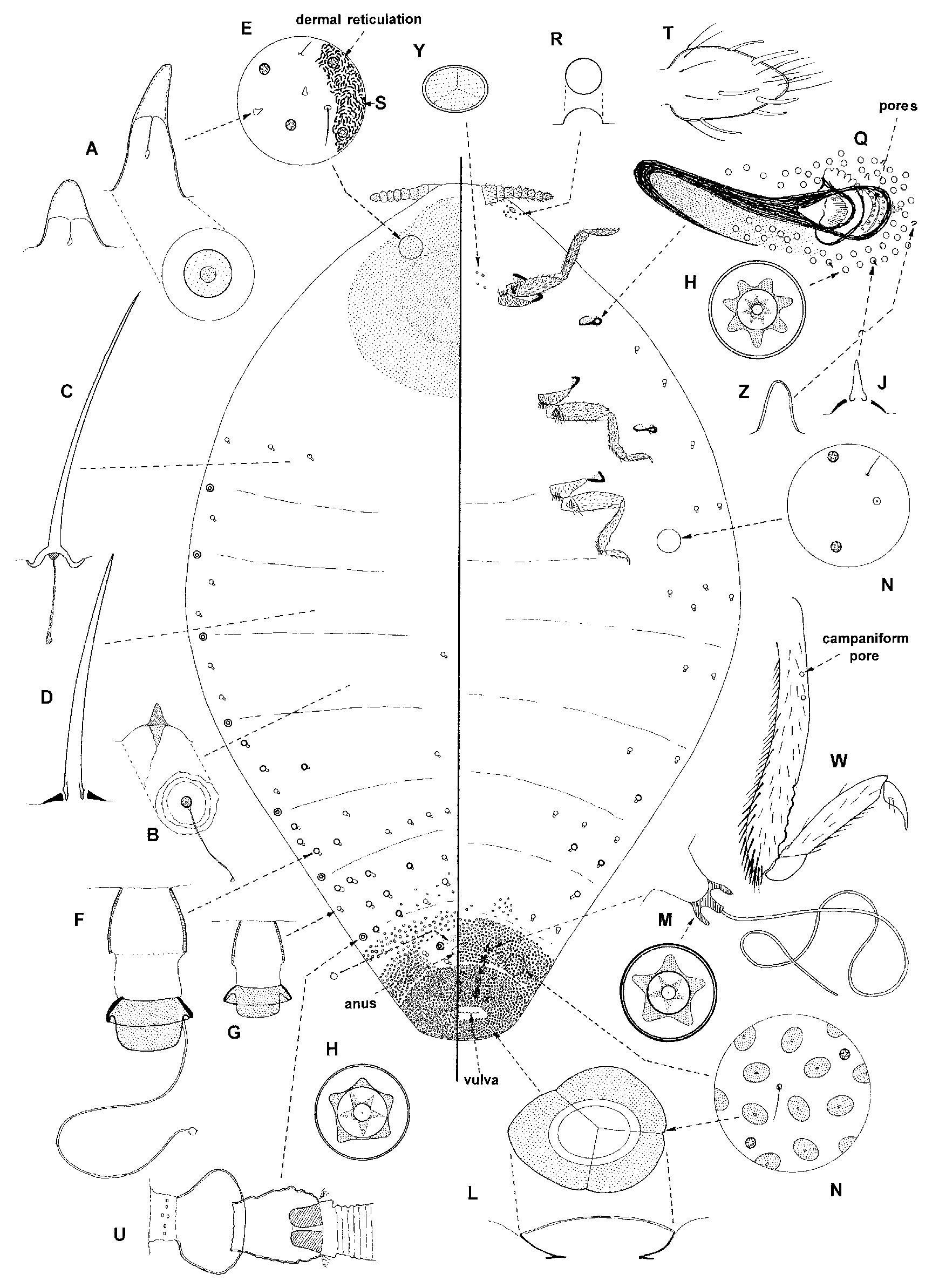
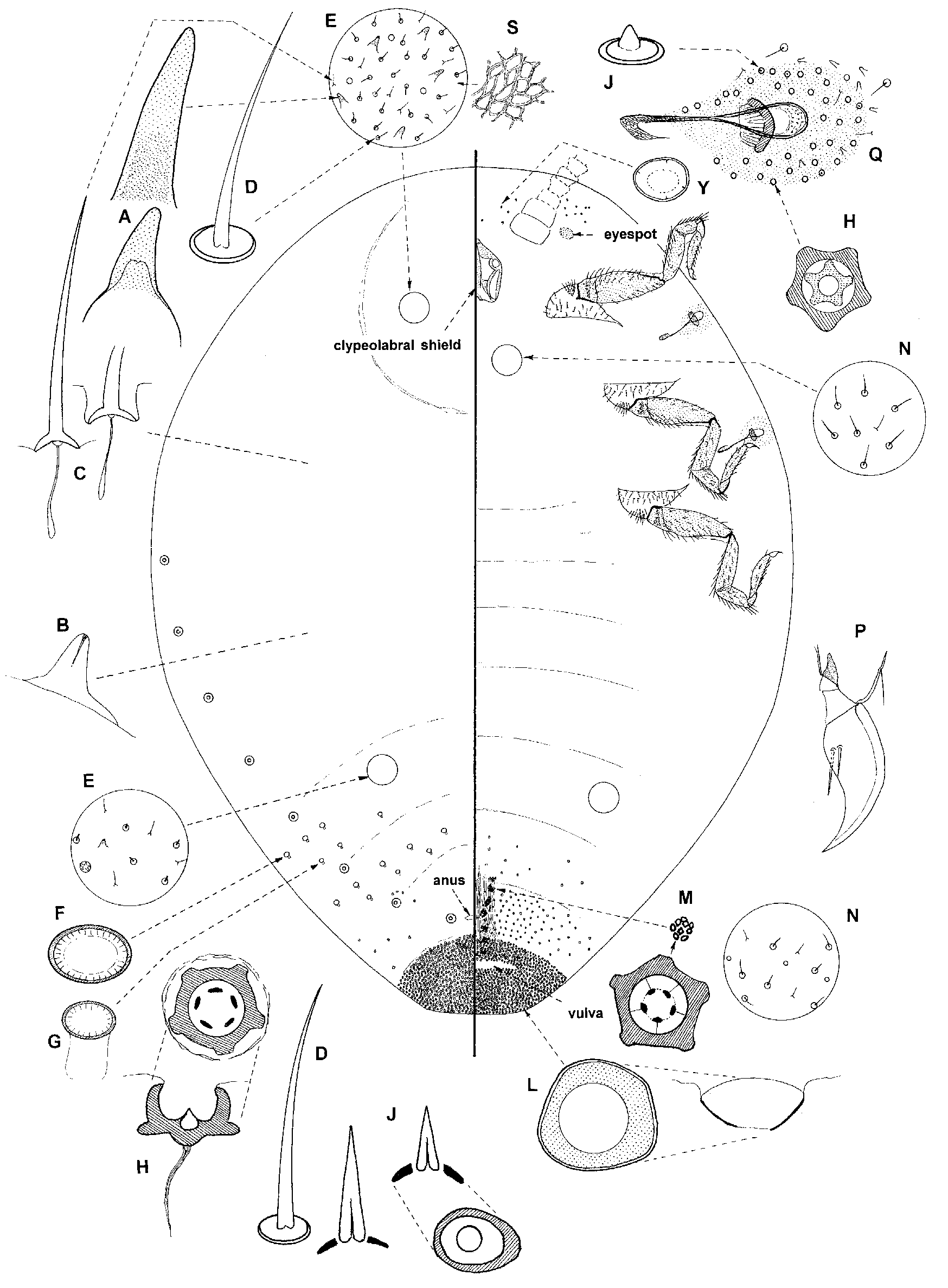
( Figs 1 View FIGURE 1 & 2 View FIGURE 2 )
Stigmacoccus asper Hempel, 1900: 400 View in CoL .
Material studied. LECTOTYPE (here designated to fix and stabilize the present concept of the name): BRAZIL, Pirassununga, on Inga sp. ( Fabaceae ), 28 April 1900, Hempel #17 ( USNM): 1/1adΨ in fair condition; as previous, PARALECTOTYPES: 4/4adΨΨ ( USNM) in fair to poor condition.
Other material. PANAMA, Mendoza, shore of Lake Gatun, no host, May 1934, Bishopp and Zetek ( USNM): 1/2 adΨΨ (in fair to good condition). COLOMBIA, Villavicencio, on Inga sp., 1972, Friedmann and Koster ( USNM): 3/3adΨΨ, mainly poor – one young (pharate?) specimen with mouthparts and derm poorly sclerotised). VENEZUELA, Maracay, on Cassia sp., 4.viii.1962, M.J. Way ( BMNH): 1/1adΨ + cyst (poor).
Note: description made mainly from Panama material; details from type series italicised in brackets.
Unmounted material. Unknown.
Mounted material. Length 6–10 (8.5–9.0) mm, width 4.7–7.0 (5.0–7.0) mm, bluntly pointed posteriorly; mature adult with a roundish area of sclerotisation on head; when unsclerotised (young individuals), derm on dorsum of head with a reticulate pattern. Antennae 8 segmented (8 or 9); legs fairly well developed but placed rather far forwards and all tibia slightly malformed but only strongly bent on young (pharate?) individual; each thoracic spiracle surrounded by a group of loculate pores; more anterior pairs of abdominal spiracles with a concentration of loculate pores nearby; hairs sparse, hair-like setae rather abundant on head; large spinose pores common on head of unsclerotised (pharate?) specimen but these not usually visible when dorsum of head sclerotised; loculate pores sparse throughout, without associated hairs on derm near margins; tubular pores few, of basically 2 sizes, almost entirely restricted to margins and submargins of abdomen and sometimes thorax; absent from head.
Dorsum. Mature specimens with a roundish area of sclerotisation on head, most heavily sclerotised medially, becoming less so laterally; total width of sclerotised area about 2.8 (2.0) mm; surface of derm with a dense pattern of minute reticulations (or dermal thickenings?). Setae rather abundant on head and anterior thorax, otherwise sparse; hairs and hair-like setae both mainly 13–25 ųm long (hair-like setae 12–16 µ m; hairs 16–26 µ m) but hair-like setae up to 40 ųm long on head where hairs possibly absent. Dermal pores of 5 types present: (i) loculate pores each 6–10 (6–10) ųm wide, without associated hairs on derm around pore margin; sparse throughout, most abundant posteriorly on abdomen and around spiracles, but few present medially on anterior abdominal segments both dorsally and ventrally; (ii) spinose pores of perhaps 2 sizes: small membranous pores with apex becoming lightly sclerotised on older individuals, each 4–6 ųm tall; about 6–20 ųm wide, with a distinct, short inner ductule and generally with some lightly sclerotised derm around each pore: sparse throughout; larger spinose pores, significantly taller than small spinose pores, of rather variable size, largest about 12–22 ųm tall and 8–12 ųm wide at base, with a short inner ductule: abundant in area of sclerotisation on head, absent elsewhere where replaced by small spinose pores; (iii) small convex pore, each about 3–5 ųm wide: perhaps 5–10 present near each eyespot; (iv) minute simple pores in a broad band around anal opening, and (v) large oval, apparently segmented pore, each about 8–23 (10–20) ųm wide at widest point; upper surface usually divided into 3 segments (occasionally up to 6) on older specimens, septa unclear on younger specimens but ventral pore very obvious; abundant around anus on abdominal segment VIII and on segment VII; also a few sometimes present laterally on VI. Tubular pores present sparsely on margins and submargins of abdominal and thoracic segments and also medially on abdominal segments IV–VIII (much less frequent); totally absent from head; of 2 sizes, those marginally significantly larger, total length 36–40 ųm, width of outer ductule 14–18 ųm; smallest ducts each 33–35 ųm long, outer ductule 8–15 ųm wide (tubular pores scarce, large near margin about 12 µ m wide, smaller pores about 8–9 µ m wide). Anus as in generic description. Eyespots each about 30–35 ųm wide. Abdominal spiracles as in generic description: outer atrium about 29 ųm long (20–28 µ m); broad middle atrium about 30 ųm (33–38 µ m) long and 70 ųm (75 µ m) wide; third, inner atrium 60–65 ųm (70–80 µ m) long and 60–65 ųm (70–80 µ m) wide, with a narrower exit into trachea; total length about 125 ųm (130 µ m); anterior 3–6 abdominal spiracles each with a concentration of loculate pores around opening.
Venter. Setae similar in size to and about as abundant as those on dorsum but some hairs up to 60 ųm long and hair-like setae up to 45 ųm long; stout, spine-like setae present laterad to each thoracic spiracle, each about 7 ųm tall and 7 ųm wide at base. Pores mainly as on dorsum: (i) loculate pores few or absent from both margins of vulva but abundant around each thoracic spiracle; (ii) small spinose pores and larger spinose pores probably absent; (iii) small convex pores absent; (vi) large segmented pores as on dorsum but more widespread, present throughout segments VII and VIII and also with a few medially on segments V and VI (VI only?); smaller segmented pores near mouthparts not always detected but 4–7 ųm wide when located (present, but hidden beneath sclerotised dorsum, very few, each 7–9 µ m wide). Tubular ducts as for dorsum but perhaps less frequent or absent (few). Antennae 8 (8 or 9) segmented, each 0.88 (0.95–1.0) mm long; scape each with many setae but no pores; pedicel narrower, sclerotised along proximal margin, wider than long, with many setae plus about 9 (6 or 7) campaniform sensilla; segments III to preapical segment all about 100–110 ųm long, with a broad band of setae and about one antennal bristle per segment; apical segment longer than broad, about 145 ųm long (130–140 µ m), with many setae + about 10 large and several smaller bristles. Mouthparts absent except on (pharate?) specimen, where clypeolabral shield present, facing anteriorly; length of clypeolabral shield about 475 ųm. Thoracic spiracles: width of peritremes about 80–85 (85–104) ųm, length of muscle plate + spiracle 370–375 (310–375) ųm; peritreme with or without rows of minute pores (with 2–3 rows); each spiracle with a loose group of 30 or more loculate pores on derm around lateral margins of peritreme. Legs: all placed rather far forward and with slightly malformed tibia; metathoracic legs: lengths (ųm): coxa 533–590 (457–480); trochanter + femur 640–750 (730–750); tibia 620–665 (640–685); tarsus 320–395 (320– 340); claw 120 (100–105); coxae with about 12–18 spinose setae on ventral surface near articulation with trochanter; each side of trochanter with 8–16 (10–17) campaniform sensilla; tibia either strongly bent about 2/ 3rds along of length (particularly on young individuals?) or showing some sign of deformation along dorsal margin but only slightly bent; each tibia with spur-like setae along ventral margin, those distally about 40–60 ųm; also with 4 (2–4) campaniform sensilla about 1/4–1/3rd along length; tarsus with many setae, those along ventral and lateral margins spur-like; tarsi each with 1 campaniform sensilla; claws with 1 or 2 (1 or 2) digitules on each margin; claw without a denticle. Vulva without a dense band of loculate pores along margins; vaginal wall with groups of 2–25 more heavily sclerotised loculate pores, each group on a bulge-like extension into haemocoel, in more or less 3 longitudinal rows; each loculate pore with a very long inner ductule emerging from central pore and extending into haemocoel, each ductule at least 65–80 ųm long (not always visible).
Comment. The main differences between these lots of material were: (i) presence or absence of small pores in each thoracic peritreme, (ii) longer tarsi on non-type material, (iii) non-type material had a sclerotised area at point where tracheae fused with spiracle (absent from the type specimens), and (iv) rather larger and more abundant tubular pores on the non-type material. No other differences could be found and all of this material is here considered to represent S. asper . At the present time, most material appears to have been collected off Inga sp. ( Fabaceae ).
The adult female of S. asper differs in several important characters from those of the other two species. In particular it has: (i) an area of quite dense sclerotisation covering dorsum of head on older adults; (ii) larger spinose pores abundant on dorsum of head (only really visible on young adults), (iii) derm of head with a reticulate pattern, and (iv) tubular pores much less widespread, with none present on head or medially on thorax. These differences appear to be related to the sclerotisation on the head. The reason for this sclerotisation is unknown. In all other respects, however, S. asper is similar to the other two species.
| USNM |
Smithsonian Institution, National Museum of Natural History |
No known copyright restrictions apply. See Agosti, D., Egloff, W., 2009. Taxonomic information exchange and copyright: the Plazi approach. BMC Research Notes 2009, 2:53 for further explanation.
|
Kingdom |
|
|
Phylum |
|
|
Class |
|
|
Order |
|
|
SuperFamily |
Coccoidea |
|
Family |
|
|
Genus |
Stigmacoccus asper Hempel
| Hodgson, Chris, Gamper, Heather, Bogo, Amauri & Watson, Gillian 2007 |
Stigmacoccus asper
| Hempel 1900: 400 |