
Goniopsarites fronticonvexus, Meng, Rui, Wang, Menglin & Wang, Yinglun, 2014
|
publication ID |
https://doi.org/ 10.11646/zootaxa.3866.1.4 |
|
publication LSID |
lsid:zoobank.org:pub:6202FA35-AF83-4F98-82EE-F089EA7ACC35 |
|
DOI |
https://doi.org/10.5281/zenodo.6143287 |
|
persistent identifier |
https://treatment.plazi.org/id/8E3287F1-C22F-F95E-FF6C-1E32FE07FAFE |
|
treatment provided by |
Plazi |
|
scientific name |
Goniopsarites fronticonvexus |
| status |
sp. nov. |
Goniopsarites fronticonvexus View in CoL sp. nov.
( Figs 1–27 View FIGURES 1 – 8 View FIGURES 9 – 17 View FIGURES 18 – 25 View FIGURES 26 – 27 )
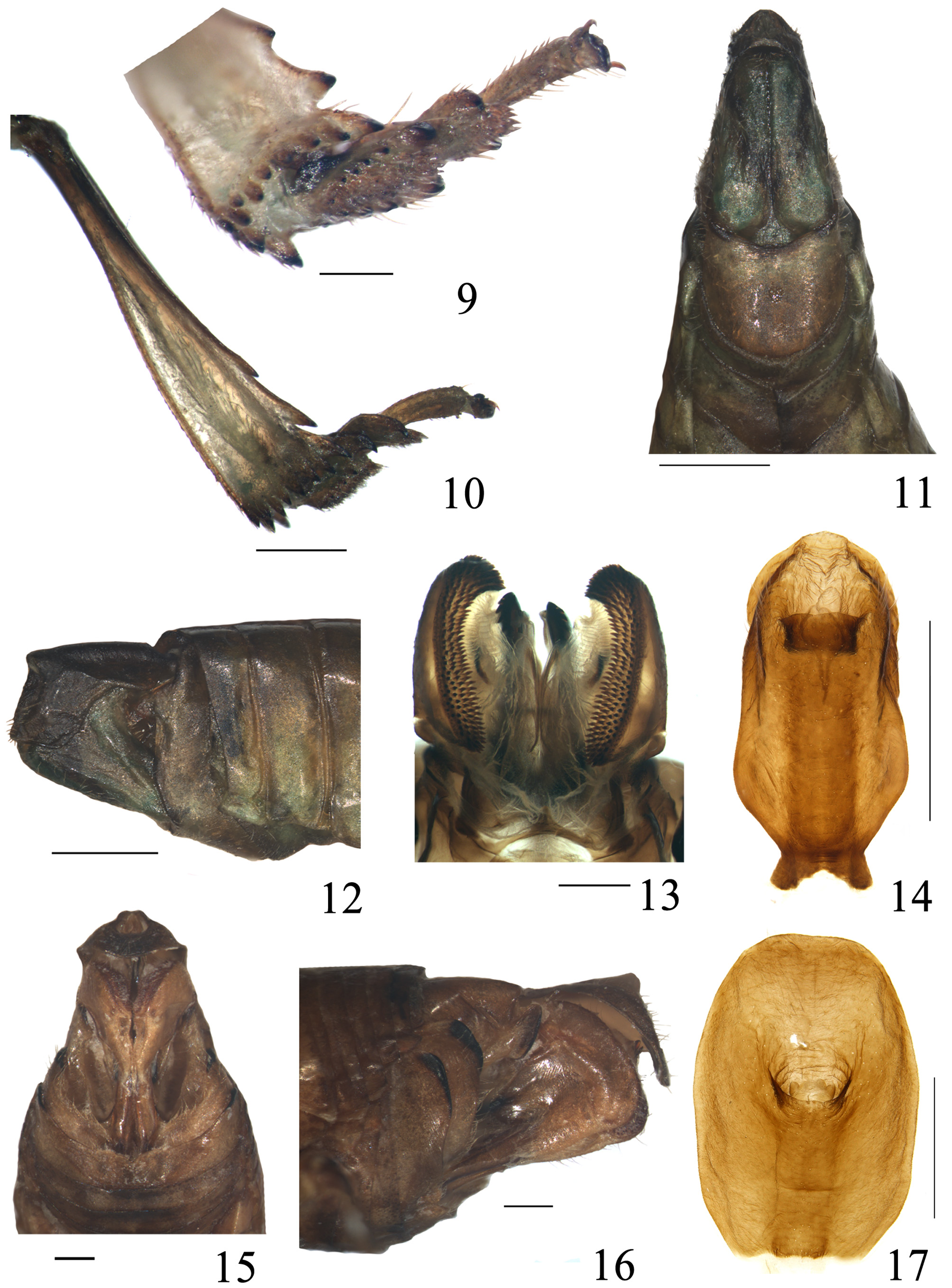
Description. Length, male (N=2) (including tegmen): 11.0–11.5 mm, length of tegmen: 9.6–10.0 mm; female (N=1) (including tegmen):13.2 mm, length of tegmen: 12.0 mm.
General color infuscate. Vertex fuscous. Frons pitchy, with a pallide-flavens transverse bar near upper margin, transverse stramineous fascia near frontoclypeal suture, and two yellow blotches near lateral margins above antennae; Y-shaped sublateral keel partly yellow. Clypeus pale cinereous with three yellow speckles at base, median carina fusco-rufous, median portion with parallel oblique pale yellow striae. Ocelli yellow. Eyes pitchy. Antenna fusco-piceous. Gena yellow with a large black speckle below antenna and a relatively small black spot in front of it. Pronotum fuscous, lateroventral pronotal lobes pitchy. Mesonotum fusco-piceous at disc, pale fulvous laterally. Tegmina translucent, tawny, with large irregularly fusco-piceous macula at basal half, and pale crineous triangular macula from apical angle to claval suture at distal half; precostal area with eight yellow spots; longitudinal veins reddish-brown or dark brown, transverse veinlets ochraceous. Wings caesious to pale fuscous, veins fuscescent. Legs testaceous with black fascia and annulus. Abdomen fusco-piceous on dorsum and ventrite, dark green laterally. Genital styles green-black. Male anal tube pitchy.
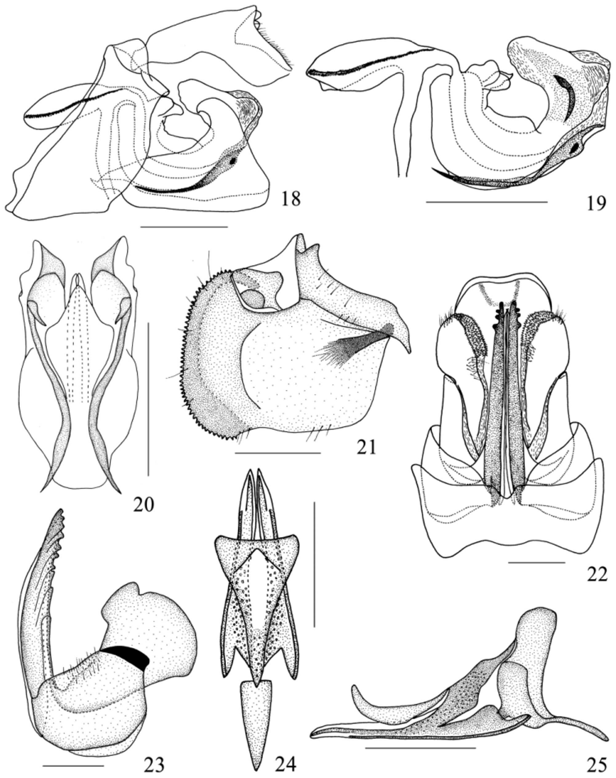
Male terminalia. Genital styles elongate in ventral view ( Fig. 11 View FIGURES 9 – 17 ), expanding distally, subtriangular in lateral view, with an oblique eminence on outer surface in lateral view ( Fig. 12 View FIGURES 9 – 17 ), dorsal margin concave, ventral margin almost straight, apical margin slightly oblique and straight. Capitulum broad and narrowing distally with apex terminating in a small hook ( Fig. 18 View FIGURES 18 – 25 ). Aedeagus deeply U-shaped in lateral view ( Fig. 19 View FIGURES 18 – 25 ). Dorso-lateral phallobase lobes strongly sclerotized, with a pair of short blade-shaped processes on lateral side; dorsal margin with large eminences extending to middle from base, ventral margin widely convex distally. Ventral phallobase lobe much shortened, bifurcated near middle. Phallus with a pair of long slender ventral processes arising laterally at middle ( Fig. 19 View FIGURES 18 – 25 ). Pygofer with posterior margin slightly convex, anterior margin oblique and shallowly concave, ventral margin straight ( Figs 11 View FIGURES 9 – 17 , 18 View FIGURES 18 – 25 ).
Female terminalia. Gonoplac rectangular in lateral view, almost as broad as long, bearing a number of minute denticles (about 5 rows) along dorsal margin to ventral margin in posterior view; median area depressed, with longitudinal carina subparallel to apical margin; third gonoplac lobes fused medially ( Figs 13, 15, 16 View FIGURES 9 – 17 , 22 View FIGURES 18 – 25 ). Posterior connective lamina of gonapophyses IX elongated, triangular-shaped in dorsal view; median field elevated with fused lobes in shape of “reversed triangle” ( Figs 24, 25 View FIGURES 18 – 25 ). Gonospiculum bridge large, flattened laterally, spadeshaped ( Fig. 24 View FIGURES 18 – 25 ). Gonocoxa VIII approximately square with slightly protruding hind margin ( Fig. 23 View FIGURES 18 – 25 ). Endogonocoxal process lance-shaped, narrowing apically, not furcated. Anterior connective lamina of gonapophyses VIII narrow and long, tapering distally, with 9 teeth (8 teeth with keels) ( Fig. 23 View FIGURES 18 – 25 ). Sternum VII deeply concave at middle of hind margin ( Figs 15 View FIGURES 9 – 17 , 22 View FIGURES 18 – 25 ).
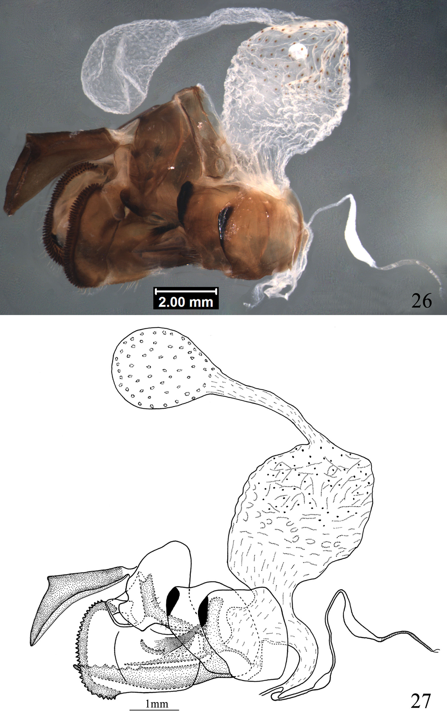
Female internal reproductive system: Distrysian. Bursa copulatrix well-developed with two pouches; first pouch (BC1) much larger than second pouch (BC2), wall of BC1 with easily visible cells and sclerotized ornamentation; BC2 membranous with weakly visible cells and unsclerotized minutely tuberculate ornamentation; BC1 and BC2 are connected by a relatively thin membranous duct. Vagina with posterior vagina and anterior vagina, posterior vagina relatively dumpy, with a pair of semicircular processes on dorsal side; anterior vagina thin, receiving anteriorly spermatheca and ventrally common oviduct near apex. Common oviduct relatively thin. Spermatheca well-developed, comprised of five parts: orificium receptaculi (or) dilated, ductus receptaculi (dr) thin and elongate, distinctly inflated scrotiform at middle, diverticulum ductus (dvd) evidently crescentiform, pars intermedialis (spp) relatively thin with delicate spiral fold and glandula apicalis (ga) distinctly divided into two longish ducts ( Figs 26, 27 View FIGURES 26 – 27 ).
Type material. Holotype: male, China, Hainan Province, Limuling Mountain, 670m, 13 August 2009, coll. Manqiang Wang and Rui Meng.
Paratypes: 1 male, China, Guangdong Province, Shenzhen City, Neilingting Island; 13 June 2002, coll. Fenglong Jia; 1 female, China, Hainan Province, Jianfengling Mountain, 28 July 1983, coll. Jianguo Long.
Diagnosis. The new species differs from the closely related Goniopsara mystica ( Melichar, 1899) in the frons forward protrude at lower part, widest at upper margin (frons flat, widest under the level of antenna in G. mystica ); hind tibia with two lateral spines (hind tibia with three lateral spines in G. mystica ).
Etymology. The specific epithet is derived from combination of Latin “frons” and “convexus”, referring to the frons forward protruding.

No known copyright restrictions apply. See Agosti, D., Egloff, W., 2009. Taxonomic information exchange and copyright: the Plazi approach. BMC Research Notes 2009, 2:53 for further explanation.
|
Kingdom |
|
|
Phylum |
|
|
Class |
|
|
Order |
|
|
Family |
|
|
Genus |