
Pisacha kwangsiensis Chou et Lu, 1977
|
publication ID |
https://doi.org/ 10.11646/zootaxa.3866.1.4 |
|
publication LSID |
lsid:zoobank.org:pub:6202FA35-AF83-4F98-82EE-F089EA7ACC35 |
|
DOI |
https://doi.org/10.5281/zenodo.6143305 |
|
persistent identifier |
https://treatment.plazi.org/id/8E3287F1-C226-F957-FF6C-1F55FBF9FF36 |
|
treatment provided by |
Plazi |
|
scientific name |
Pisacha kwangsiensis Chou et Lu, 1977 |
| status |
|
Pisacha kwangsiensis Chou et Lu, 1977 View in CoL
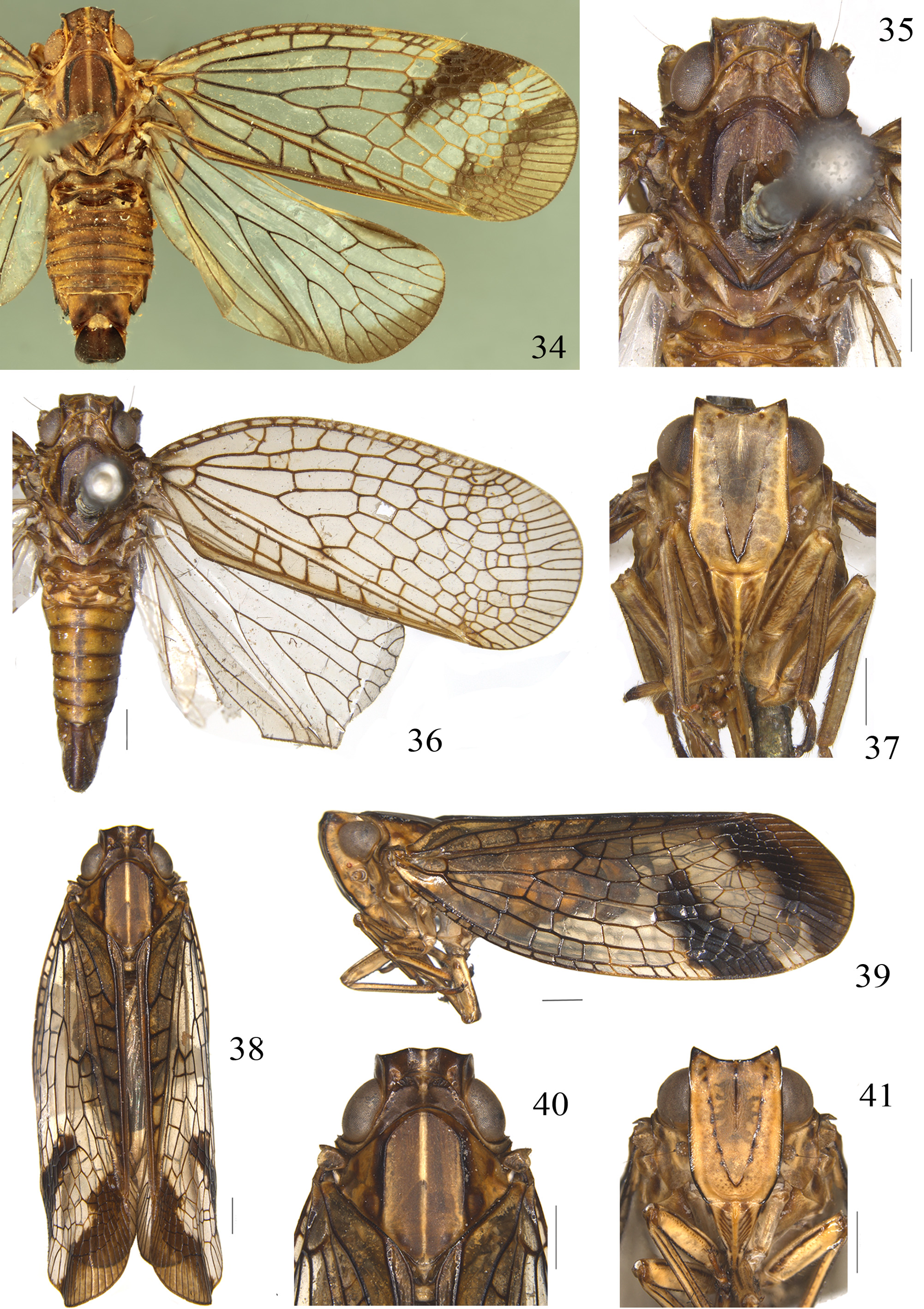
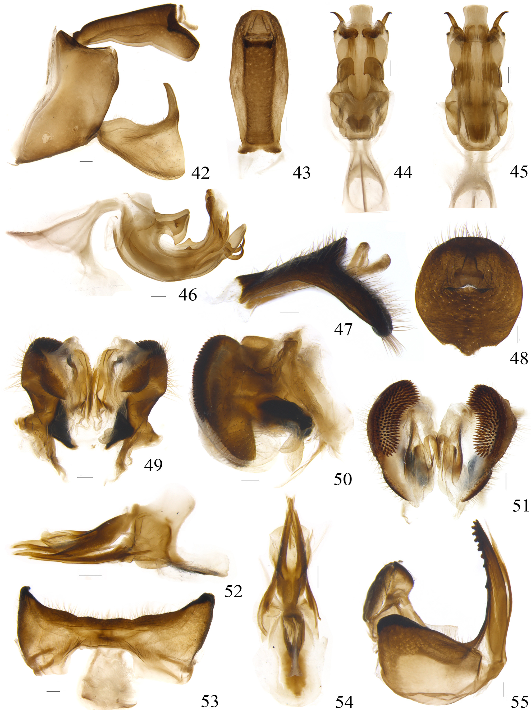
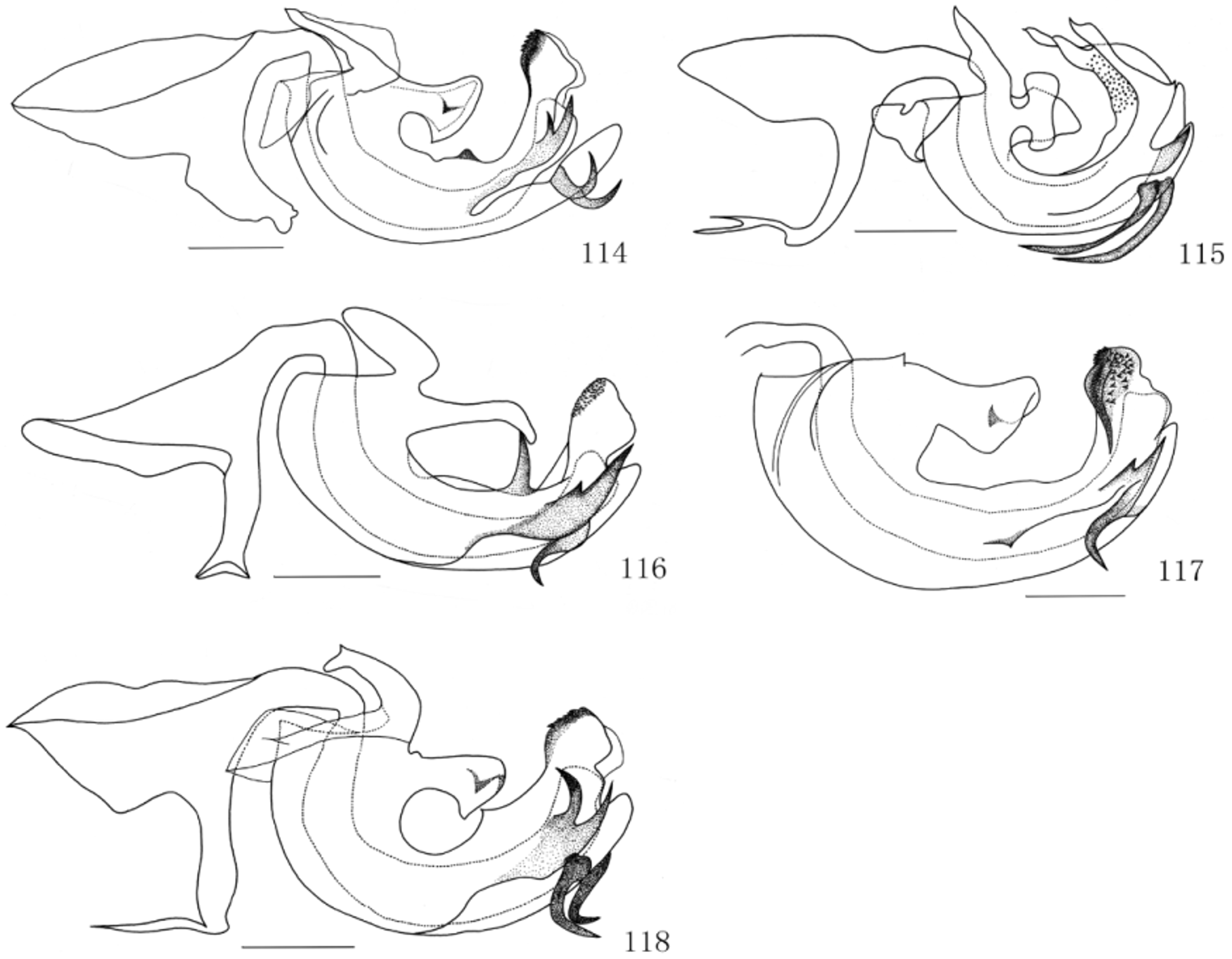
( Figs 35–37 View FIGURES 34 – 41 , 42–55 View FIGURES 42 – 55 , 114 View FIGURES 114 – 118 , 119 View FIGURES 119 – 123 )
Pisacha kwangsiensis Chou et Lu, 1977: 317 View in CoL ; 1985: 101.
Supplementary description. Head with eyes slightly narrower than pronotum ( Fig. 35 View FIGURES 34 – 41 ). Vertex 3.6 times wider than long in middle line, anterior margin acutely convex medianly, and posterior margin deeply concave; median carina wide, offwhite ( Figs 35, 36 View FIGURES 34 – 41 ). Frons longer than wide in widest part about 1.6 times; upper margin deeply concave, median carinae short, presented at apical fourth, pale yellow, “V” lateral carinae pale black and darkened at base, with about 5 obscure black spots along lateral carinae ( Fig. 37 View FIGURES 34 – 41 ). Clypeus triangular, median carina pale yellow ( Fig. 37 View FIGURES 34 – 41 ). Pronotum about 2.0 times longer than vertex in middle line; anterior margin convex between eyes, almost reaching the level of tops of eyes ( Fig. 35 View FIGURES 34 – 41 ). Mesonotum large, 2.8 times as long as vertex and pronotum in middle line ( Fig. 35 View FIGURES 34 – 41 ). Tegmina transparent, clavus with 7–10 transverse veinlets between CuP and Pcu veins, 0–3 transverse vein between two claval veins ( Fig. 36 View FIGURES 34 – 41 ). Spinulation formula of hind leg 13–12–2.
Male terminalia. Anal tube with ventral margin sinuate in lateral view; 2.5 times longer than widest part at middle, apical margin slightly convex ( Figs 42, 43 View FIGURES 42 – 55 ). Dorso-basal processes of phallobase hammer-shaped, apicoventral angles sharp in lateral view. Dorso-lateral phallobase lobes with a pair of small triangular processes on dorsal margins at middle, dorsum with about 60 small spikes on each side surface; lateral apical bifurcate processes with upper branches about half length of the under branches. Ventral phallobase lobe strongly sclerous, apical margin nearly straight (Figs 45,119). Phallus with a pair of deeply curved hooks backward directed dorsally near apex ( Figs 46 View FIGURES 42 – 55 , 114 View FIGURES 114 – 118 ). Pygofer with posterior margin slightly convex near dorsal third ( Fig. 42 View FIGURES 42 – 55 ).
Female terminalia. Anal tube with apical margin obtusely convex ( Fig. 48 View FIGURES 42 – 55 ). Gonoplac bearing about 6–7 rows of denticles in posterior view ( Fig. 51 View FIGURES 42 – 55 ). Anterior connective lamina of gonapophyses VIII with 10 teeth ( Fig. 55 View FIGURES 42 – 55 ). Sternum VII with posterior margin slightly concave at middle ( Fig. 53 View FIGURES 42 – 55 ).
Type material examined. Holotype: male, China, Guangxi Zhuangzu Autonomous Region, Longsheng County, Sanmen Town, 20 August 1964, coll. Shengli Liu.
Paratypes: 1 female, China, Guangxi Zhuangzu Autonomous Region, Longzhou City, Nonggang National Nature Reserve, May 1980, coll. Zhuyin Wang; 1 female, China, Guangxi Zhuangzu Autonomous Region, Longzhou City, Nonggang National Nature Reserve, 20 May 1983, coll. Jikun Yang.

No known copyright restrictions apply. See Agosti, D., Egloff, W., 2009. Taxonomic information exchange and copyright: the Plazi approach. BMC Research Notes 2009, 2:53 for further explanation.
|
Kingdom |
|
|
Phylum |
|
|
Class |
|
|
Order |
|
|
Family |
|
|
Genus |
Pisacha kwangsiensis Chou et Lu, 1977
| Meng, Rui, Wang, Menglin & Wang, Yinglun 2014 |
Pisacha kwangsiensis
| Chou 1977: 317 |