
Pisacha Distant, 1906
|
publication ID |
https://doi.org/ 10.11646/zootaxa.3866.1.4 |
|
publication LSID |
lsid:zoobank.org:pub:6202FA35-AF83-4F98-82EE-F089EA7ACC35 |
|
DOI |
https://doi.org/10.5281/zenodo.6143295 |
|
persistent identifier |
https://treatment.plazi.org/id/8E3287F1-C225-F950-FF6C-1D4DFAA3FAB6 |
|
treatment provided by |
Plazi |
|
scientific name |
Pisacha Distant, 1906 |
| status |
|
Pisacha Distant, 1906 View in CoL View at ENA
Pisacha Distant, 1906: 391 View in CoL . Type speices: Pisacha naga Distant, 1906 View in CoL , by original designation. Soaemis Jacobi, 1915 . Nomen nudum.
Soaemis Jacobi, 1916: 311 . Type species: Soaemis encaustica Jacobi, 1916 View in CoL , by original designation. Synonymized by Ishihara, 1965: 207.
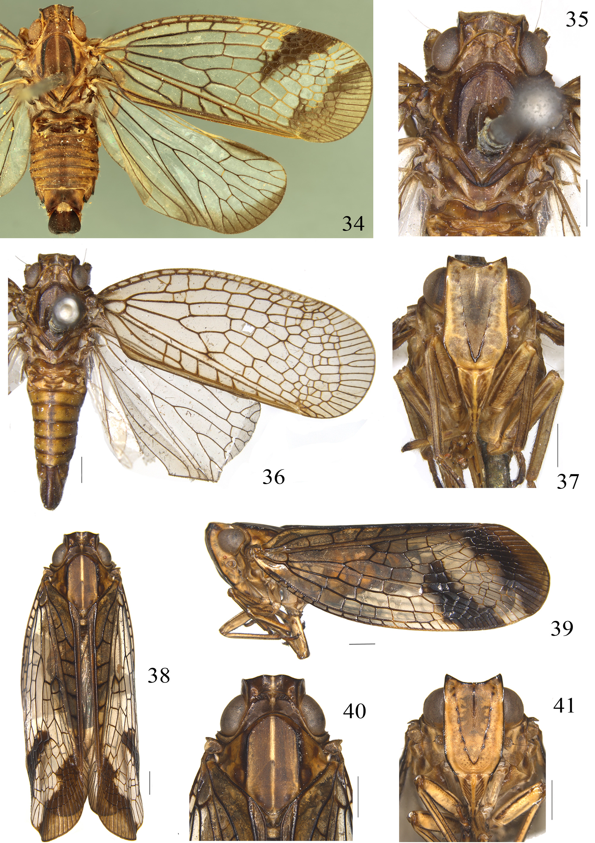
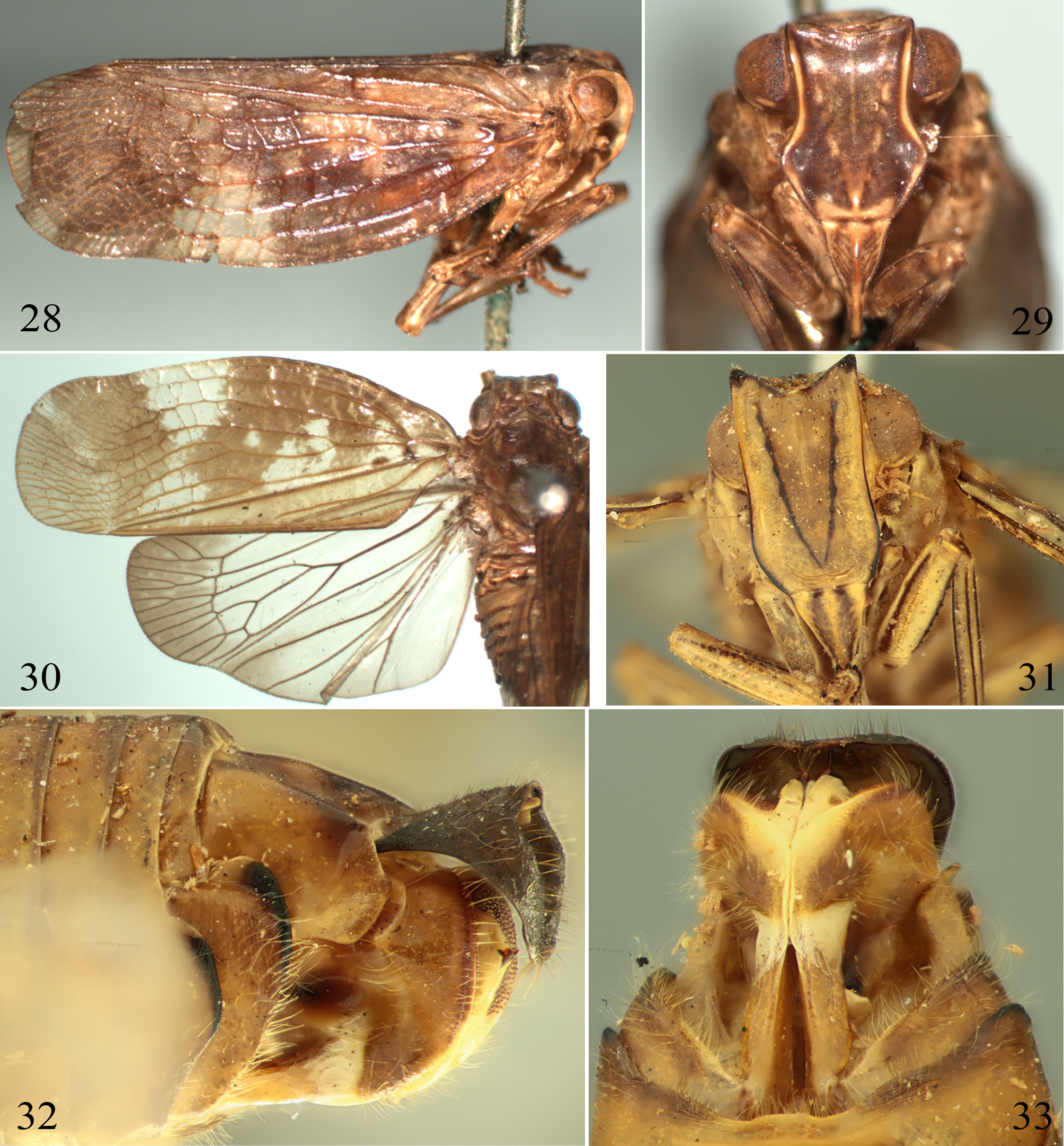
Supplementary description. Head with eyes slightly narrower than pronotum ( Figs 34, 35, 40 View FIGURES 34 – 41 , 72 View FIGURES 70 – 78 , 81, 85 View FIGURES 79 – 86 ). Vertex with median carina, disc depressed, lateral margins clearly elevated ( Figs 34, 35, 40 View FIGURES 34 – 41 , 72 View FIGURES 70 – 78 , 81, 85 View FIGURES 79 – 86 ). Frons long, lateral margins nearly parallel, slightly expanded below the level of antennae, then tapered to frontoclypeal suture; median carina disappeared near apical one-fourth to half of frons ( Figs 31 View FIGURES 28 – 33 , 37, 41 View FIGURES 34 – 41 , 73 View FIGURES 70 – 78 , 82, 86 View FIGURES 79 – 86 ). Clypeus narrow, triangular, with distinct yellow median carina, and with fuscous fascia on each side of mendian carina ( Figs 31 View FIGURES 28 – 33 , 37, 41 View FIGURES 34 – 41 , 73 View FIGURES 70 – 78 , 82, 86 View FIGURES 79 – 86 ). Frontoclypeal suture nearly straight. Rostrum elongate surpassing post trochanters, subapical segment slightly longer than apical segment. Pronotum with median carina, depressed at center and elevated laterally; anterior margin obtusely convex, almost reaching or surpassing the level of tops of eyes, posterior margin concave, both distinctly carinate, lateral lobe distinctly broadened ( Figs 34, 35, 40 View FIGURES 34 – 41 , 72 View FIGURES 70 – 78 , 81, 85 View FIGURES 79 – 86 ). Mesonotum large, almost as wide in widest part medianly as long in middle line, lateral carinae reaching or not to posterior margins ( Figs 34, 35, 40 View FIGURES 34 – 41 , 72 View FIGURES 70 – 78 , 81, 85 View FIGURES 79 – 86 ). Tegmina transparent, costal membrane narrow, Sc+R veins forked near basal cell, M vein bifurcate at basal third, Cu vein simple, between the veins with many distinct transverse veinlets at apical half, and the apical transverse veinlets forming distinct nodal line; clavus with 7–10 transverse veinlets between CuP and Pcu veins, 0–3 transverse vein between two claval veins. Legs yellow with black fascia and spots along margins, hind tibia widened at apical half and with two lateral spines.
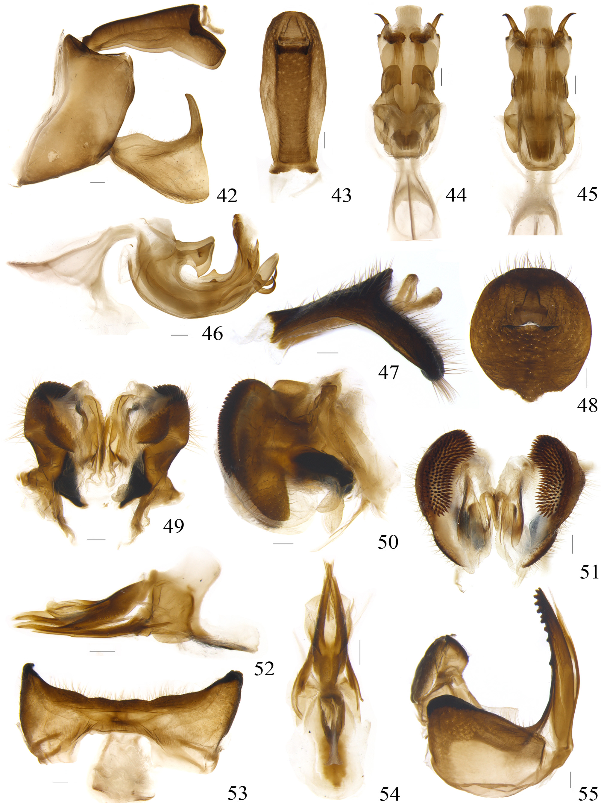
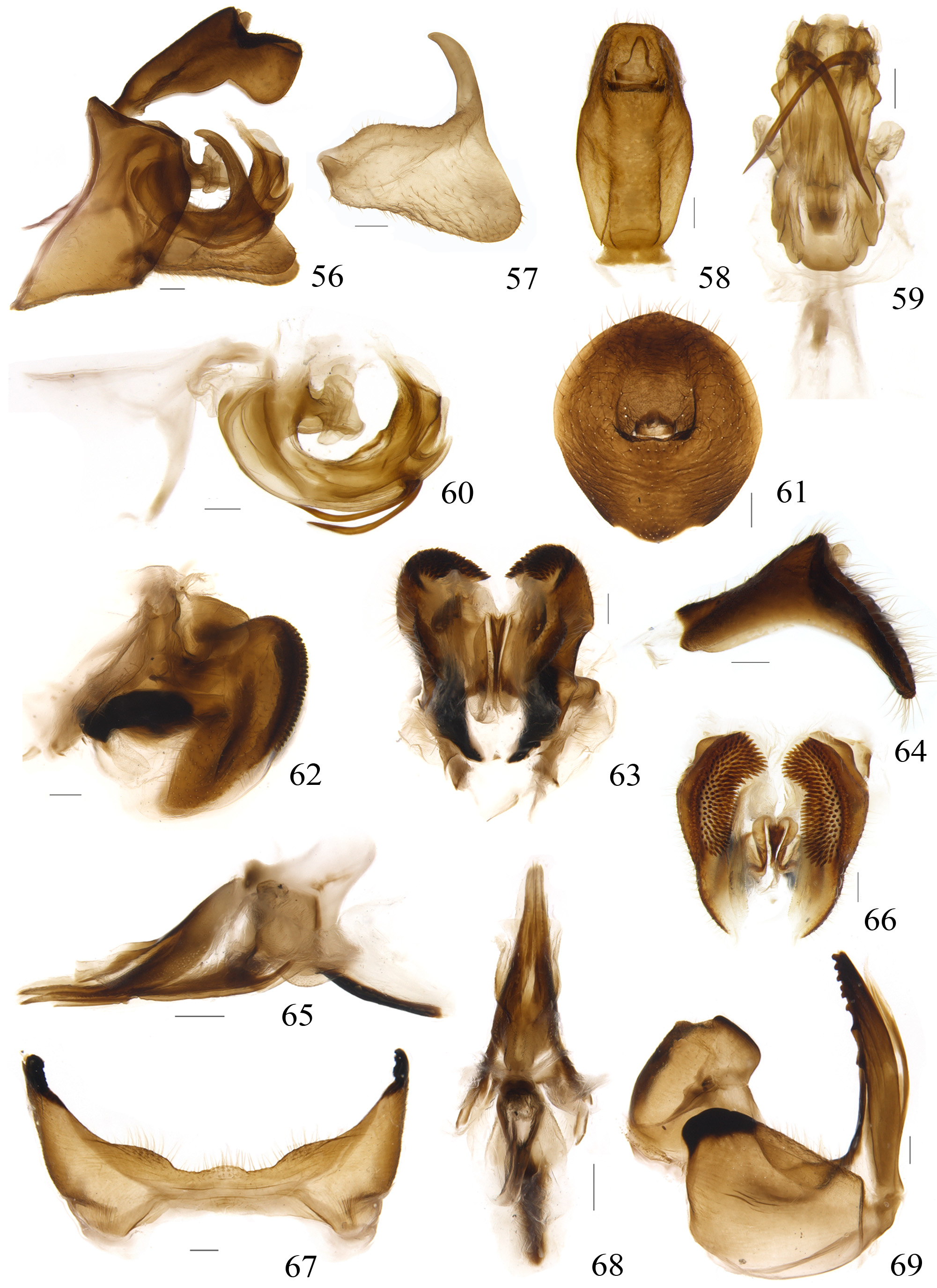
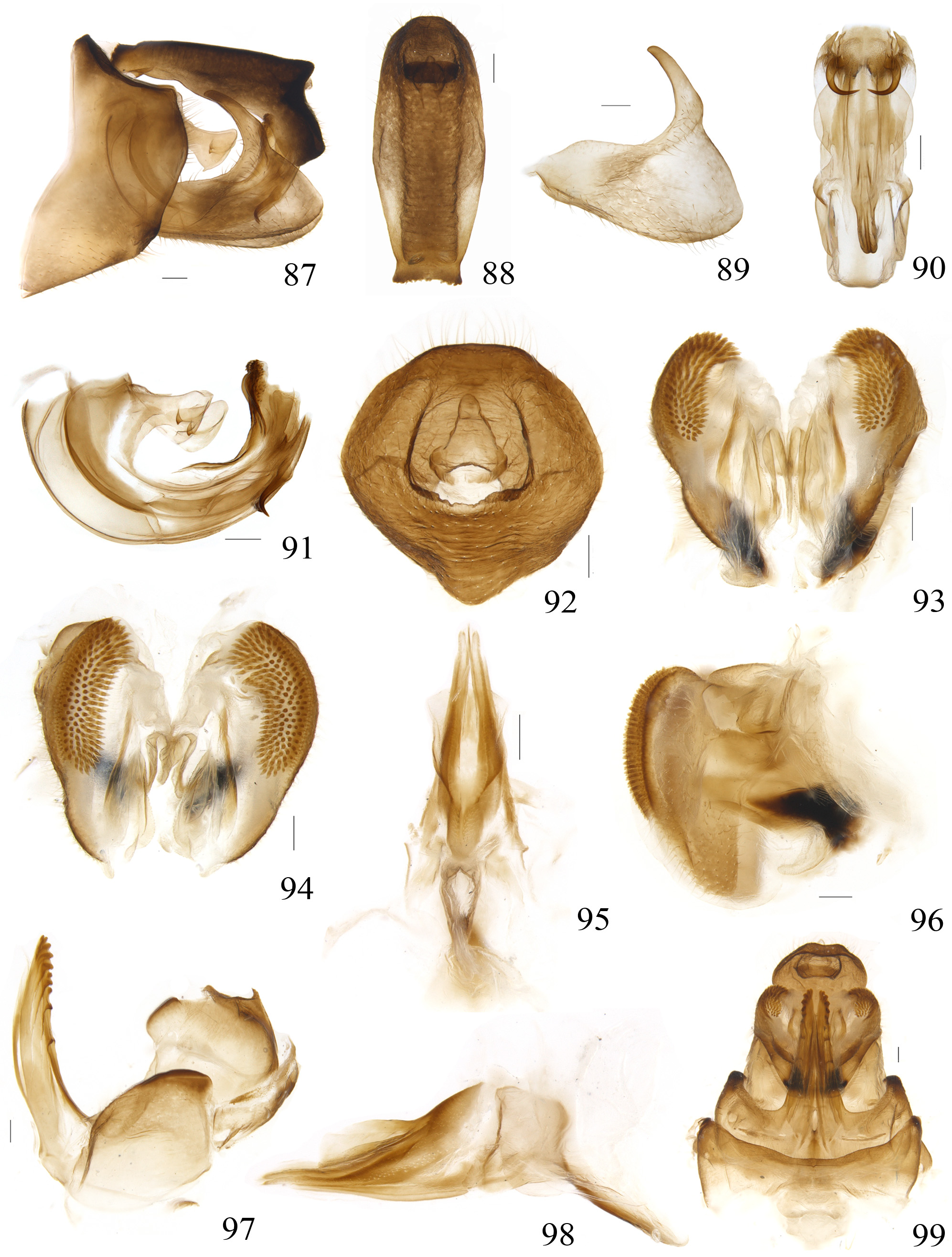
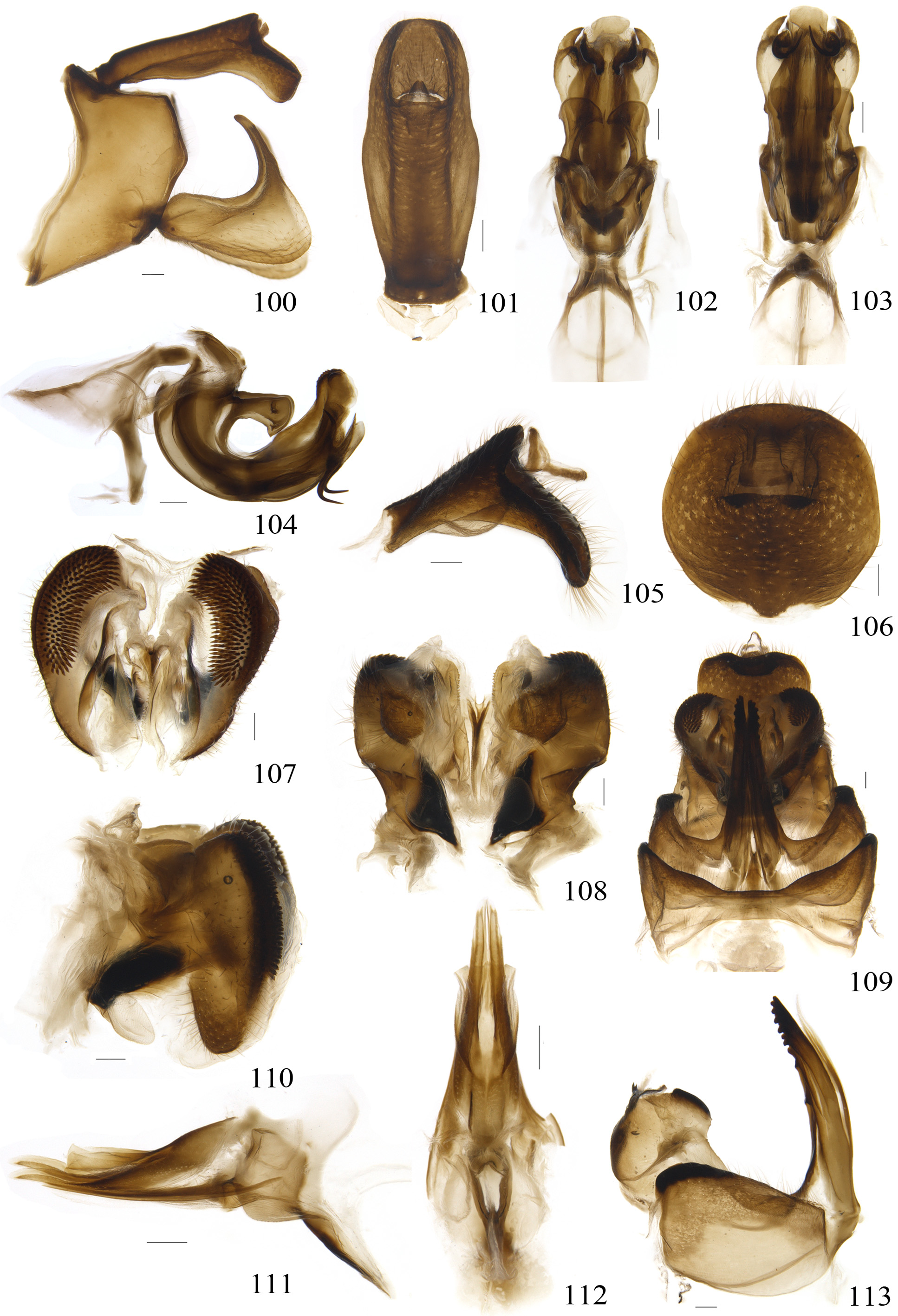
Male terminalia. Anal tube elongate, expended near middle in dorsal view ( Figs 43 View FIGURES 42 – 55 , 58 View FIGURES 56 – 69 , 77 View FIGURES 70 – 78 , 88 View FIGURES 87 – 99 , 101 View FIGURES 100 – 113 ), bent down near apical fourth, ventral margin sinuate in lateral view. Anal column short, located at point of flexure ( Figs 42 View FIGURES 42 – 55 , 56 View FIGURES 56 – 69 , 74 View FIGURES 70 – 78 , 87 View FIGURES 87 – 99 , 100 View FIGURES 100 – 113 ). Genital styles subtriangular in lateral view, expanding distally, posterior margin concave at middle, caudo-ventral angle strongly convex, ventral margin almost straight and dorsal margin slightly convex at basal third ( Figs 42 View FIGURES 42 – 55 , 57 View FIGURES 56 – 69 , 74 View FIGURES 70 – 78 , 89 View FIGURES 87 – 99 , 100 View FIGURES 100 – 113 ). Capitulum long, without teeth. Aedeagus U-shaped in lateral view, with large phallobase ( Figs 46 View FIGURES 42 – 55 , 60 View FIGURES 56 – 69 , 78 View FIGURES 70 – 78 , 91 View FIGURES 87 – 99 , 104 View FIGURES 100 – 113 ). Phallobase with dorso-basal portion prolonged into a pair of large processes. Dorso-lateral phallobase lobes spilt near apex, dorsum curved upward and bifurcate apically, with numbers of small spikes on each side surface or not; lateral forming into a pair of processes, bifurcate or not ( Figs 46 View FIGURES 42 – 55 , 60 View FIGURES 56 – 69 , 78 View FIGURES 70 – 78 , 91 View FIGURES 87 – 99 , 104 View FIGURES 100 – 113 ). Ventral phallobase lobe more or less round at median part, and nearly rectangular at apical part ( Figs 45 View FIGURES 42 – 55 , 59 View FIGURES 56 – 69 , 76 View FIGURES 70 – 78 , 90 View FIGURES 87 – 99 , 103 View FIGURES 100 – 113 ). Phallus with a pair of short or long ventral processes arising from apical part ( Figs 46 View FIGURES 42 – 55 , 60 View FIGURES 56 – 69 , 78 View FIGURES 70 – 78 , 91 View FIGURES 87 – 99 , 104 View FIGURES 100 – 113 ). Pygofer short and wide, anterior margin concave medianly, clearly longer than posterior margin, ventral margin oblique ( Figs 42 View FIGURES 42 – 55 , 56 View FIGURES 56 – 69 , 74 View FIGURES 70 – 78 , 87 View FIGURES 87 – 99 , 100 View FIGURES 100 – 113 ).
Female terminalia. Anal tube nearly oval in dorsal view ( Figs 48 View FIGURES 42 – 55 , 61 View FIGURES 56 – 69 , 92 View FIGURES 87 – 99 , 106 View FIGURES 100 – 113 ), bent down at midlength in lateral view ( Figs 47 View FIGURES 42 – 55 , 64 View FIGURES 56 – 69 , 105 View FIGURES 100 – 113 ). Anal column relatively long, situated at point of flexure. Gonoplac with apical half wider than basal half, apical margin obtusely convex with dorsal two-third minutely denticulate, ventral one-third membranous; basal part bearing a black sclerous structure, lying on the top of proximal part of gonapophyses IX ( Figs 50 View FIGURES 42 – 55 , 62 View FIGURES 56 – 69 , 96 View FIGURES 87 – 99 , 110 View FIGURES 100 – 113 ); in posterior view, apical part bearing several rows of denticles, inner margin of membranous part bearing minutely spinules ( Figs 51 View FIGURES 42 – 55 , 66 View FIGURES 56 – 69 , 94 View FIGURES 87 – 99 , 107 View FIGURES 100 – 113 ); the third gonoplac lobes slightly sclerotized and nearly fused at central axis ( Figs 49 View FIGURES 42 – 55 , 63 View FIGURES 56 – 69 , 93 View FIGURES 87 – 99 , 108 View FIGURES 100 – 113 ). Posterior connective lamina of gonapophyses IX elongated, triangularshaped; median field sclerous, with fused lobe nearly quadrate ( Figs 52, 54 View FIGURES 42 – 55 , 65, 68 View FIGURES 56 – 69 , 95, 98 View FIGURES 87 – 99 , 111, 112 View FIGURES 100 – 113 ). Gonospiculum bridge large, spade-shaped ( Figs 52 View FIGURES 42 – 55 , 65 View FIGURES 56 – 69 , 98 View FIGURES 87 – 99 , 111 View FIGURES 100 – 113 ). Gonocoxa VIII approximately square with hind margin slightly protruding. Endogonocoxal process narrowing apically, not furcated. Anterior connective lamina of gonapophyses VIII narrow, tapering apically, with a row of teeth along outer margin ( Figs 55 View FIGURES 42 – 55 , 69 View FIGURES 56 – 69 , 97 View FIGURES 87 – 99 , 113 View FIGURES 100 – 113 ). Sternum VII with posterior margin sinuate, middle part quite lower than two sides ( Figs 33 View FIGURES 28 – 33 , 53 View FIGURES 42 – 55 , 67 View FIGURES 56 – 69 , 99 View FIGURES 87 – 99 , 109 View FIGURES 100 – 113 ).
Distribution. Indian, China ( Taiwan, Guangxi, Hainan, Zhejiang, Chongqing, Sichuan), Vietnam.


No known copyright restrictions apply. See Agosti, D., Egloff, W., 2009. Taxonomic information exchange and copyright: the Plazi approach. BMC Research Notes 2009, 2:53 for further explanation.
|
Kingdom |
|
|
Phylum |
|
|
Class |
|
|
Order |
|
|
Family |
Pisacha Distant, 1906
| Meng, Rui, Wang, Menglin & Wang, Yinglun 2014 |
Soaemis
| Ishihara 1965: 207 |
| Jacobi 1916: 311 |
Pisacha
| Distant 1906: 391 |