
Putaoa titanoverpa, Liu & Xu & Hormiga & Yin & Li, 2023
|
publication ID |
https://doi.org/ 10.11646/zootaxa.5277.3.7 |
|
publication LSID |
lsid:zoobank.org:pub:3762065B-479C-4669-AA3A-8855D5814516 |
|
DOI |
https://doi.org/10.5281/zenodo.7896113 |
|
persistent identifier |
https://treatment.plazi.org/id/88418785-5C7C-B554-BA9D-5B47FD89FE40 |
|
treatment provided by |
Plazi |
|
scientific name |
Putaoa titanoverpa |
| status |
sp. nov. |
Putaoa titanoverpa new species
(ĦṜƥgḓ)
Figures 3 View FIGURE 3 , 4 View FIGURE 4 , 5D–F View FIGURE 5 , 6C, D View FIGURE 6
Holotype ♁ ( HNU284 View Materials ): CHINA, Hunan Province, Liuyang City, Daweishan Mountain , 28°26.09'N, 114°10.09'E, 530m, 17 I 2018, Keke Liu, Luyu Wang, Guchun Zhou GoogleMaps . Paratypes: 1♁ ( HNU285 View Materials ) 4♀ ( HNU286–289 View Materials ), same data as the holotype GoogleMaps . Additional specimens examined: 8♀ ( HNU290–297 View Materials ), same data as the types GoogleMaps .
Etymology. The species epithet refers to the robust and strongly sclerotized embolus and is derived from the Greek word titan (in the sense of large size) and the Latin word verpa (penis).
Diagnosis. Males of this new species are similar to those of Putaoa huaping Hormiga and Tu, 2008 in having “U” shaped and sclerotized embolus (compare Figs 4A View FIGURE 4 , 6D View FIGURE 6 with fig. 3C in Hormiga and Tu, 2008), but can be distinguished by having the palpal tibia with one distal macroseta (cuspule-like), and the suprategulum with a lamelliform distal suprategular apophysis, while in P. huaping the palpal tibia has several distal macrosetae and the suprategulum has a stronger distal suprategular apophysis (compare Figs 4A–D View FIGURE 4 , 6C, D View FIGURE 6 with fig. 3 in Hormiga and Tu 2008). The diagnosis of the females of this new species is given under that of Putaoa annulata n. sp.
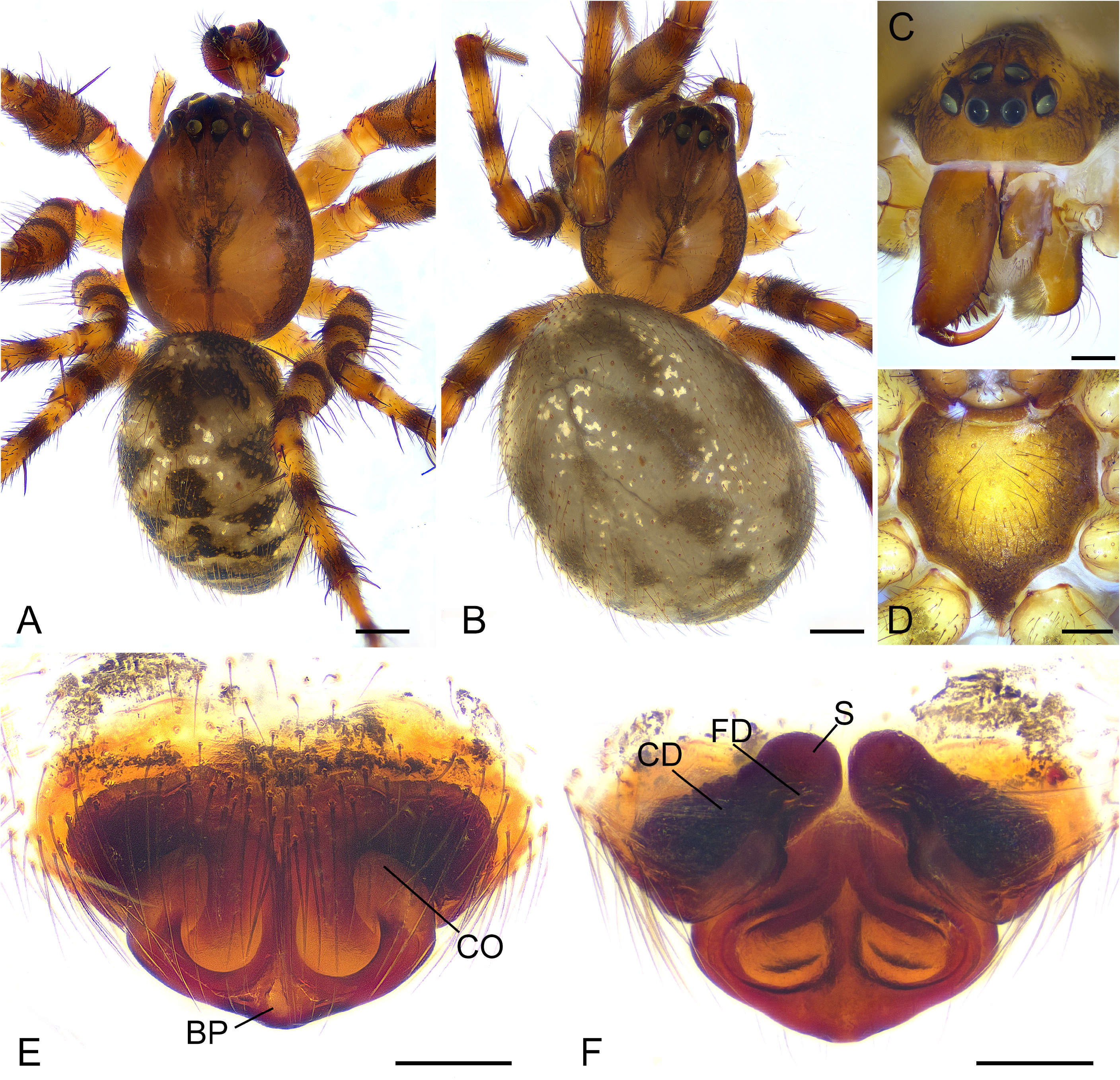
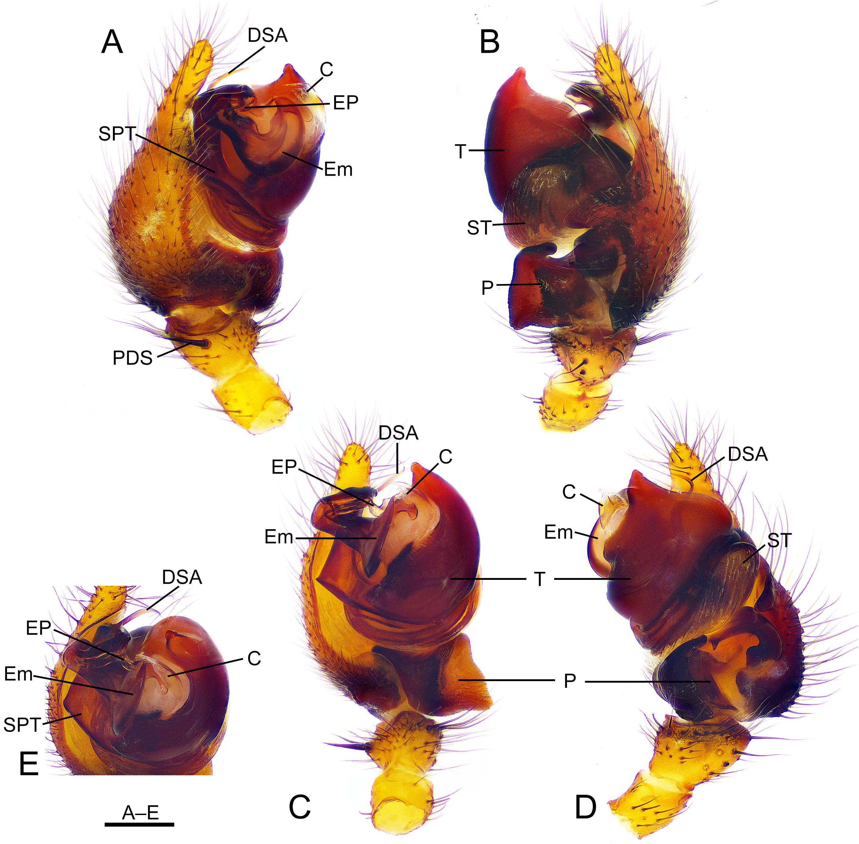
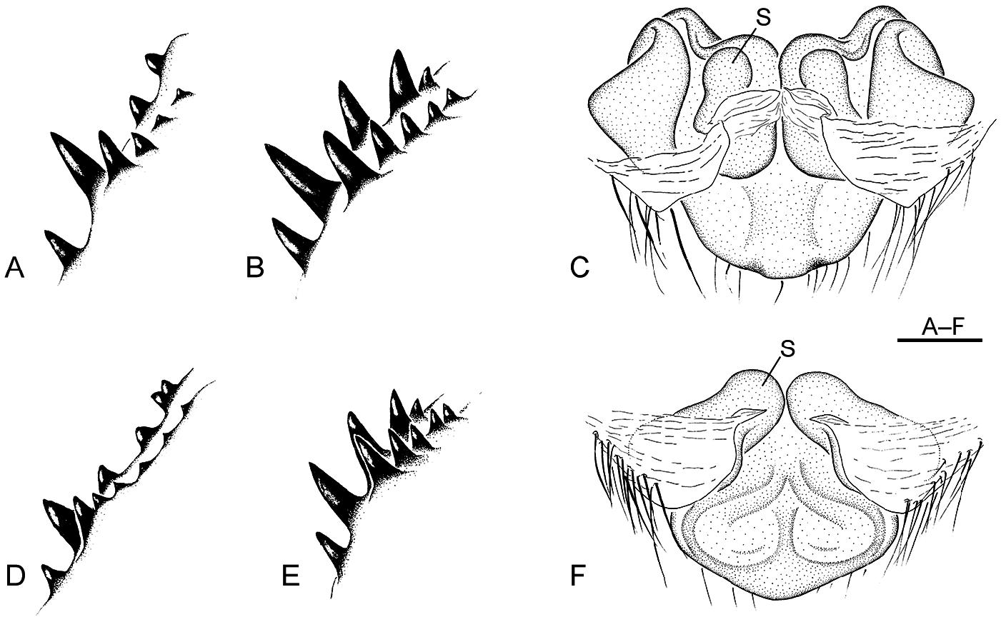
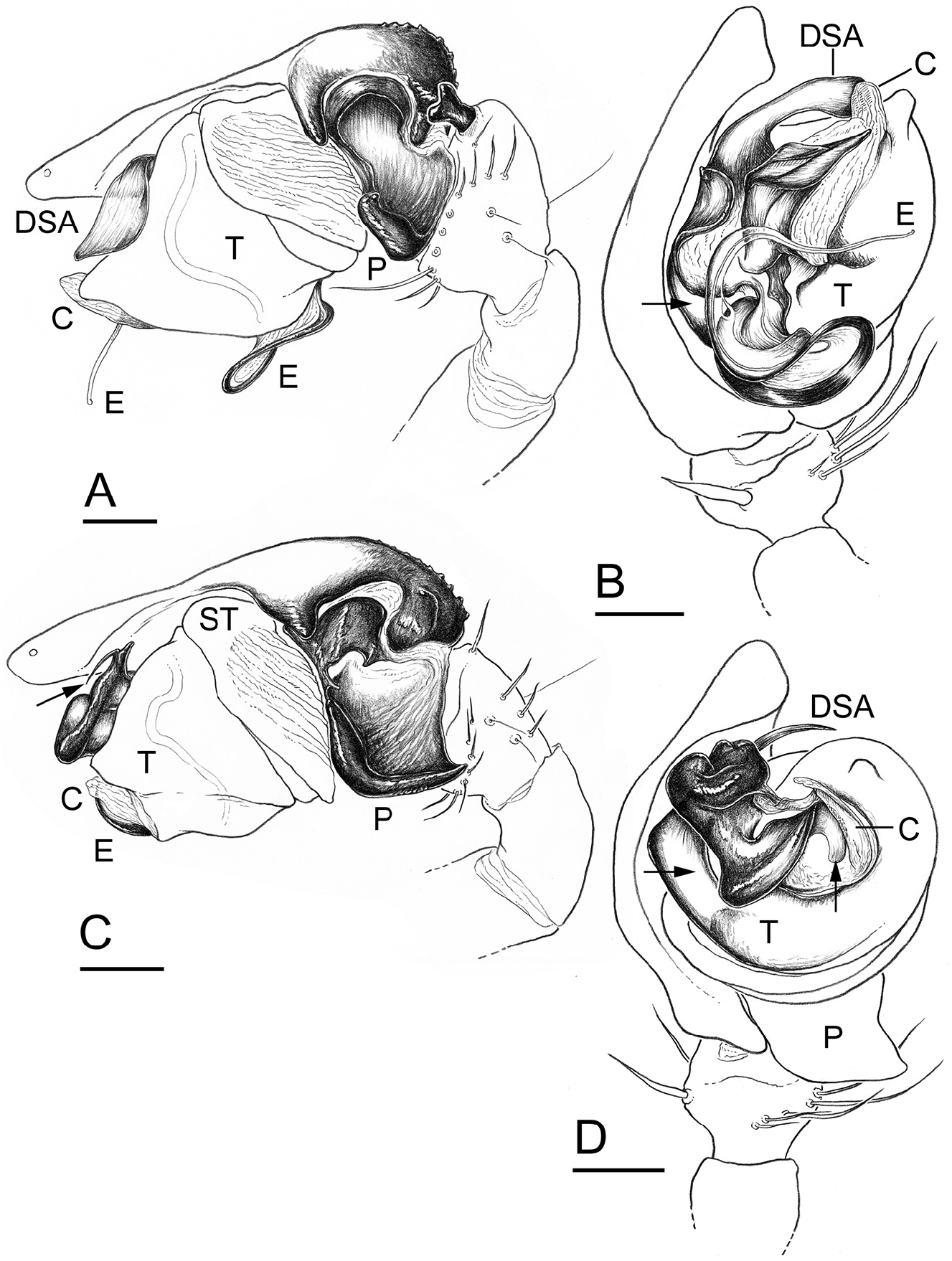
Description. Male (holotype; HNU284, Figs 3A View FIGURE 3 , 4 View FIGURE 4 ). Total length 9.68. Cephalothorax 4.33 long, 3.56 wide. Sternum 2.22 long, 1.99 wide. Abdomen 5.07 long, 3.69 wide. Anterior median eye diameter 0.17. Clypeus height 1.5 times one anterior median eye diameter. Carapace with shallow and longitudinal fovea ( Fig. 3A View FIGURE 3 ). Chelicerae with six prolateral and six retrolateral teeth ( Fig. 5D View FIGURE 5 ); stridulatory striae absent. Legs annulated with spines and with annulations alternating brown and black. Leg measurements: I 15.06 (4.25, 1.45, 3.78, 3.81, 1.77), II 13.68 (4.04, 1.34, 3.27, 3.45, 1.58), III 10.68 (3.20, 1.19, 2.47, 2.56, 1.26), IV 13.16 (3.83, 1.20, 3.18, 3.55, 1.40). All metatarsal trichobothria 0.33 approximately (TmI–IV), located on the dorsum near the retrolateral surface. Abdomen with white spots in anterior part and irregular patches dorsally. Palp: tibia short, with one dorsal and two retrolateral trichobothria and one distal seta prolaterally ( Figs 4A, C, D View FIGURE 4 , 6C, D View FIGURE 6 ). Cymbium with a conical ectal process in medial retrolateral margin ( Figs 4B View FIGURE 4 , 6C View FIGURE 6 ), apically pointed. Paracymbium glabrous, concave and bowl-like, with apical end broad and nearly square in ventral view, connected to cymbial base by means of a membrane, ventral to a sclerotized cymbial apophysis ( Figs 4B–D View FIGURE 4 , 6C View FIGURE 6 ). Tegulum large and pointed apically, with tegular base connecting to suprategulum which is divided into three processes, embolus, embolic process and distal suprategular apophysis ( Figs 4C, E View FIGURE 4 , 6D View FIGURE 6 ). The embolus “U” shaped (especially in prolateral view), developed and sclerotized, flattening in distal half part ( Figs 4A, C, E View FIGURE 4 , 6D View FIGURE 6 ). Embolic process thin and small, close to embolus apex ( Figs 4C, E View FIGURE 4 , 6D View FIGURE 6 ). Distal suprategular apophysis short and spiculate, curved in retrolateral view, extending towards end of tegulum across the gap between bulb and cymbium ( Figs 4C, D View FIGURE 4 , 6C, D View FIGURE 6 ). Conductor triangular, membranous, pointing apically end of tegulum ( Figs 4D View FIGURE 4 , 6D View FIGURE 6 ). Median apophysis absent.
Female (paratype, HNU287). Total length 9.85 ( Fig. 3B View FIGURE 3 ). Cephalothorax 4.06 long, 3.01wide. Sternum 2.27 long, 2.08 wide ( Fig. 4D View FIGURE 4 ). Abdomen 6.86 long, 5.13 wide. Anterior median eye diameter 0.18. Clypeus height 1.4 times one anterior median eye diameter. Chelicerae with five prolateral and five retrolateral teeth ( Fig. 5E View FIGURE 5 ); stridulatory striae absent. Legs annulated with spines and with annulations alternating brown and black. Leg measurements: I 15.68 (4.44, 1.50, 3.89, 3.97, 1.88), II 14.62 (4.18, 1.54, 3.60, 3.60, 1.70), III 11.57 (3.38, 1.35, 2.68, 2.73, 1.40), IV 13.96 (4.10, 1.34, 3.46, 3.55, 1.51). All metatarsal trichobothria 0.33 approximately (TmI–IV), located on the dorsum near the retrolateral surface. Similar to male in somatic features. Epigynum: basal plate slightly forming an obtuse triangle, and with the median area of posterior margin protruding caudally ( Figs 3E, F View FIGURE 3 , 5F View FIGURE 5 ). Atrium divided in two by septum. Copulatory openings visible ventrally ( Fig. 3E View FIGURE 3 ). Copulatory ducts short and thick. Spermathecae spherical, almost in contact with each other. Fertilization ducts medially oriented, arising from the middle of spermathecae ( Figs 3F View FIGURE 3 , 5F View FIGURE 5 ).
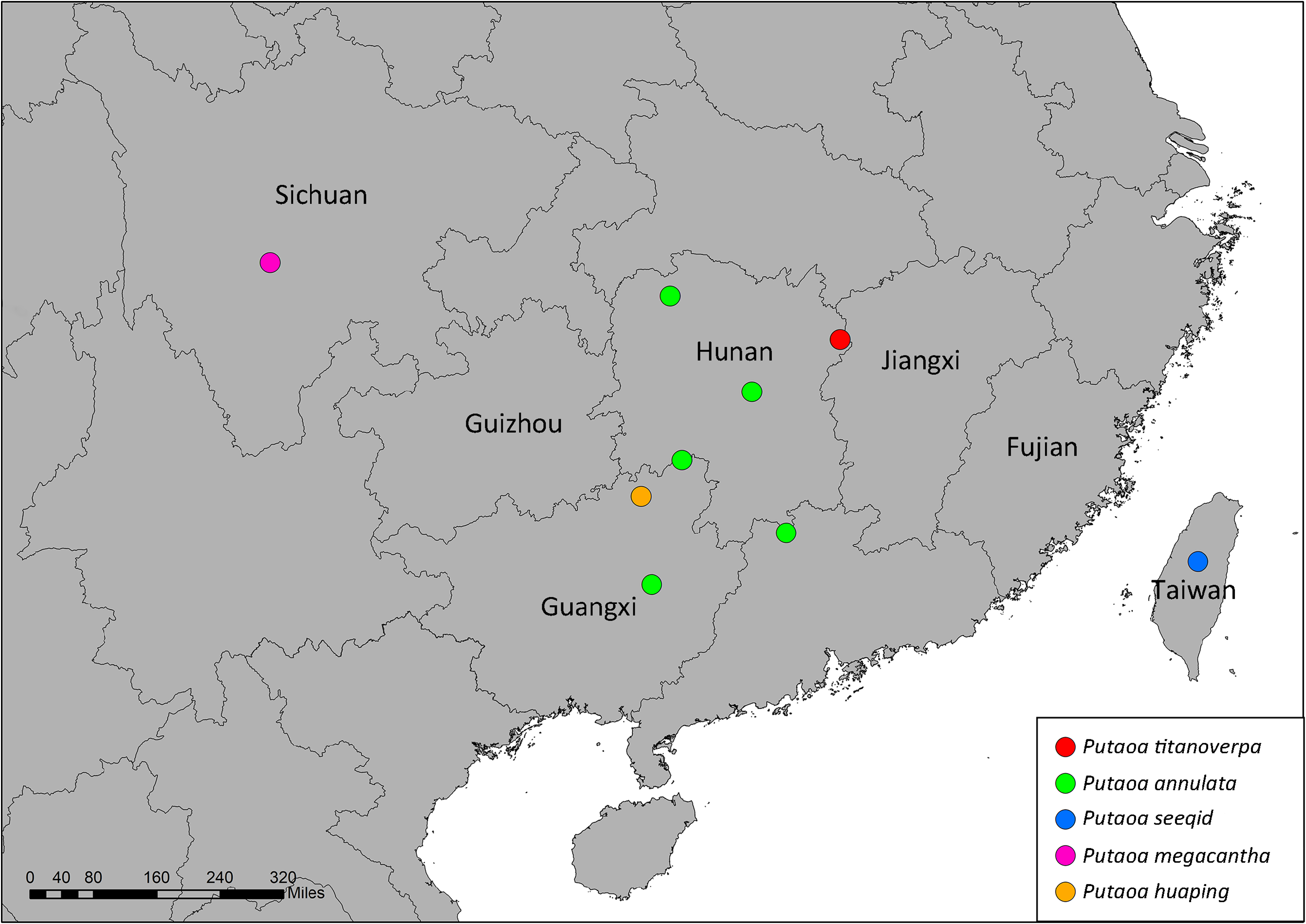
Distribution. China (Hunan) ( Fig. 8 View FIGURE 8 ).
No known copyright restrictions apply. See Agosti, D., Egloff, W., 2009. Taxonomic information exchange and copyright: the Plazi approach. BMC Research Notes 2009, 2:53 for further explanation.