
MEMBRANIPOROIDEA Busk, 1852
|
publication ID |
https://doi.org/ 10.1080/00222930701391773 |
|
persistent identifier |
https://treatment.plazi.org/id/877A7251-CC58-DE25-FE9B-26E0D2041A96 |
|
treatment provided by |
Felipe |
|
scientific name |
MEMBRANIPOROIDEA Busk, 1852 |
| status |
|
Superfamily MEMBRANIPOROIDEA Busk, 1852 View in CoL Family ELECTRIDAE d’Orbigny, 1851
Genus Electra Lamouroux, 1816 View in CoL Electra korobokkura Nikulina, 2006
( Figure 3 View Figure 3 ) Electra korobokkura Nikulina 2006, p 23 , Figures 1–5 View Figure 1 View Figure 2 View Figure 3 View Figure 4 View Figure 5 .
Material examined
MIN, colony on abraded metal plate (NHM 2006.2.27.1), colony on rock (NHM 2006.2.27.2). Additional material: 149 specimens. Also examined for comparison: Electra arctica ( Borg, 1931) , ZIRAS 36 /50535, colony on a pebble sorted from a crab trap Middle Fishery Refrigerator Trawler Rodino, 57 ° 36.29N, 156 ° 09.09E, Sea of Okhotsk , Western Kamchatka shelf, about 32 km from cape Hayryuzova , depth 78–81 m, coll. 12 September 1992 by A. V. Grischenko GoogleMaps .
Description
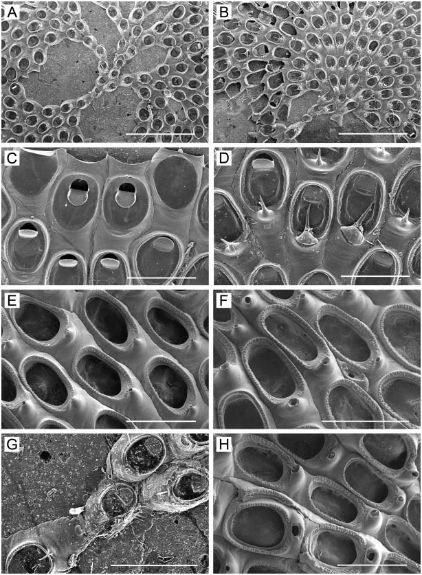
Colony encrusting, yellowish when alive, forming a thin, irregular network, the largest observed about 2.5 cm across; zooids arranged in anastomosing uniserial branches that expand to lobes two to four zooids across, sometimes more ( Figure 3A–C View Figure 3 ). Zooids oblong, thin-walled, widest in middle, 0.40–0.60 mm long (0.49¡ 0.05 mm), 0.22–0.35 mm wide (0.28¡ 0.03 mm), rounded distally, narrower proximally; separated by a deep groove along lateral margins when anastomosed; lateral walls smooth, vertical to sloping when uniserial; transverse boundary between zooids indistinct. Mural rim raised, sharp. Cryptocyst a narrow, granulated sloping shelf below marginal rim ( Figure 3D View Figure 3 ). Opesia oval to elliptical, 0.25–0.35 mm long (0.31¡ 0.03 mm), 0.14–0.20 mm wide (0.16¡ 0.02 mm), occupying 50–70% of zooidal length. Frontal membrane thin, transparent. Operculum ( Figure 3D View Figure 3 ) semicircular, calcified, of sharply contrasting white colour. Gymnocyst smooth, narrow distally and laterally, elongate and tapering proximally, semicircular in transverse section, sometimes with weak transverse rugae. Proximal to opesia, gymnocyst often rises into a short, calcified spinous process ( Figure 3D View Figure 3 ). Zooids interconnect via a few minute pores in the basal half of distal wall. Avicularia, lateral spines, ovicells, and hibernacules absent. Ancestrula not seen.
Remarks
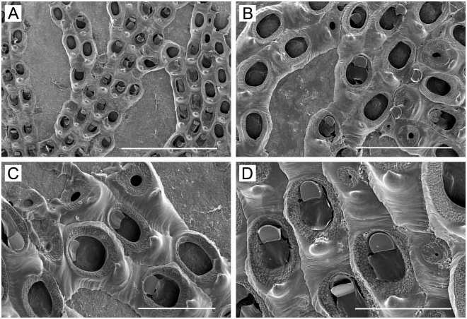
Nikulina (2006) recently described this species from Akkeshi Bay. Among congeners, Electra korobokkura most closely resembles Electra arctica ( Borg, 1931) in growth form, as both can form narrowly multiserial branches in portions of colonies. For comparison with E. korobokkura ( Figure 3 View Figure 3 ), we illustrate a subtidal specimen of what we consider to be E. arctica , from the western Kamchatka shelf, Sea of Okhotsk ( Figure 4 View Figure 4 ). This specimen illustrates all the characters considered diagnostic by Hansen (1962), who briefly reviewed E. arctica : well-developed gymnocyst ( Figure 4B–D View Figure 4 ); enlarged base of proximal spine ( Figure 4C, D View Figure 4 ); well-developed cryptocyst with a crenulated border ( Figure 4D View Figure 4 ); more heavy calcification than in other Electra species ; operculum heavily calcified, with the proximal border straight (in our specimen, there is intra-colony variation, with some opercula having a concave proximal border) ( Figure 4D View Figure 4 ); the opesial opening restricted or reduced by a closure plate ( Figure 4C View Figure 4 ) in some zooids; and a tendency to form kenozooids ( Figures 4B–D View Figure 4 ) of reduced size among autozooids. Compared to E. arctica , E. korobokkura has a greater tendency to form uniserial branches (compare Figure 3 View Figure 3 with Figure 4 View Figure 4 ). Compared to Electra arctica , zooids of Electra korobokkura appear less heavily calcified; the proximal gymnocyst has the transverse rugae weaker or lacking; the cryptocyst is narrower and much less heavily granulated, leaving a larger opesial opening in relation to overall zooid size; the operculum is less heavily calcified; and the basal chamber of the proximal spine is smaller. Furthermore, the closure plates of kenozooids have a smooth surface in E. korobokkura ( Nikulina 2006, Figures 2 View Figure 2 , 3B View Figure 3 , 4 View Figure 4 ) whereas in E. arctica the closure plates have a broad, granulated component of cryptocystal calcification surrounding the opening ( Figure 4C, D View Figure 4 ). Zooids of the E. arctica specimen we illustrate ( Figure 4 View Figure 4 ) are 0.42– 0.73 mm long (0.57¡ 0.07 mm) by 0.22–0.48 mm wide (0.34¡ 0.06 mm), and the opesia is 0.20–0.33 mm long (0.26¡ 0.03 mm) by 0.12–0.21 mm wide (0.18¡ 0.02 mm). Thus, while the zooids of E. arctica are larger than those of E. korobokkura , the opesia of the former is roughly the same size or even smaller than that of the latter.
Distribution
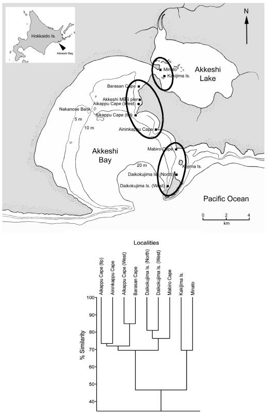
Akkeshi Bay is the only known locality. Some previous records of nominal E. arctica in the northwestern Pacific (e.g. Mawatari 1956, 1974; Mawatari and Mawatari 1981b) may have included specimens of this species.


| V |
Royal British Columbia Museum - Herbarium |
No known copyright restrictions apply. See Agosti, D., Egloff, W., 2009. Taxonomic information exchange and copyright: the Plazi approach. BMC Research Notes 2009, 2:53 for further explanation.
|
Kingdom |
|
|
Phylum |
|
|
Class |
|
|
Order |
MEMBRANIPOROIDEA Busk, 1852
| Grischenko, Andrei V., Dick, Matthew H. & Mawatari, Shunsuke F. 2007 |
Electra korobokkura
| Nikulina 2006 |
Electra korobokkura
| Nikulina 2006: 23 |
Electra
| Lamouroux 1816 |