
Phidolopora elongata ( Smitt, 1868 )
|
publication ID |
https://doi.org/ 10.1080/00222930701391773 |
|
persistent identifier |
https://treatment.plazi.org/id/877A7251-CC2F-DE5F-FE61-274CD3D51808 |
|
treatment provided by |
Felipe |
|
scientific name |
Phidolopora elongata ( Smitt, 1868 ) |
| status |
|
Phidolopora elongata ( Smitt, 1868) View in CoL
( Figure 41 View Figure 41 )
Retepora cellulosa forma notopachys var. elongata Smitt 1868, p 36 , Plate 28, Figures 226–332. Retepora wallichiana: Hincks 1877, p 107, Plate 11, Figures 9–13 View Figure 9 View Figure 10 View Figure 11 View Figure 12 View Figure 13 ; 1884, p 55.
Retepora elongata: Androsova 1958, p 117 View in CoL , Figure 107; Kluge 1961, p 142; 1962, p 527, Figure 368.
Phidolopora elongata: Hansen 1962, p 45 View in CoL ; Cuffey and Turner 1987, p 67; Kubanin 1997, p 125; Grischenko and Ivanyushina 2002, p 32, Text figure 2.
Phydolopora elongata [sic]: Gontar 1980, p 13; 1990, p 133; 1992, p 188; 1993, p 202; 1996, p 46; Gontar and Denisenko 1989, p 357; Grischenko 1997, p 188.
? Retepora pacifica Robertson 1908, p 310 , Plate 24, Figures 81–84.
? Retepora pacifica: O’Donoghue and O’Donoghue 1923, p 189 .
? Phidolopora pacifica: Canu and Bassler 1923, p 154 View in CoL , Plate 39, Figures 1–7 View Figure 1 View Figure 2 View Figure 3 View Figure 4 View Figure 5 View Figure 6 View Figure 7 ; O’Donoghue and O’Donoghue 1925, p 106; 1926, p 118; Osburn 1952, p 448, Plate 53, Figures 1 View Figure 1 , 2 View Figure 2 ; Soule et al. 1995, p 277, Plate 106A–E.
Material examined
ANC, 10 colony fragments (NHM 2006.2.27.107), three colony fragments (NHM 2006.2.27.108), two young colonies on rock (NHM 2006.2.27.109) Additional material: three specimens; holotype, SMNH-1316, Svalbard, Hinlopen Strait , Waigatsöarna .
Description
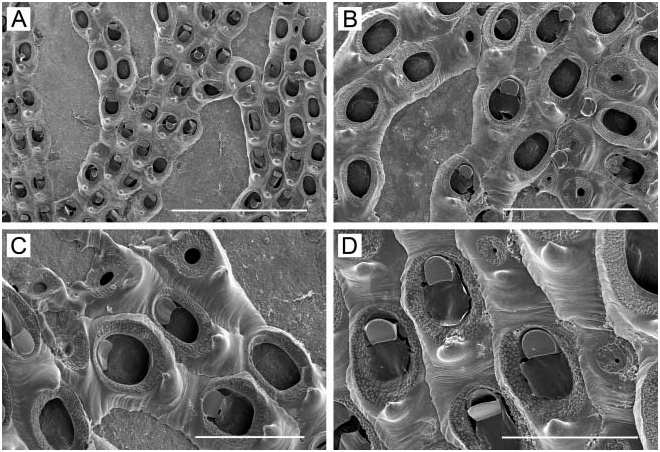
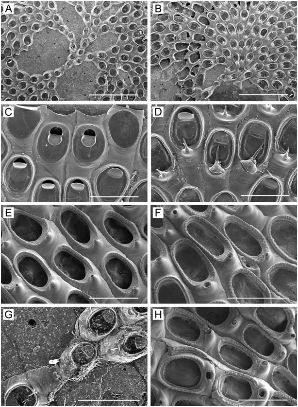
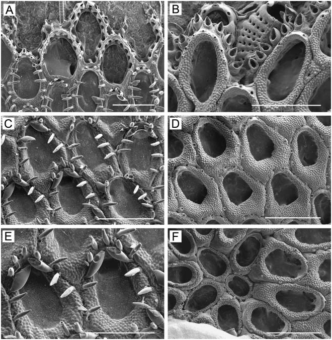
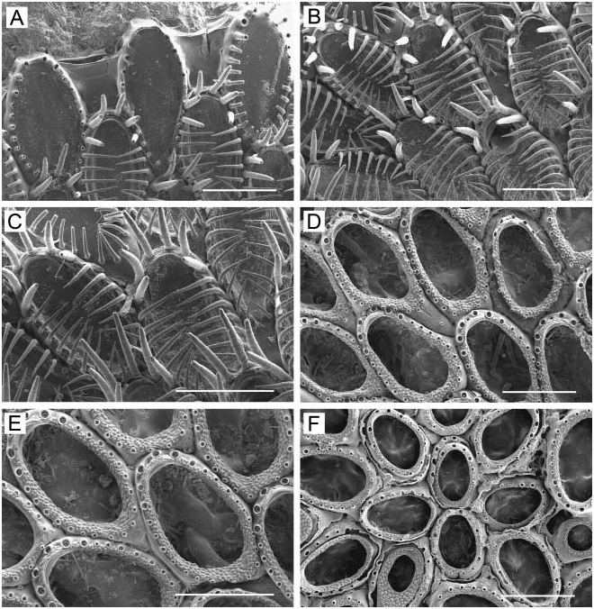
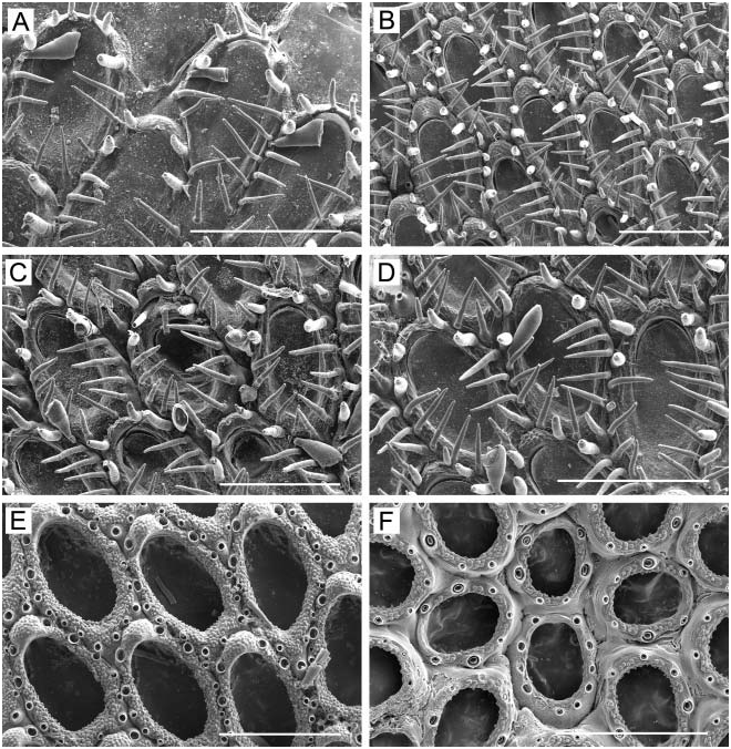
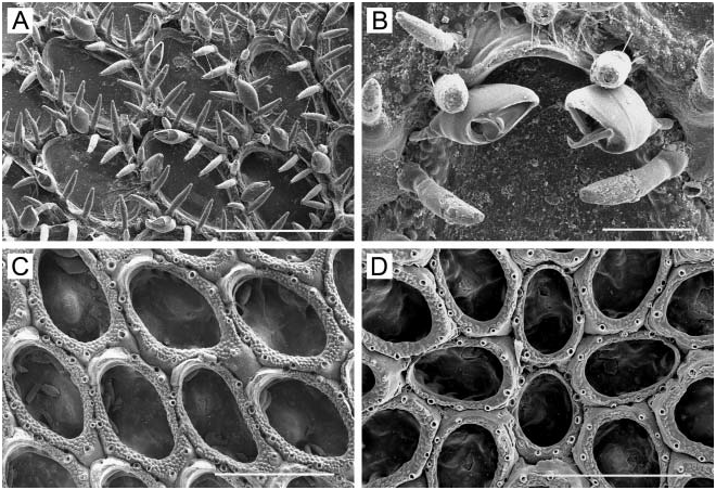
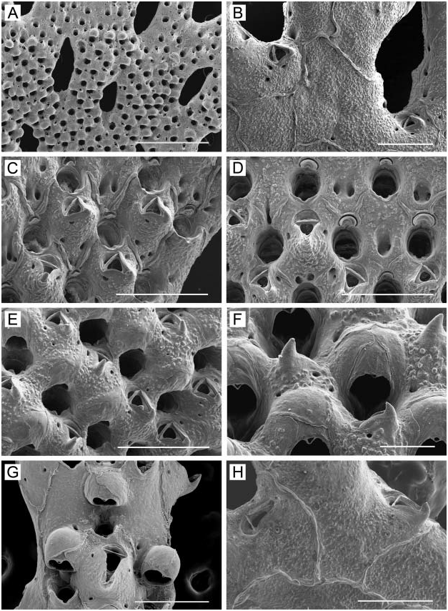
Colony ( Figure 41A View Figure 41 ) erect, orange in colour when alive, consisting of fenestrate bilaminar sheets, complexly folded and fused into rigid three-dimensional meshwork, largest observed 11×8× 4.5 cm in dimensions, rising from rounded base attached to substratum by kenozooids. Branches range from three to 11 zooids wide between fenestrulae. All feeding zooids open on one surface of a sheet; opposite surface consists of kenozooids. Fenestrulae elongate-oval or irregularly rhombic in shape, 0.7–1.9 long by 0.3–0.9 mm wide. Zooids ( Figure 41C View Figure 41 ) hexagonal, rhombic, or oval, 0.48–0.70 mm long (0.59¡ 0.07 mm), 0.24–0.38 mm wide (0.31¡ 0.04 mm). Newly budded zooids delineated by raised vertical walls; in older regions of colony, boundaries between zooids occluded by secondary calcification or represented by fine sutures. Frontal wall slightly convex to inflated in young zooids, markedly convex in mature zooids, finely granulated, imperforate except for two or three pores along each proximolateral margin. Primary orifice ( Figure 41D View Figure 41 ) long-semicircular, with beaded rim, 0.10–0.13 mm long (0.11¡ 0.01 mm), 0.09–0.13 mm wide (0.11¡ 0.01 mm); proximal margin straight, with shallow but distinct U-shaped sinus flanked by condylar shelves bearing low, blunt condyles. With age, primary orifice submerged in peristome; secondary orifice with median pseudosinus. Two short, hollow, ephemeral oral spines with enlarged bases frequently present at distolateral edges of primary orifice in marginal zooids. Large frontal avicularium ( Figure 41C–E View Figure 41 ), 0.14– 0.20 mm long, with raised, beak-shaped rostrum, occupies central part of frontal wall in many zooids; cross-bar complete, rostral opesia triangular to oval in shape; mandible longtriangular, with acute tip, directed proximally or proximolaterally; avicularian chamber broad, conical, very prominent in immature zooids, inflated in ovicellate zooids ( Figure 41D View Figure 41 ) by general thickening of frontal wall owing to secondary calcification, coarsely granulated, with two or three small pores flanking its base. Single transversely orientated avicularium occupies proximal abfrontal axils of fenestrulae ( Figure 41B View Figure 41 ), 0.15– 0.18 mm long, with short, equilaterally triangular mandible; one or two pairs of circular pores flank rostrum. Ovicell ( Figure 41F View Figure 41 ) hyperstomial, spherical, smooth, often forming a hood overhanging orifice, 0.18–0.23 mm long (0.20¡ 0.01 mm), 0.20–0.25 mm wide (0.22¡ 0.01mm), with incompletely calcified ectooecium having fine concentric striae; proximal edge of ovicell often with slight median denticle. Ovicell rapidly immersed ( Figure 41E View Figure 41 ) by secondary calcification from distal and lateral zooids covering most of its surface, with fine sutures delineating calcification from different zooids. Dorsal surface of colony ( Figure 41B View Figure 41 ) shows outlines of kenozooids of irregular form and size, recognizable by sutures indicating raised vertical walls; kenozooids inflated, roughly granulated, with a few sparse circular pores on the surface; avicularia lacking dorsally except for those in axils of fenestrulae. Ancestrula and early astogeny not observed.
Remarks
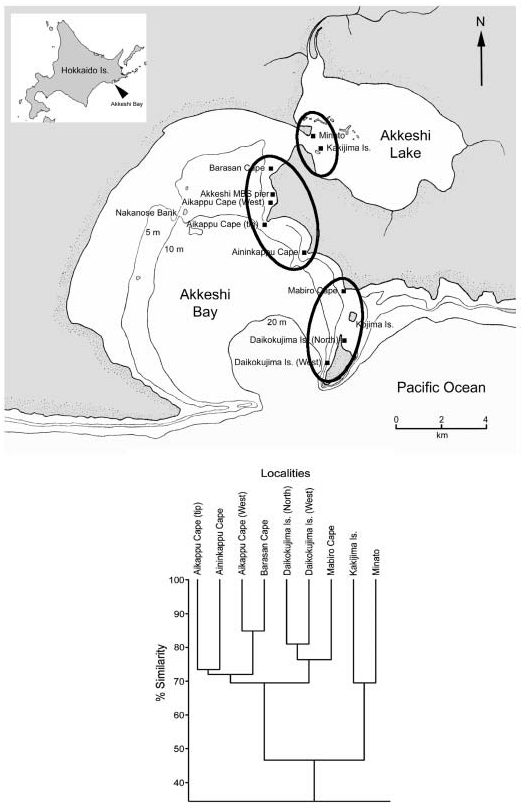
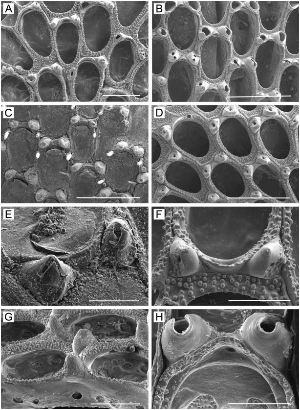
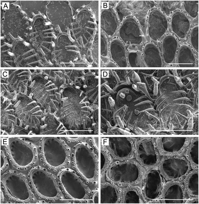
Specimens from Akkeshi Bay are similar in many characters to the type specimen of P. elongata (SMNH-1316, many fragments) ( Figure 41G, H View Figure 41 ). Both have rhombic zooids with a submerged, rounded primary orifice having a shallow median sinus and encircled by peristome bearing a proximal pseudosinus. Large frontal avicularia are similar in form and position; the ovicells have, on the proximal margin, a median denticle that is often incompletely closed. There are some differences; specimens from Akkeshi Bay have 3–11 zooids comprising branches between fenestrulae, the type of P. elongata three to seven. This may be ecophenotypic variation, as depth-dependent variation in branch width occurs in P. elongata from the Commander Islands region; colonies from depths of 1–25 m have twice the number of zooids per branch than those from depths of 65–165 m (A. V. Grischenko, unpublished data). Another is that the abfrontal avicularian mandibles are larger with a long-triangular mandible in the type of P. elongata , versus smaller, with an equilateral mandible in specimens from Akkeshi.
A congener, P. pacifica ( Robertson, 1908) , is known from the eastern Pacific, reported from Alaska, Puget Sound, and the coast of California ( Robertson 1908); British Columbia ( O’Donoghue and O’Donoghue 1923, 1925, 1926); and from the Santa Barbara Channel to Mexico and the Galapagos Islands ( Soule et al. 1995). Soule et al. (1995) illustrated this species for the first time using SEM. Phidolopora pacifica appears to be similar to P. elongata in the shape of the orifice and the ovicell with a median denticle, which was also illustrated in line drawings by Robertson (1908) and Osburn (1952). However, P. pacifica seems to form more pronounced lappets flanking the peristomial pseudosinus, and the abfrontal avicularium appears to differ somewhat in shape and orientation. Unfortunately, Robertson’s types of P. pacifica were mixed with other specimens, and neotypes will eventually have to be selected; in any case, we here consider this species as valid. Records of P. pacifica from Mexico and the Galapagos Islands ( Osburn 1952; Soule et al. 1995) need re-examination.
Many large, erect colonies of P. elongata were found growing in horizontal crevices in solid rock at the base of a cliff overhang at Aininkappu Cape; these were inhabited by a number of benthic organisms, including ascidians, sabellid and spirorbid polychaetes, sponges, crustaceans, sipunculids, holothurians, nemerteans, and ophiuroids. A similar hermatypic growth form of this species, supporting a rich associated fauna, occurs below cliffs in the Commander Islands ( Grischenko and Ivanyushina 2002).
Distribution
Phidolopora elongata View in CoL is considered a circumpolar Arctic-Boreal species; Kluge (1962, 1975) and Gontar and Denisenko (1989) gave many distributional records. In the northern Pacific, it has been previously reported from the Bering Sea near the Commander Islands ( Kluge 1961; Grischenko 1997; Grischenko and Ivanyushina 2002), along the Kuril Islands ( Gontar 1980, 1993), and in Tatar Strait in the northern Sea of Japan ( Kluge 1961). Akkeshi Bay is the southernmost known locality for this species in the western North Pacific.


| V |
Royal British Columbia Museum - Herbarium |
No known copyright restrictions apply. See Agosti, D., Egloff, W., 2009. Taxonomic information exchange and copyright: the Plazi approach. BMC Research Notes 2009, 2:53 for further explanation.
|
Kingdom |
|
|
Phylum |
|
|
Class |
|
|
Order |
|
|
Family |
|
|
Genus |
Phidolopora elongata ( Smitt, 1868 )
| Grischenko, Andrei V., Dick, Matthew H. & Mawatari, Shunsuke F. 2007 |
Phydolopora elongata
| Grischenko AV 1997: 188 |
| Gontar VI & Denisenko NV 1989: 357 |
| Gontar VI 1980: 13 |
Phidolopora elongata:
| Grischenko AV & Ivanyushina EA 2002: 32 |
| Kubanin AA 1997: 125 |
| Cuffey RJ & Turner RF 1987: 67 |
| Hansen KB 1962: 45 |
elongata:
| Kluge GA 1961: 142 |
| Androsova EI 1958: 117 |
Phidolopora pacifica:
| Soule DF & Soule JD & Chaney HW 1995: 277 |
| Osburn RC 1952: 448 |
| O'Donoghue CH & O'Donoghue E 1925: 106 |
| Canu F & Bassler RS 1923: 154 |
Retepora pacifica
| Robertson A 1908: 310 |