
Microporella luellae, Grischenko & Dick & Mawatari, 2007
|
publication ID |
https://doi.org/ 10.1080/00222930701391773 |
|
persistent identifier |
https://treatment.plazi.org/id/877A7251-CC1E-DE6F-FE6D-24F9D3421F04 |
|
treatment provided by |
Felipe |
|
scientific name |
Microporella luellae |
| status |
sp. nov. |
Microporella luellae View in CoL new species
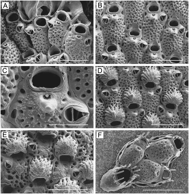
( Figure 34 View Figure 34 )
Diagnosis
Frontal wall inflated, uniformly perforated with numerous small pores. Primary orifice rounded-semicircular, with straight proximal margin. Two widely separated oral spines. Single avicularium situated anywhere along lateral margin, mandible triangular, pointed distolaterally or laterally, cross-bar complete. Ascopore crescentic, with denticulate edge, located on elevated oval prominence, just distal to prominent conical umbo. Ovicell raised, hemispherical, imperforate centrally, often umbonate, with radial ribs and pores around base. Ancestrula tatiform with 12 spines, budding a pair of zooids distolaterally.
Etymology
Named in honour of Luella Taranto, who greatly helped with collecting during July 2004.
Material examined
Holotype: DIN, four colony fragments (NHM 2006.2.27.93). Paratypes: DIW, two colony fragments (NHM 2006.2.27.94); BAC, ancestrular colony on bivalve shell (NHM 2006.2.27.52). Additional material: 1065 specimens.
Description
Colony encrusting, unilaminar, coherent, irregularly circular, up to 4 cm across; bright orange, pink, or beige in colour when alive. Zooids ( Figure 34A, B, D View Figure 34 ) hexagonal, oval, or somewhat irregular in shape, 0.50–0.73 mm long (0.57¡ 0.06 mm), 0.27–0.43 mm wide (0.36¡ 0.04 mm), separated by deep groove and fine, meandering suture line. Frontal wall moderately to markedly convex, finely granulated, uniformly perforated from margin to margin with small, round pores, except in suboral area. Orifice ( Figure 34C View Figure 34 ) broadly semicircular, with rounded proximolateral corners, 0.09–0.11 mm long (0.10¡ 0.01 mm), 0.12–0.16 mm wide (0.15¡ 0.01 mm), proximal margin straight. No condyles. Two widely separated, short, hollow oral spines along distolateral corners of orifice in immature zooids; some zooids have only one spine or lack them altogether. Ascopore ( Figure 34C View Figure 34 ) crescentic, with a finely denticulate edge, not closed by connecting rays; close to proximal border of orifice, separated from it by a distance about equivalent to distance across short axis of ascopore; located on elevated oval prominence; proximal side of prominence developed into small, smooth, conical umbo. Zooids have a single avicularium ( Figure 34A–E View Figure 34 ), rarely a pair of them, and many zooids lack them altogether; avicularium lateral or proximolateral to ascopore, raised from frontal wall, directed laterally or distolaterally, end of rostrum with narrow channel, cross-bar complete; mandible sharp, elongate-triangular with setiform tip; avicularian chamber with smooth surface. Ovicell ( Figure 34D, E View Figure 34 ) hemispherical, prominent, 0.21–0.26 mm long (0.23¡ 0.01 mm), 0.25– 0.32 mm wide (0.29¡ 0.02 mm), radially ribbed, finely granulated, often umbonate, imperforate except for outer margin; proximal margin with a smooth rim or upturned lip. Lateral wall of zooids with two or three distal and two distolateral basal pore chambers. Ancestrula ( Figure 34F View Figure 34 ) tatiform, elongate-oval, 0.35 mm long, 0.23 mm wide, with large, elliptical opesia, 0.18 mm long, 0.15 mm wide, surrounded by 12 spines; ancestrula buds two zooids distolaterally. Periancestrular zooids with six or seven long, tubular oral spines.
Remarks
The genus Microporella has been well studied around Hokkaido ( Mawatari and Mawatari 1981b; Mawatari et al. 1991), including a recent revision incorporating new taxonomic characters ( Suwa and Mawatari 1998). Eight species have previously been reported from Hokkaido: M. orientalis Harmer, 1957 ; M. echinata Androsova, 1958 ; M. neocribroides Dick and Ross, 1988 ; M. borealis Suwa and Mawatari, 1998 ; M. elegans Suwa and Mawatari, 1998 ; M. formosa Suwa and Mawatari, 1998 ; M. pulchra Suwa and Mawatari, 1998 ; and M. trigonellata Suwa and Mawatari, 1998 . It is surprising that previous investigators did not detect M. luellae , which we found to be exceptionally abundant in Akkeshi Bay.
Microporella luellae View in CoL is similar in many characters to M. neocribroides Dick and Ross, 1988 View in CoL , previously reported intertidally from Densin-Hama, Muroran, southern Hokakido. The two widely separated oral spines distinguish M. luellae View in CoL and M. neocribroides View in CoL from all other congeners reported around Hokkaido. However, in M. luellae View in CoL , the ascopore is always crescentic, with a denticulate edge, and is located on an elevated oval prominence. Except for developing and heavily calcified zooids, the proximal side of this prominence is developed into a small, conical umbo. In M. neocribroides View in CoL , the ascopore is transversely elliptical and covered with a cribriform plate with 10–20 round pores ( Suwa and Mawatari 1998, p 899, Figure 2 View Figure 2 ; Dick et al. 2005, p 3753, Figure 19D View Figure 19 ); only occasionally is there an umbonate process proximal to the ascopore. Another difference is that the ovicell is often umbonate in M. luellae View in CoL , but rarely so in M. neocribroides View in CoL .
Distribution
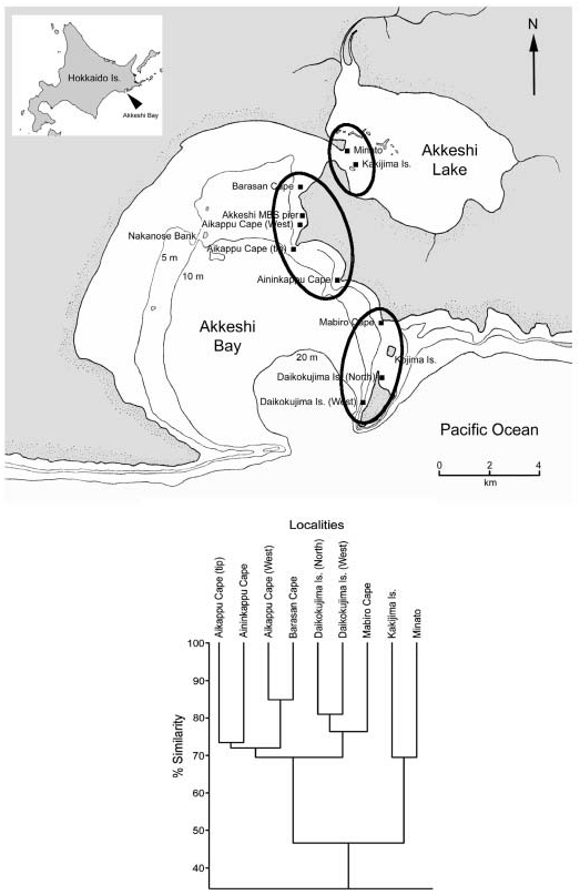
Microporella luellae is presently known only from Akkeshi Bay.

No known copyright restrictions apply. See Agosti, D., Egloff, W., 2009. Taxonomic information exchange and copyright: the Plazi approach. BMC Research Notes 2009, 2:53 for further explanation.
|
Kingdom |
|
|
Phylum |
|
|
Class |
|
|
Order |
|
|
Family |
|
|
Genus |
Microporella luellae
| Grischenko, Andrei V., Dick, Matthew H. & Mawatari, Shunsuke F. 2007 |
Microporella luellae
| Grischenko & Dick & Mawatari 2007 |
M. luellae
| Grischenko & Dick & Mawatari 2007 |
M. luellae
| Grischenko & Dick & Mawatari 2007 |
M. luellae
| Grischenko & Dick & Mawatari 2007 |
M. neocribroides
| Dick and Ross 1988 |
M. neocribroides
| Dick and Ross 1988 |
M. neocribroides
| Dick and Ross 1988 |
M. neocribroides
| Dick and Ross 1988 |