
Schizoporella japonica Ortmann, 1890
|
publication ID |
https://doi.org/ 10.1080/00222930701391773 |
|
persistent identifier |
https://treatment.plazi.org/id/877A7251-CC0F-DE7F-FE71-21B8D4111DDA |
|
treatment provided by |
Felipe |
|
scientific name |
Schizoporella japonica Ortmann, 1890 |
| status |
|
Schizoporella japonica Ortmann, 1890 View in CoL
( Figure 27 View Figure 27 ) Schizoporella unicornis var. japonica Ortmann 1890, p 49 , Plate 3, Figure 35 View Figure 35 . Schizoporella japonica: Dick et al. 2005, p 3742 , Figures 15A–H View Figure 15 , 16A–D View Figure 16 (illustration of holotype).
Schizoporella unicornis: Okada 1929, p 20 View in CoL , Text figure 7; Osburn 1952, p 317, Plate 37, Figures 1 View Figure 1 , 2 View Figure 2 ; Powell 1970, p 1849, Figures 2 View Figure 2 , 3 View Figure 3 ; McCain and Ross 1974, p 13, Figure 2c, d View Figure 2 ; Ross and McCain 1976, p 164, Figures 1–6 View Figure 1 View Figure 2 View Figure 3 View Figure 4 View Figure 5 View Figure 6 ; Mawatari and Mawatari 1981b, p 51; Kubota and Mawatari 1985b, p 201, Figure 3A–E View Figure 3 ; Liu et al. 2001, p 596, Plate 48; Soule et al. 1995, p 204.
Material examined
KAI, colony on rock (NHM 2006.2.27.39), colony on rock (NHM 2006.2.27.85); BAC, ancestrular colony detached from bivalve shell (NHM 2006.2.27.86), extensive colony detached from bivalve shell (NHM 2006.2.27.87), extensive colony on bivalve shell (NHM 2006.2.27.88). Additional material: 80 specimens .
Description
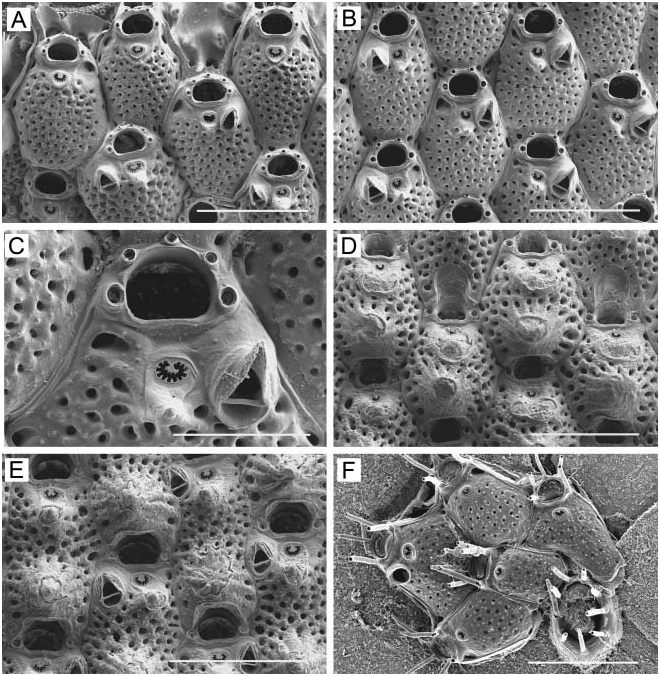
Colony encrusting, unilaminar, occasionally with bilaminar overgrowth of one portion of colony by another, forming extensive irregularly circular patches up to 5.5 cm across, red to bright orange when alive. Zooids ( Figure 27A, B View Figure 27 ) rectangular to hexagonal, 0.52–0.73 mm long (0.63¡ 0.05 mm), 0.30–0.43 mm wide (0.38¡ 0.03 mm), separated by shallow grooves. Frontal wall weakly to moderately convex, uniformly perforated from margin to margin with small circular pores except in suboral area, with seven to nine larger areolar pores along each lateral margin. Frontal pores become infundibular with development of calcification in mature zooids. Usually frontal wall rises into a small conical median umbo proximal to orifice. Orifice ( Figure 27B, E View Figure 27 ) broader than long, 0.11–0.15 mm long (0.13¡ 0.01 mm), 0.15–0.17 mm wide (0.16¡ 0.01 mm); sinus broadly U-shaped, flattened on bottom; conspicuous condylar shelves bearing blunt condyles. Oral avicularia ( Figure 27B View Figure 27 ) situated lateral or proximolateral to orifice, close to condylar shelf; mandible elongate-triangular, with acute tip, directed distolaterally to distally, cross-bar complete; chamber comparatively small, narrow, smooth, with one to three minute pores laterally around base. Oral avicularia usually single ( Figure 27B View Figure 27 ), often absent ( Figure 27C View Figure 27 ), occasionally paired. Additionally, some colonies have zooids with large frontal avicularia ( Figure 27D, E View Figure 27 ) similar in form to oral avicularia, but with a highly raised, smooth chamber. Position of frontal avicularia variable; they can lie close to orifice, but a little more proximal than oral avicularium, on opposite side; just proximal to oral avicularium on same side; along zooidal lateral margin; or centrally on frontal surface. Large avicularia close to orifice point distolaterally; those in central or lateral region of frontal wall point distally or laterally. Large frontal avicularia developed predominantly in mature zones of colony, among zooids with complete ovicells. Ovicell ( Figure 27C, D View Figure 27 ) hemispherical, prominent, 0.30–0.35 mm long (0.32¡ 0.02 mm), 0.29–0.36 mm wide (0.33¡ 0.02 mm), lying on frontal wall of daughter zooid and partially overhanging zooidal orifice; rugose, with heavily calcified, finely granulated radiating ribs, evenly perforated by numerous small pores, with larger round to slit-like pores around base. Occasionally ovicell has a small, knob-like central umbo. Ovicellate zooids can be sparsely distributed among non-fertile zooids or concentrated as a reproductive band within colony. Zooids intercommunicate via three to five distal and six lateral basal pore chambers. Ancestrula ( Figure 27F View Figure 27 ) oval, imperforate, 0.33 mm long, 0.28 mm wide, with eight spines around D-shaped orifice (0.13 mm long, 0.15 mm wide) with straight proximal margin. Ancestrula buds three small zooids distally; surrounded by seven zooids.
Remarks
Ortmann (1890) reported nominal S. unicornis ( Johnston, 1844) from Japan, and additionally erected a new variety, S. unicornis var. japonica . Dick et al. (2005) recently examined Ortmann’s type specimens and elevated variety japonica to species rank, as S. japonica .
Distribution
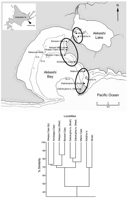
In the western Pacific, S. japonica (reported as S. unicornis ) extends from China ( Liu et al. 2001) northward to Hokkaido Island, where it has previously been recorded at Akkeshi, Muroran, and Shirikishinai on the Pacific side ( Mawatari and Mawatari 1981b) and at Oshoro Bay on the Sea of Japan ( Kubota and Mawatari 1985b).
Schizoporella japonica View in CoL was introduced on Pacific oysters ( Crassostrea gigas View in CoL ) from Japan to the Pacific coast of North America during the 20th century; it is now widely distributed from San Francisco, California to southeastern Alaska ( Powell 1970; McCain and Ross 1974; Ross and McCain 1976; Dick et al. 2005). As mentioned by Dick et al. (2005), the actual range of S. japonica View in CoL may be much more extensive, since this species could have been introduced on oysters to other parts of the world as well.
Family STOMACHETOSELLIDAE Canu and Bassler, 1917 View in CoL


| BAC |
Beijing Agricultural College |
No known copyright restrictions apply. See Agosti, D., Egloff, W., 2009. Taxonomic information exchange and copyright: the Plazi approach. BMC Research Notes 2009, 2:53 for further explanation.
|
Kingdom |
|
|
Phylum |
|
|
Class |
|
|
Order |
|
|
Family |
|
|
Genus |
Schizoporella japonica Ortmann, 1890
| Grischenko, Andrei V., Dick, Matthew H. & Mawatari, Shunsuke F. 2007 |
Schizoporella unicornis:
| Liu X & Yin X & Ma J 2001: 596 |
| Soule DF & Soule JD & Chaney HW 1995: 204 |
| Kubota K & Mawatari SF 1985: 201 |
| Mawatari S & Mawatari SF 1981: 51 |
| Ross JRP & McCain KW 1976: 164 |
| McCain KW & Ross JRP 1974: 13 |
| Powell NA 1970: 1849 |
| Osburn RC 1952: 317 |
| Okada Y 1929: 20 |