
Parkermavella orientalis (Androsova and Gontar, 1982)
|
publication ID |
https://doi.org/10.1080/00222930701391773 |
|
persistent identifier |
https://treatment.plazi.org/id/877A7251-CC0D-DE70-FF43-2499D24C1935 |
|
treatment provided by |
Felipe |
|
scientific name |
Parkermavella orientalis |
| status |
|
Parkermavella orientalis View in CoL (Androsova and Gontar in Gontar 1982), new combination
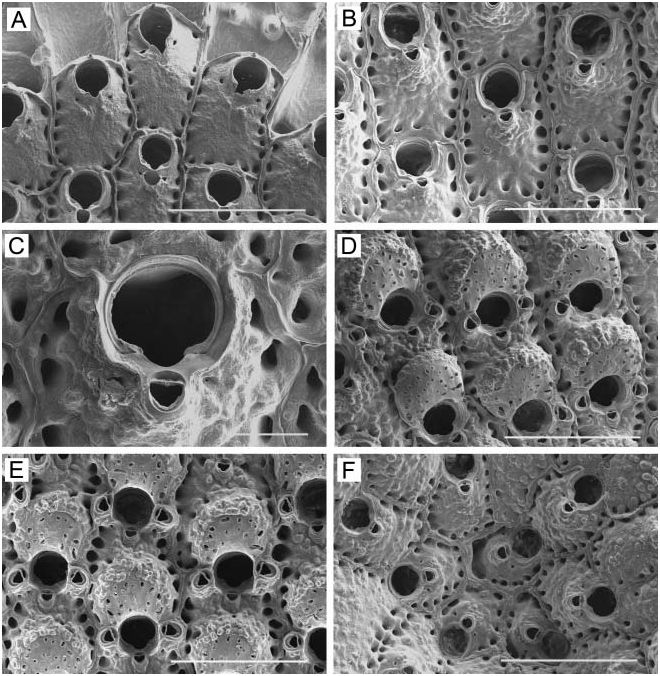
( Figure 26 View Figure 26 ) Schizomavella auriculata orientalis Androsova and Gontar in Gontar 1982, p 549,
Figure 2-2 View Figure 2 .
Material examined
ANC, colony on rock (NHM 2006.2.27.31), intact colony (NHM 2006.2.27.84); ACW, colony on rock (NHM 2006.2.27.83). Additional material: 38 specimens.
Description
Colony unilaminar, encrusting, coherent, forming circular patches up to 3 cm in diameter, bright beige or light orange in colour when alive. Zooids ( Figure 26A, B View Figure 26 ) irregularly hexagonal, barrel-shaped, or rectangular, 0.35–0.53 mm long (0.42¡ 0.05 mm), 0.28– 0.45 mm wide (0.36¡ 0.04 mm), separated by fine suture lines between raised adjacent vertical walls. Frontal wall moderately convex to inflated; vitreous, smooth in young zooids, nodulose in mature zooids; imperforate centrally with five to nine conspicuous areolar pores along each lateral margin, separated by short ridges. Primary orifice ( Figure 26C View Figure 26 ) subcircular, 0.10–0.13 mm long (0.11¡ 0.01 mm), 0.10–0.14 mm wide (0.12¡ 0.01 mm); proximal margin with shallow, U-shaped median sinus flanked by broad condylar shelves bearing blunt condyles. Newly budded zooids ( Figure 26A View Figure 26 ) have one or two short, hollow ephemeral spines distal to primary orifice. Secondary orifice cormidial, formed proximally by sharp, elevated flanges of peristome extending from sides of suboral avicularian chamber to distal curvature formed by raised, thickened margin of succeeding zooid; circular in outline in immature zooids, transversely oval in ovicellate zooids; in mature zooids, lateral flanges connect with proximolateral corners of ovicell. Immediately proximal to orificial sinus is a small suboral avicularium ( Figure 26B View Figure 26 ) with complete cross-bar and semicircular mandible directed proximally at an angle to frontal plane; avicularian chamber narrow, crescentic, prominent in immature zooids, becoming immersed and covered by nodules in ovicellate zooids, flanked by a pair of small pores. Ovicellate zooids additionally have paired lateral oral avicularia ( Figure 26D, E View Figure 26 ) directed proximolaterally at an angle to frontal plane, somewhat larger than median avicularium, with complete cross-bar; mandible subspatulate, slightly elongate; rostral opesia triangular distal to cross-bar; lateral avicularia occasionally single or lacking altogether; rarely only one or two lateral avicularia present, with median suboral avicularium absent. Ovicell ( Figure 26D, E View Figure 26 ) hemispherical, recumbent on following zooid, broad, 0.18–0.25 mm long (0.21¡ 0.02 mm), 0.22–0.27 mm wide (0.25¡ 0.01 mm); with concave proximal margin; smooth on top, perforated with 25–30 irregular pores; with age, covered around periphery with heavy nodular secondary calcification. Zooids intercommunicate via uniporous septula. Ancestrula ( Figure 26F View Figure 26 ) similar in form to autozooid, reduced in size, 0.27 mm long, 0.20 mm wide, irregularly hexagonal, with quite convex frontal wall and raised vertical walls; orifice circular, 0.08 mm long, 0.08 mm wide; surrounded by seven zooids.
Remarks
Gordon and d’Hondt (1997) established the genus Parkermavella for Schizomavella -like species that differ from Schizomavella in having an imperforate frontal shield and only marginal areolae. Characters of Parkermavella include a proximal oral sinus; articulated oral spines distally; one or more adventitious avicularia near the orifice or elsewhere on the frontal surface; and a prominent or subimmersed ovicell with smooth ectooecial calcification, many perforations that may be rimmed, and secondary calcification sometimes encroaching around the distal margin. The species of Androsova and Gontar treated here lacks frontal perforation and has most of the other characters of Parkermavella , and therefore belongs in that genus.
Gontar (1982) originally described this species as subspecies orientalis of Schizomavella auriculata ( Hassall, 1842) . However, the nominal subspecies never has more than a single median avicularium associated with the orifice, and also differs in orifice shape and in having numerous frontal pores; it is distributed in the northeastern Atlantic from Scotland to Gibraltar ( Hayward and Ryland 1999). On the basis of these differences in morphology and range, we here elevate Gontar’s subspecies to species rank as P. orientalis (Androsova and Gontar, 1982) .
Parkermavella orientalis View in CoL is very similar to S. triavicularia Soule, Soule, and Chaney, 1995 View in CoL , described from the Santa Barbara Channel, which likewise has a single median suboral avicularium in non-ovicellate zooids and an additional pair of lateral oral avicularia in ovicellate zooids. Parkermavella orientalis View in CoL differs from the latter in several characters: (1) it lacks frontal pores, with well-developed areolar pores instead; (2) developing zooids near the colony margin have one or two ephemeral distal oral spines, whereas S. triavicularia View in CoL has three spines; (3) the median suboral avicularium is closer to the suboral sinus than in S. triavicularia View in CoL ; and (4) ovicellate zooids have dimorphic suboral avicularia, with the lateral avicularia larger than the median one, and with more elongate mandibles; the median and lateral avicularia are similar in size and form in mature zooids of S. triavicularia View in CoL .
The remarkable overall similarity of S. triavicularia View in CoL and P. orientalis View in CoL dispels misgivings one might have in accepting genera with and genera without a perforate frontal shield in the same family. Schizomavella triavicularia View in CoL , which is evenly perforated, is otherwise so similar to S. orientalis View in CoL , which is not, that there is little doubt the two are closely related; the similarity extends to both species having a pair of small pores flanking the avicularian chamber. Although convergence is a possibility, we conclude that the secondary loss of frontal perforation in Parkermavella View in CoL is a more likely explanation. The small pores that flank the avicularian chamber are actually primary perforations in the frontal shield, as indicated by their presence in the forming shield in marginal zooids ( Figure 26A View Figure 26 ), and in this sense, the frontal pores can be viewed as having been severely reduced in number, rather than lost entirely.
Distribution
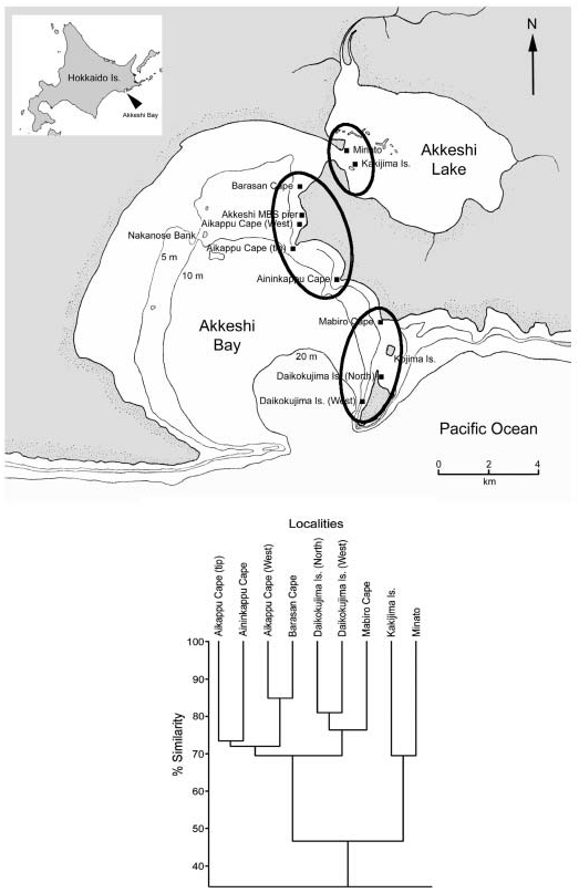
This species was originally described from Crabovaya Bay, Shikotan Island, Kuril Islands, and also recorded from Zelenyy Island, Habomai Islands (Small Kuril Achipelago). Akkeshi Bay is the third known locality.
No known copyright restrictions apply. See Agosti, D., Egloff, W., 2009. Taxonomic information exchange and copyright: the Plazi approach. BMC Research Notes 2009, 2:53 for further explanation.
|
Kingdom |
|
|
Phylum |
|
|
Class |
|
|
Order |
|
|
Family |
|
|
Genus |
Parkermavella orientalis
| Grischenko, Andrei V., Dick, Matthew H. & Mawatari, Shunsuke F. 2007 |
Parkermavella
| Gordon and d'Hondt 1997 |
S. triavicularia
| Soule, Soule, and Chaney 1995 |
S. triavicularia
| Soule, Soule, and Chaney 1995 |
S. triavicularia
| Soule, Soule, and Chaney 1995 |
S. triavicularia
| Soule, Soule, and Chaney 1995 |
S. triavicularia
| Soule, Soule, and Chaney 1995 |
Schizomavella triavicularia
| Soule, Soule, and Chaney 1995 |