
Troporhysipolis Quicke, Belokobylskij & Butcher
|
publication ID |
https://doi.org/10.3161/00034541Anz2016.66.2.003 |
|
DOI |
https://doi.org/10.5281/zenodo.14849269 |
|
persistent identifier |
https://treatment.plazi.org/id/86006125-FFC0-6865-75F9-6B43FA71FB47 |
|
treatment provided by |
Plazi |
|
scientific name |
Troporhysipolis Quicke, Belokobylskij & Butcher |
| status |
gen. nov. |
Troporhysipolis Quicke, Belokobylskij & Butcher , gen. nov.
Etymology. From “tropic”, the climatic belt where this taxon lived, and the generic name “ Rhysipolis ”. Gender: masculine.
Diagnosis. Cyclostome. Labial palp 4-segmented, maxillary palp 6-segmented. Occipital carina complete. Hypostomal and occipital carinae reaching mandible base separately. Epicnemial carina complete. Propodeum areolate. Fore wing vein cu-a (nervulus) anterfurcal and second subdiscal (brachial) cell swollen.
Subfamilies and genera Species Specimen voucher Provenance GenBank 28S Number CO1 Doryctinae Dendrosoter protuberans JM920 Turkey EF645736 View Materials EF645775 View Materials Doryctes erythromelas JM601 USA: Texas, College Station GQ374709 View Materials GQ374627 View Materials Heterospilus prosopidis — ex culture AY935469 View Materials AY935396 View Materials Hypodoryctes sibiricus none & JM 981 Finland & no data AJ302895 View Materials DQ498965 View Materials Syngaster lepidus — Australia AJ245698 View Materials DQ498963 View Materials Hormiinae Hormius moniliatus za201|203 Greenland — KF604624 View Materials Hormius Janzen09 — Costa Rica: A. C. Guanacaste — HQ549093 View Materials Hormius Janzen08 — Costa Rica: A. C. Guanacaste — HQ548789 View Materials Hormius Janzen04 — Costa Rica: A. C. Guanacaste — HQ549032 View Materials Hormius Janzen02 — Costa Rica: A. C. Guanacaste — HQ548710 View Materials Hormius Janzen10 — Costa Rica: A. C. Guanacaste — HQ548755 View Materials Hormius Janzen01 — Costa Rica: A. C. Guanacaste — HQ549024 View Materials Hormius sp.1 AL0161? Malaysia AY935455 View Materials AY935385 View Materials Hormius sp.2 PNG00680177 PNG: Madang, Wanang — FN662432 View Materials Hormius sp.3 PNG00405874 PNG: East Sepik — FN662431 View Materials Hormius sp.4 CNIN954 Mexico: Chamela — KX058580 View Materials Hormius sp.5 BF00473 Hungary — KX058581 View Materials Hormius sp.6 BJS2011 — JF979798 View Materials | 932 — Hormius sp.7 JM582 Madagascar: Mahajanga Prov. AY935455 View Materials AY935385 View Materials Parahormius JM576 Cameroun: Mt. Coupe AY935456 View Materials AY935386 View Materials Lysiterminae Acanthormius sp.1 JM692 Madagascar AJ302883 View Materials AY935381 View Materials Acanthormius sp.2 BCLDQ00238 Thailand: Khao Yai N.P. — KM067236 View Materials Acanthormius sp.3 QL2013?China — KF385867 View Materials Atritermus pedestris NHM675950 Madagascar: Mahajanga, Baie de Bali N.P. DQ414401 View Materials — Aulosaphes ? DQBKK0001 Thailand — KX058582 View Materials Aulosaphoides — Thailand KX058606 View Materials — Carinitermus NHM671037 Madagascar DQ414402 View Materials — Katytermus Hym-08 Japan: Honshu, Kobe, Rokko Mts EU854406 View Materials EU979624 View Materials Lysitermus sp.1 AL0220 Uganda: Western Prov., Kibale KM067238 View Materials — Lysitermus sp.2 BF000511 Nigeria: Ibadan KM067177 View Materials JF963503 View Materials Lysitermus sp.3 AAP0959 Mexico — HQ201061 View Materials
Accession
The new genus is morphologically similar to Rhysipolis Haliday , but differs in the form of the second subdiscal (brachial) cell (widened medially or posteriorly), vein 1cu-a (nervulus) anterfurcal and vein 2CUb (parallel) arising distinctly before middle of distal margin of second subdiscal (brachial) cell.
Type species. Clinocentrus antefurcalis Granger, 1949 View in CoL .
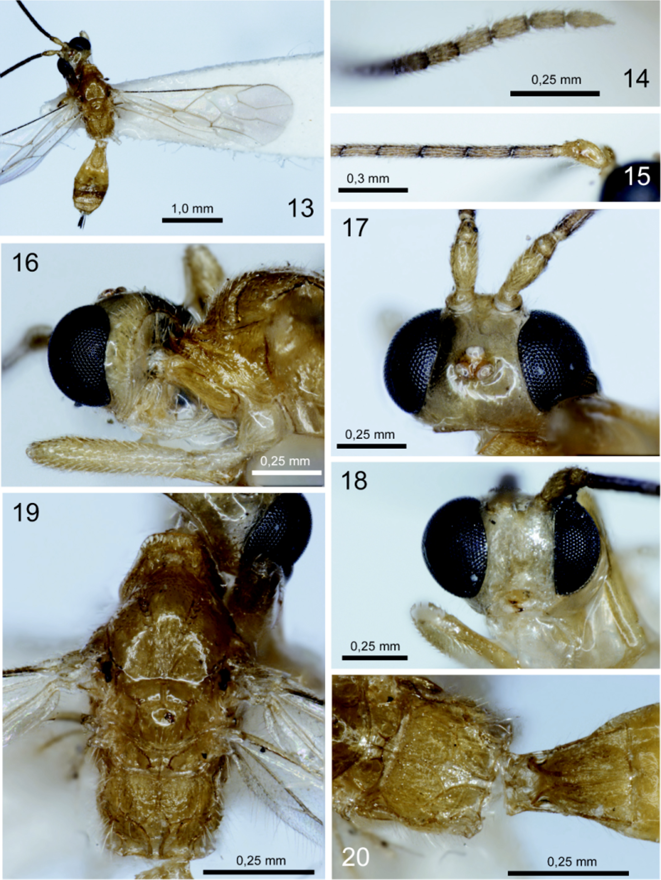
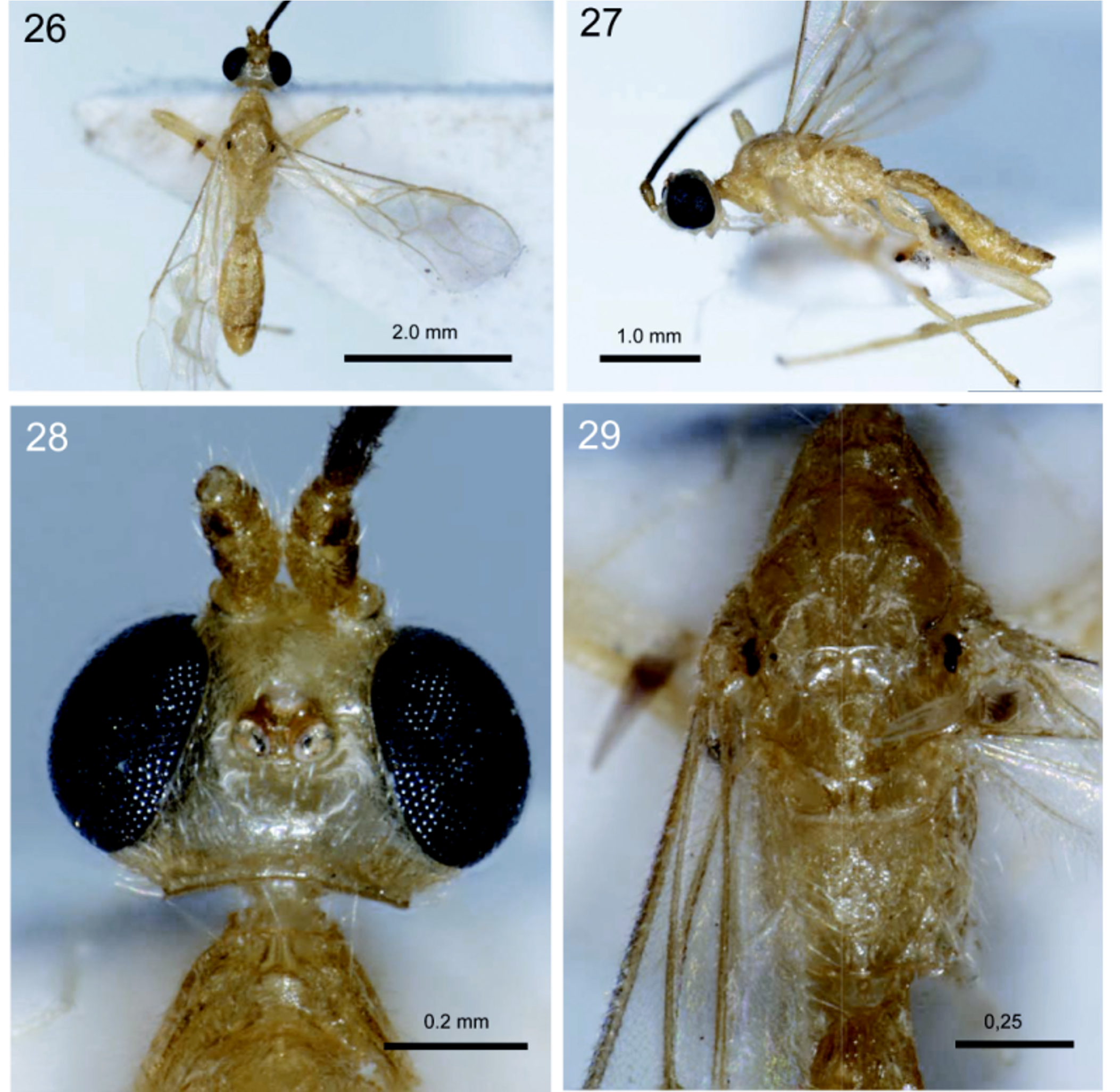
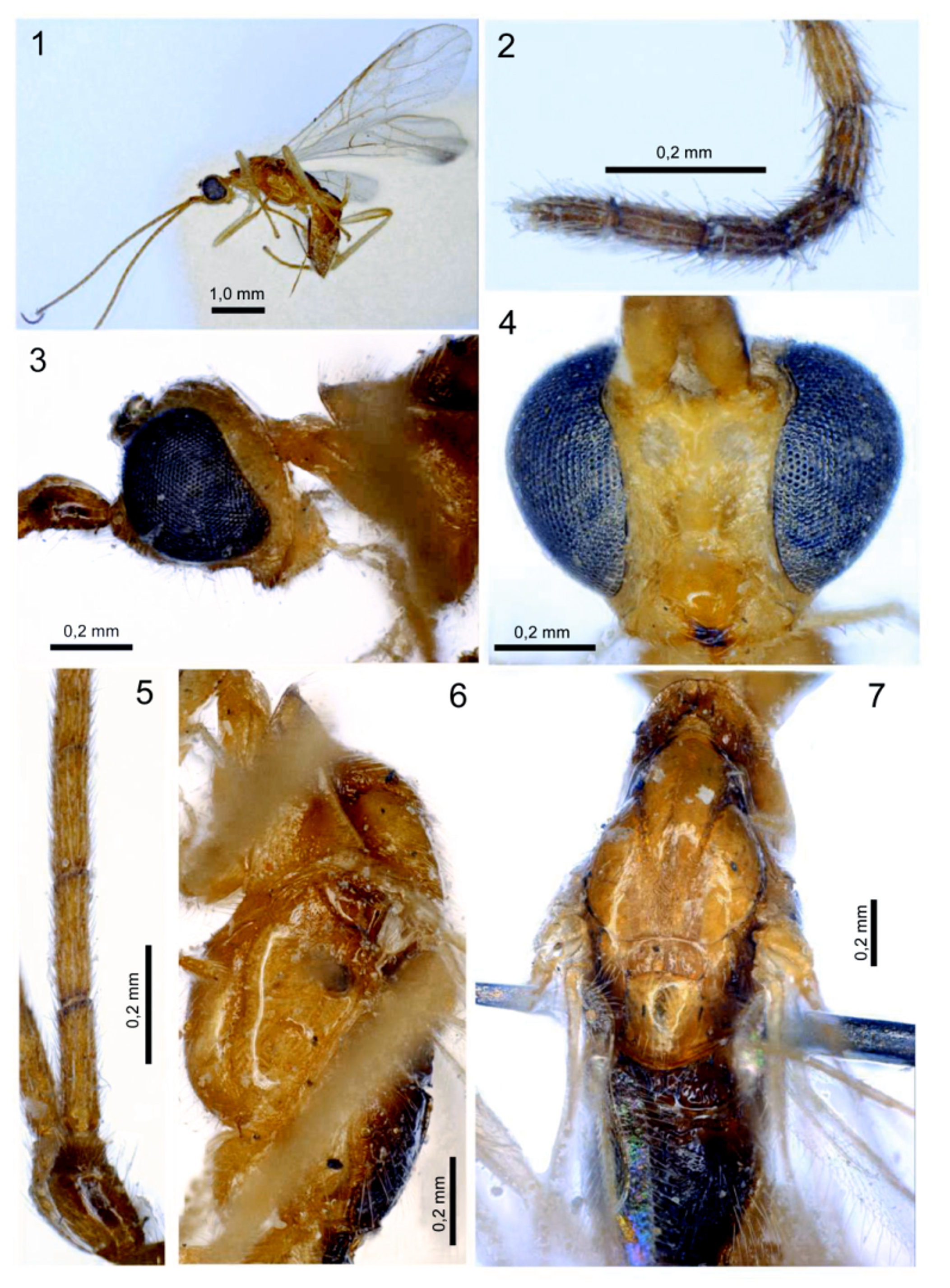
Description. Head transverse ( Figs 17 View Figures 13–20 , 28 View Figures 26–29 ). Vertex smooth. Ocelli enlarged, arranged in triangle with base weakly larger than its sides or in almost equilateral triangle ( Figs 17 View Figures 13–20 , 28 View Figures 26–29 ). Occipital carina distinct, complete dorsally and ventrally, ventrally not joining with hypostomal carina and reaching lower margin of head capsule near mandible separate from hypostomal carina. Eyes glabrous, distinctly and widely emarginated opposite antennal sockets. Face convex, without pointed tooth between antennal sockets, without carina between antennal socket and eye ( Figs 4 View Figures 1 – 7 , 18 View Figures 13–20 ). Clypeal suture entirely distinct. Hypoclypeal depression mediumsized and subrounded. Malar suture absent. Mandible short, thick basally and strongly narrowed apically. Maxillary palp 6-segmented; labial palp 4-segmented, second labial segment thickened, third segment long. Scape without spine, its dorsal margin weakly longer than ventral margin (lateral view). First flagellomere approximately as long as second flagellomere or weakly longer. Apical flagellomere pointed distally and with distinct apical spine ( Figs 2 View Figures 1 – 7 , 14 View Figures 13–20 ).
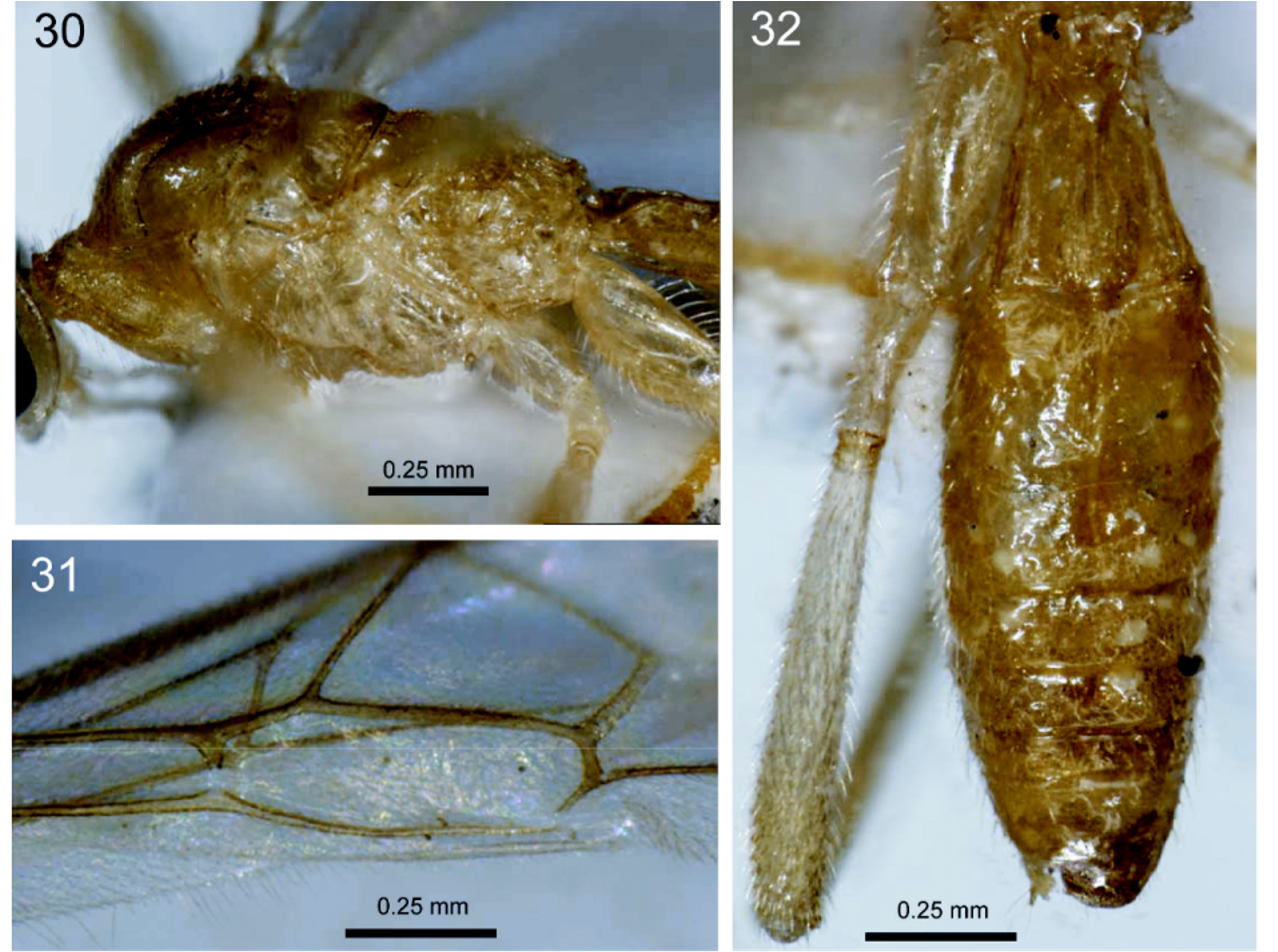
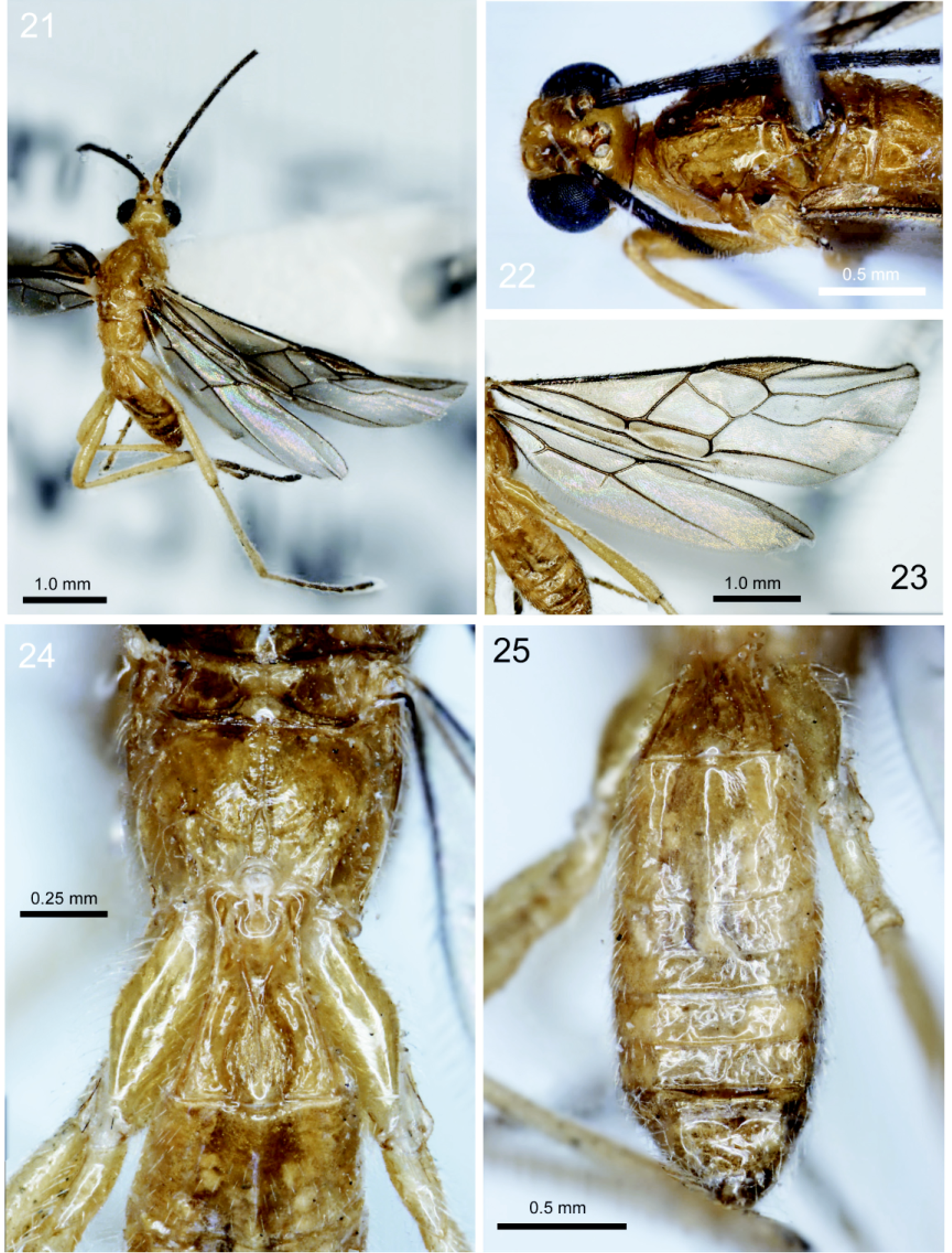
Mesosoma ( Figs 5, 6 View Figures 1 – 7 , 19 View Figures 13–20 , 30 View Figures 30 – 32 ). Pronotum without pronope, its anterior flange wide. Propleuron with large posterodorsal flange. Notauli complete, deep anteriorly and shallow posteriorly. Mesoscutum mostly smooth, distinctly roundly elevated above pronotum. Scuto-scutellar suture distinct medially and fine or very fine laterally. Scutellar sulcus (prescutellar depression) deep and long, with high median carina; submedial carinae variable. Scutellum convex, without lateral carinae and subposterior depression. Metanotum long, with distinct dorsomedian carina. Epicnemial (prepectal) carina distinct and complete. Precoxal sulcus (sternaulus) long, narrow, weakly sinuate, more or less distinctly separated from median coxa. Metapleural flange short and wide, rounded apically. Propodeum with areas delineated by distinct carinae ( Figs 20 View Figures 13–20 , 24 View Figures 21–25 ).
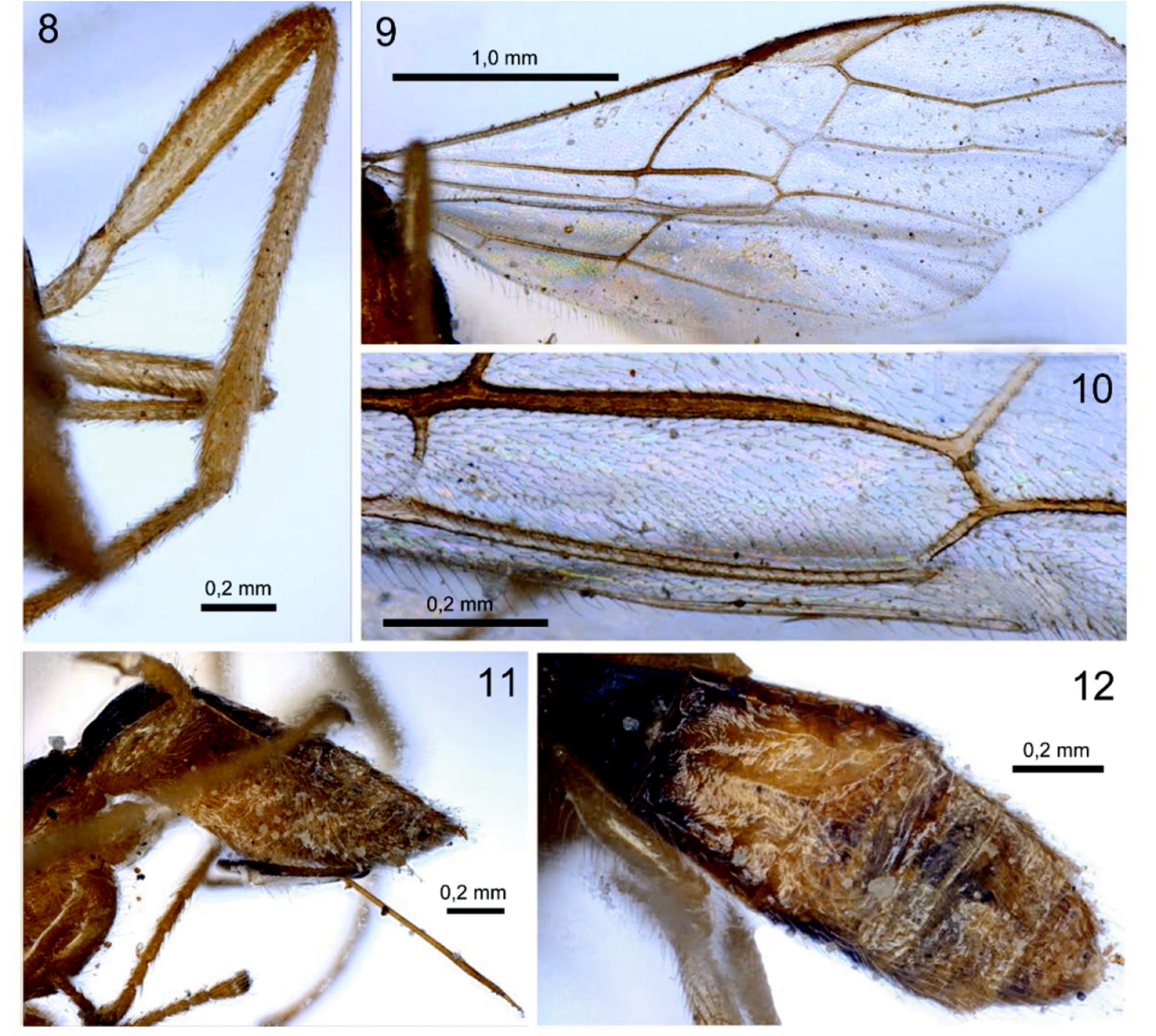
Wings ( Figs 9 View Figures 8–12 , 23 View Figures 21–25 ). Vein r-rs (radial) arising from or almost from middle of pterostigma. Parastigma long and distinctly separated from pterostigma. Marginal (radial) cell not shortened. Second submarginal (second radiomedial) cell long and narrowed towards apex. Vein m-cu (recurrent) distinctly antefurcal. Veins 1-M (basal) and m-cu (recurrent) weakly divergent posteriorly or subparallel. Vein 1cu-a (nervulus) distinctly antefurcal. First discal (discoidal) cell wide, petiolate anteriorly. First subdiscal (brachial) cell long, wide, weakly swollen, sometimes weakly widened towards apex ( Figs 12 View Figures 8–12 , 31 View Figures 30 – 32 ). Vein M+CU (medio-cubital) entirely straight. Vein CU1a (parallel) vein arising weakly before or almost from middle of distal margin of first subdiscal (brachial) cell. Vein CU1b (brachial) distinctly oblique towards base of wing. In hind wing, Vein M+CU (first abscissa of medio-cubital) 0.7–0.8 times as long as vein 1M (second abscissa of medio-cubital). Vein m-cu (recurrent) present, but short or very short.
Legs. All femora narrow ( Figs 8 View Figures 8–12 , 32 View Figures 30 – 32 ). Hind coxa long and narrow, longer than propodeum ( Fig. 24 View Figures 21–25 ). Tarsal segments long and rather slender. Hind tibial spurs subequal, straight and entirely setose. Claws simple, short, distinctly curved apically.
Metasoma ( Figs 11, 12 View Figures 8–12 , 25 View Figures 21–25 , 32 View Figures 30 – 32 ). First tergite distinctly and linearly widened towards apex, with large dorsope, with distinctly convergent but not fused and not complete basal carinae ( Figs 20 View Figures 13–20 , 24 View Figures 21–25 ). Tergites behind first one weakly sclerotized, smooth or shagreened. Second suture very shallow, narrow, almost straight. Second tergites with separated laterotergites. Fourthseventh tergites distinctly exposed behind third tergite. Ovipositor ( Figs 1 View Figures 1 – 7 , 11 View Figures 8–12 ) straight, rather long, more or less thickened subapically, with distinct dorsal notch.
Key to species of Troporhysipolis gen. nov.
1. Antenna with more than 37 flagellomeres; prescutellar depression with median and two or more lateral carinae ( Fig. 7 View Figures 1 – 7 ); fore wing vein cu-a (nervulus) antefurcal by approximately 0.2–0.5 of distance between veins 1-M (basal) and cu-a (nervulus) ( Fig. 10 View Figures 8–12 ); propodeum, first tergite entirely and at least second tergite laterally more or less distinctly but always darker than rest of body ( Figs 1, 6, 7 View Figures 1 – 7 , 12 View Figures 8–12 ). [Afrotropical]................. T. antefurcalis (Granger) View in CoL –. Antenna with fewer than 37 flagellomeres; prescutellar depression with only median carina ( Figs 19 View Figures 13–20 , 29 View Figures 26–29 ); fore wing vein cu-a (nervulus) antefurcal by approximately 0.9–1.2 of distance between veins 1-M (basal) and cu-a (nervulus) ( Figs 13 View Figures 13–20 , 23 View Figures 21–25 , 31 View Figures 30 – 32 ); propodeum and first-third tergites unicolorous with remainder of body ( Figs 14 View Figures 13–20 , 24–26 View Figures 21–25 View Figures 26–29 ) [ Papua New Guinea].................................... 2
2. Wings infumate with dark brown venation ( Fig. 23 View Figures 21–25 ); notauli uniting well before scutellar sulcus forming a single mid-longitudinal groove ( Fig. 22 View Figures 21–25 ); median area of metanotum large, without mid-longitudinal carina; propodeum with mid-longitudinal carina present on anterior 0.7 and posteriorly dividing to give rise to a narrow lozenge-shaped areola ( Fig. 24 View Figures 21–25 ); fore wing vein m-cu (recurrent) not especially thickened, more or less straight ( Fig. 23 View Figures 21–25 ); hind tarsus black ( Fig. 21 View Figures 21–25 )........ T. markshawi sp. nov.
–. Wings hyaline with pale brownish yellow venation ( Figs 13 View Figures 13–20 , 26 View Figures 26–29 ); notauli remaining separate to posterior of mesoscutum where they are separated by a mid-longitudinal groove and coarse sculpture ( Figs 19 View Figures 13–20 , 29 View Figures 26–29 ); median area of metanotum small, with weak but distinct mid-longitudinal ridge or carina ( Fig. 29 View Figures 26–29 ); propodeum with mid-longitudinal carina present on anterior 0.3–0.5 and posteriorly dividing to give wider, more angularly shaped areola ( Figs 20 View Figures 13–20 , 29 View Figures 26–29 ); fore wing vein m-cu (recurrent) distinctly thickened and curved ( Figs 13 View Figures 13–20 , 31 View Figures 30 – 32 ); hind tarsus yellow ( Figs 26, 27 View Figures 26–29 )............................. 3
3. Frons with a distinct though weak mid-longitudinal sulcus ( Fig. 28 View Figures 26–29 ) [see also barcode characters in Table 2 View Table 2 ]................ T. molecularis sp. nov.
–. Frons essentially flat, without any mid-longitudinal sulcus ( Fig. 17 View Figures 13–20 )...... T. brenthiaphagus sp. nov.
No known copyright restrictions apply. See Agosti, D., Egloff, W., 2009. Taxonomic information exchange and copyright: the Plazi approach. BMC Research Notes 2009, 2:53 for further explanation.
|
Kingdom |
|
|
Phylum |
|
|
Class |
|
|
Order |
|
|
Family |