
Glyptothrips Hood
|
publication ID |
https://doi.org/ 10.11646/zootaxa.5375.1.2 |
|
publication LSID |
lsid:zoobank.org:pub:2B77503D-20D9-45AD-92BB-8935AFB44C5F |
|
DOI |
https://doi.org/10.5281/zenodo.10248573 |
|
persistent identifier |
https://treatment.plazi.org/id/8500A75F-E072-FFB3-FF09-FD05FBF621CD |
|
treatment provided by |
Plazi |
|
scientific name |
Glyptothrips Hood |
| status |
|
Glyptothrips Hood, 1912: 116 View in CoL . Type species Glyptothrips flavescens Hood, 1912 View in CoL , by monotypy.
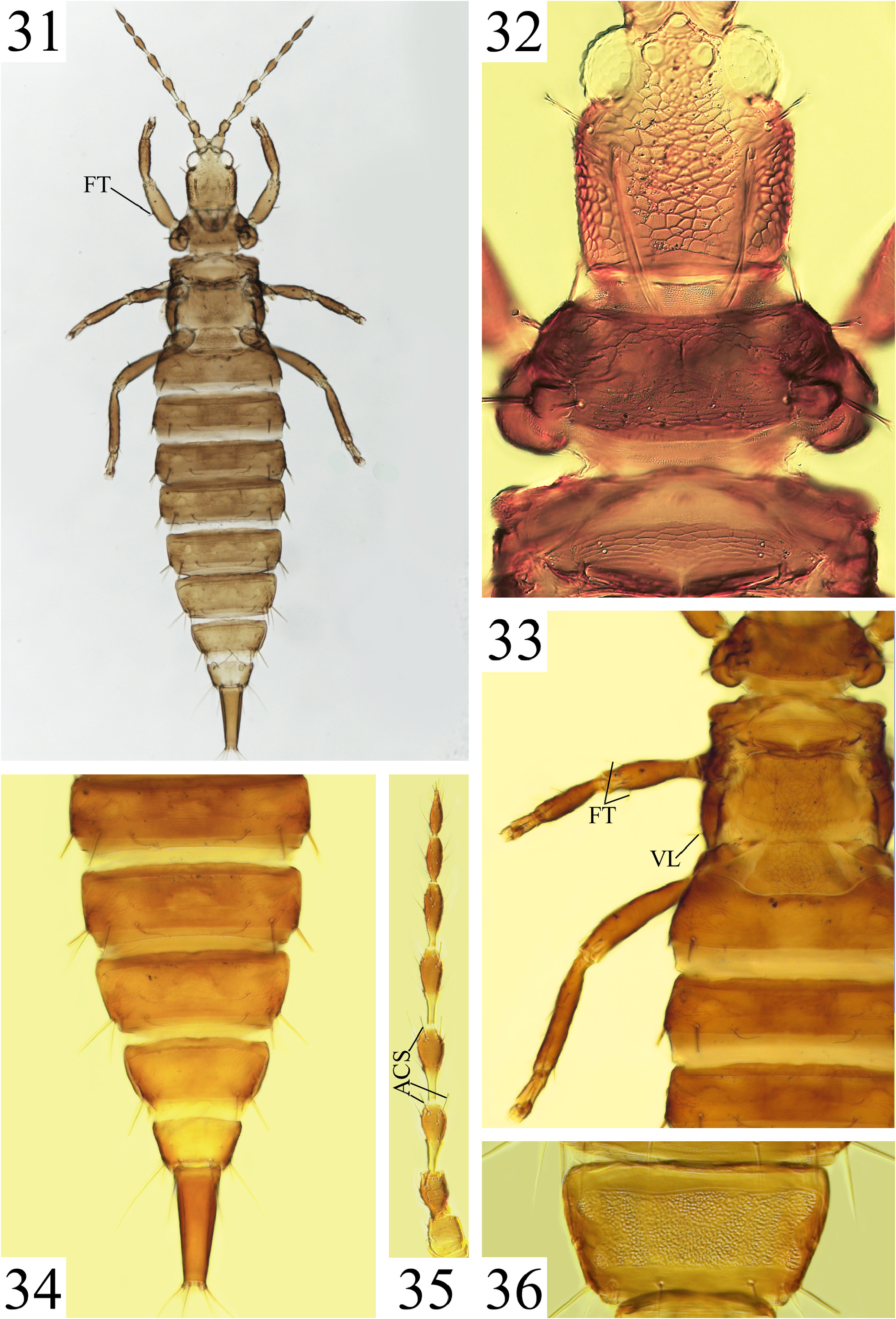
There is no unique character state to diagnose this genus, but the following combination of character states seem useful to distinguish a member of the genus from other Glyptothripini : (1) body strongly reticulate ( Figs 6 View FIGURES 5–8 , 15 View FIGURES 14–18 , 58 View FIGURES 57–60 ), except the tube which has at most some weak sculpture basally ( Figs 16 View FIGURES 14–18 , 45 View FIGURES 41–46 , 59 View FIGURES 57–60 ); (2) pronotal AM reduced ( Figs 20 View FIGURES 19–24 , 32 View FIGURES 31–36 , 69 View FIGURES 66–73 ); (3) pterothoracic ventrolateral setae usually thick and capitate ( Figs 21 View FIGURES 19–24 , 33 View FIGURES 31–36 , 62 View FIGURES 61–65 ), except in G. arkansanus View in CoL , G. bucca View in CoL , G. flavescens View in CoL and G. reticulatus View in CoL . In addition, all observed Glyptothrips species share the following: head incut behind globose/moruloid eyes ( Figs 26 View FIGURES 25–27 , 48 View FIGURES 47–50 ); PO setae and all pronotal setae except AM well-developed with dilated tips ( Figs 15 View FIGURES 14–18 , 32 View FIGURES 31–36 ); antennal segments II–IV with at least one pair of capitate setae (except in G. bucca View in CoL ) ( Fig. 35 View FIGURES 31–36 ); prosternal basantra present; tube with straight, tapering sides ( Figs 7 View FIGURES 5–8 , 11 View FIGURES 9–13 ); fore tarsi armed with a tooth (except in G. arkansanus View in CoL ) ( Figs 42 View FIGURES 41–46 , 48 View FIGURES 47–50 ); fore wings without duplicated cilia.
Character state variation amongst included species:
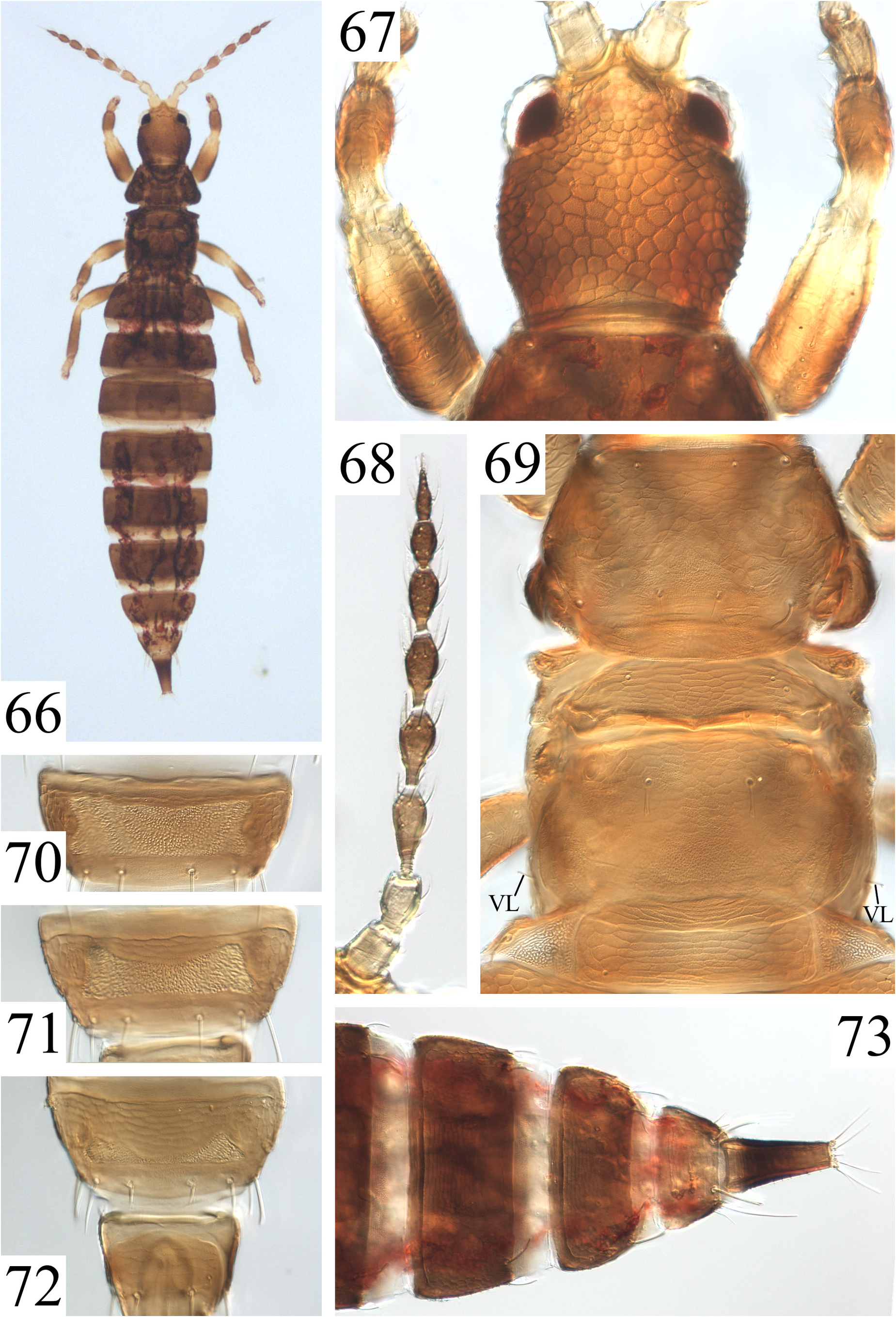
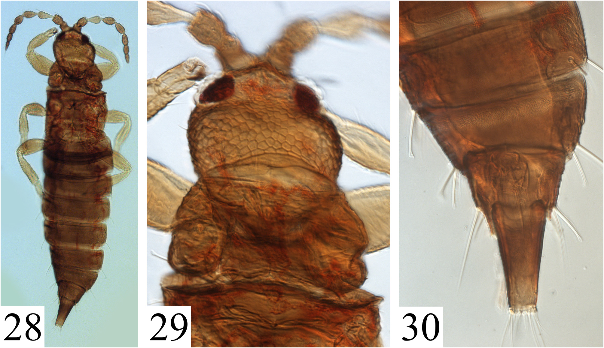
Colouration: Body structures vary in colour, which is heavily influenced by how the specimen was prepared for slide mounting: many species were described from individuals not macerated in NaOH prior to mounting. Several type specimens were also mounted initially in Hoyers, a mounting media not as durable as Canada Balsam, and much colour information could be modified or lost (e.g. Figs 28–30 View FIGURES 28–30 ). Some patterns remain useful for identifications. Original descriptions state some species are yellowish (e.g. G. interior — Fig. 41 View FIGURES 41–46 and G. silvaticus — Fig. 61 View FIGURES 61–65 ) or darker brown (e.g. G. claviger — Fig. 14 View FIGURES 14–18 and G. subcalvus — Fig. 66 View FIGURES 66–73 ). However, some macerated specimens identified structurally as G. subcalvus have a lighter, yellowish colour (e.g. Figs 69–72 View FIGURES 66–73 ).
Antennae are frequently of similar colour to the head, with distal antennal segments progressively darker, and darker antennal segments (especially IV–VI) often have the basal areas and/or pedicels lighter ( Figs 25 View FIGURES 25–27 , 35 View FIGURES 31–36 , 43 View FIGURES 41–46 ). In G. arkansanus the antennae are mostly yellow with only the last antennal segment slightly darker ( Fig. 5 View FIGURES 5–8 ); and G. subcalvus has the first two antennal segments very pale yellow, much lighter than the head ( Figs 66, 68 View FIGURES 66–73 ).
Head and thorax are commonly the same colour ( Figs 9 View FIGURES 9–13 , 41 View FIGURES 41–46 ), or the thorax is partially or fully darker than the head ( Figs 47 View FIGURES 47–50 , 57 View FIGURES 57–60 ). The abdomen is frequently paler or gets progressively paler distally ( Fig. 31 View FIGURES 31–36 ). Some species have a darker antecostal ridge and/or spot on tergites II–VII ( G. divergens — Fig. 19 View FIGURES 19–24 ; G. interior — Fig. 41 View FIGURES 41–46 ; G. saltuarius — Fig. 57 View FIGURES 57–60 ; G. silvaticus — Fig. 61 View FIGURES 61–65 ). Some species have two longitudinal pale lines dividing the tergites into thirds ( G. arkansanus — Figs 5–7 View FIGURES 5–8 ), and the posteromedian area of the tergite may be much paler. Similar lines are present but not as obvious in G. flavescens ( Fig. 26 View FIGURES 25–27 ), and in G. subcalvus the tergal median third seems to be darker than the rest of the abdomen ( Fig. 66 View FIGURES 66–73 ). The tube is also variable between species, but more commonly a similar ( Figs 7 View FIGURES 5–8 , 27 View FIGURES 25–27 ) or darker colour to the previous abdominal segments ( Figs 22 View FIGURES 19–24 , 65 View FIGURES 61–65 ). The basal and/or apical areas of the tube are frequently lighter or darker than the middle of the tube ( Figs 45 View FIGURES 41–46 , 73 View FIGURES 66–73 ).
Head: all Glyptothrips species have the head fully covered by strong reticulation, both on dorsal and ventral surfaces ( Figs 6 View FIGURES 5–8 , 20 View FIGURES 19–24 , 67 View FIGURES 66–73 ). The genae vary from almost straight ( G. divergens — Fig. 20 View FIGURES 19–24 ; G. hylaeus — Fig. 38 View FIGURES 37–40 ), to strongly curved ( G. bucca — Fig. 10 View FIGURES 9–13 ; G. subcalvus — Fig. 67 View FIGURES 66–73 ). Observed head length x greatest width ratio from original descriptions vary from 1 ( G. floridensis ) to 1.5 ( G. longiceps ). However, this character should be used with caution; it can be affected by slide mounting, and ranges can overlap between species ( Table 2 View TABLE 2 ).
...Continued on the next page
¹All given ratios and measures were obtained from original descriptions and/or the individuals listed in the “observed specimens” section under each species in the manuscript. Intraspecific variation has not been studied for the majority of the species due to the limited number of individuals available.
Most species in the genus have small and acute postocellar, occipital and head lateral setae, but G. floridensis is illustrated by Stannard (1955) with the occipital and many lateral setae as long and capitate as the PO. The occipital setae have dilated tips also in G. claviger ( Fig. 15 View FIGURES 14–18 ) and G. interior ( Fig. 42 View FIGURES 41–46 ), but neither of these species has the head lateral setae thickened or capitate. Length of PO varies from very short and inconspicuous ( G. divergens — Fig. 20 View FIGURES 19–24 ), to as long or longer than the compound eye ( G. silvaticus — Fig. 63 View FIGURES 61–65 ) ( Table 2 View TABLE 2 ).
Antennae: the type species, G. flavescens , has only 7 antennal segments with antennal segments VII and VIII fused ( Fig. 26 View FIGURES 25–27 ). G. arkansanus approximates this condition in the genus, with antennal segments VII–VIII broadly joined but separated by a suture. All other Glyptothrips species have VII and VIII clearly separated ( Figs 12 View FIGURES 9–13 , 23 View FIGURES 19–24 , 35 View FIGURES 31–36 ). The most common sense cone formula in Glyptothrips is 3 sense cones on antennal segment III and 4 on IV. However, reductions in the number of sense cones are known in G. bucca , G. flavescens , G. interior , G. reticulatus and G. subcalvus ( Table 2 View TABLE 2 ).
The shape of antennal segments is also variable within the genus, from very short and globose in G. flavescens ( Fig. 26 View FIGURES 25–27 ) and G. arkansanus ( Fig. 5 View FIGURES 5–8 ), to elongate in G. divergens ( Fig. 23 View FIGURES 19–24 ) and G. fuscipes ( Fig. 35 View FIGURES 31–36 ). Some species are also intermediate between these extremes, such as G. bucca , G. subcalvus (short but not globose antennal segments), G. hylaeus or G. silvaticus .
Prothorax: most species have the pronotum reticulate ( Figs 6 View FIGURES 5–8 , 20 View FIGURES 19–24 ), but the sculpture can be less obvious in some species ( Fig. 69 View FIGURES 66–73 ). The epimeral sutures are usually incomplete. The position of AA setae is at or very close to the pronotum anterior angles in most Glyptothrips species, but in at least G. flavescens ( Fig. 26 View FIGURES 25–27 ) and G. floridensis this pair of setae arises close to AM.
Pterothorax: meso- and metanotum are both clearly sculptured, usually with equiangular reticles without any internal markings ( Figs 6 View FIGURES 5–8 , 21 View FIGURES 19–24 ). However, at least G. flavescens and some specimens of G. claviger have “tubercles” in the metanotum, in place of some of the sculpture lines ( Fig. 26 View FIGURES 25–27 ). The ventrolateral setae are thick and capitate in most Glyptothrips ( Figs 21 View FIGURES 19–24 , 62 View FIGURES 61–65 ), with the only exceptions listed in the proposed diagnosis above.
Legs: all legs usually have similar sizes and sculpturing, and are covered in many acute setae ( Figs 5–6 View FIGURES 5–8 , 14–15 View FIGURES 14–18 ). The fore tibia does not have tubercles. In G. fuscipes , all femora and the fore tibiae have at least one or two elongate, thick and capitate setae ( Figs 31, 33 View FIGURES 31–36 ); most other Glyptothrips species lack such setae, although a short but capitate seta has been seen in the fore femora of some specimens of G. claviger , G. longiceps , G. reticulatus , G. silvaticus ( Fig. 63 View FIGURES 61–65 ) and G. subcalvus .
Wings: there are multiple wing forms within the genus, with macropterous ( Fig. 5 View FIGURES 5–8 ), micropterous ( Fig. 15 View FIGURES 14–18 ) and apterous ( Fig. 55 View FIGURES 51–56 ) specimens known. Five species ( G. arkansanus , G. flavescens , G. reticulatus , G. saltuarius and G. subcalvus ) are known to have two or more wing morphs. The degree of wing development is frequently associated to changes in some character states, especially when comparing macropterae (eyes sometimes larger, ocelli present, pelta not as wide) with apterae (eyes sometimes reduced, ocelli reduced or absent, pelta wide and rectangular or oval in shape) ( Mound 1977, 2005; Mound & Marullo 1996; personal observations).
Abdomen: the pelta is highly variable, and at least partially associated with the degree of wing development. The most common shape seems to be a broad oval or rectangular plate ( Figs 10 View FIGURES 9–13 , 26 View FIGURES 25–27 , 44 View FIGURES 41–46 , 69 View FIGURES 66–73 ), however some species may have a triangular, trapezoidal ( Figs 15 View FIGURES 14–18 , 36 View FIGURES 31–36 ) or hat-shaped ( Fig. 6 View FIGURES 5–8 ) pelta. Tergites II–IX are sculptured, with the median posterior area weaker or sometimes smooth ( Figs 6 View FIGURES 5–8 , 44 View FIGURES 41–46 , 58 View FIGURES 57–60 ). The three main setal pairs on tergite IX are well developed, with S2 reduced in males ( Fig. 45 View FIGURES 41–46 ). Setae S1 are most frequently dilated or capitate ( Figs 45 View FIGURES 41–46 , 73 View FIGURES 66–73 ), while S2 are more frequently acute or blunt ( Table 2 View TABLE 2 ). The tube is mostly smooth, with weak lines of sculpture basally ( Figs 7 View FIGURES 5–8 , 16 View FIGURES 14–18 , 39 View FIGURES 37–40 ), but never with strong reticulation as in Chamaeothrips Hood or Eschatothrips species. The tube is usually shorter than the head or almost as long as the head; only two species ( G. divergens and G. flavescens ) have recorded specimens where the tube is slightly longer than the head ( Figs 19 View FIGURES 19–24 , 25 View FIGURES 25–27 ).


No known copyright restrictions apply. See Agosti, D., Egloff, W., 2009. Taxonomic information exchange and copyright: the Plazi approach. BMC Research Notes 2009, 2:53 for further explanation.
|
Kingdom |
|
|
Phylum |
|
|
Class |
|
|
Order |
|
|
Family |
Glyptothrips Hood
| Lindner, Mariana Flores, Ferrari, Augusto, Lima, Élison Fabrício Bezerra & Cavalleri, Adriano 2023 |
Glyptothrips
| Hood, J. D. 1912: 116 |