
Genyocerus Motschulsky
|
publication ID |
https://doi.org/ 10.11646/zootaxa.1576.1.3 |
|
publication LSID |
lsid:zoobank.org:pub:C0ED40AA-840D-4950-B8D6-7CE87870D64B |
|
persistent identifier |
https://treatment.plazi.org/id/842B6D7D-FFFD-FFD8-FF67-9C005399B9EC |
|
treatment provided by |
Felipe |
|
scientific name |
Genyocerus Motschulsky |
| status |
|
Genyocerus Motschulsky View in CoL
Genyocerus Motschulsky, 1858: 68 View in CoL . Type species, by monotypy, G. albipennis Motschulsky, 1858 View in CoL .
Diacavus Schedl, 1939: 363 View in CoL . Type species, by original designation, Diapus frontalis Strohmeyer, 1914 View in CoL . Synonymy: Wood 1969: 118.
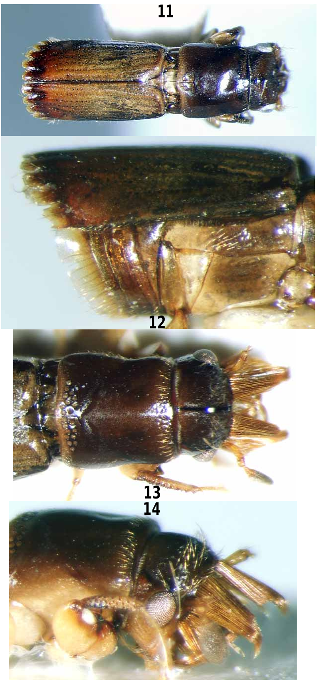
Length 1.8–5.4 mm. 3.0–4.2 times as long as wide. Colour reddish brown to black, underside and appendages usually paler, elytral disc sometimes partly testaceous. Vestiture sparse except on female frons. Head with a very short rostrum, frons angularly separated from vertex, flattened, sometimes with a longitudinal keel or keels in the male; young females with one to several brushes or pencils of long hairs on frons and sometimes also on epistoma, often largely concealing frontal surface but lost later during gallery construction. Eyes oval or egg–shaped, not extending onto ventral surface of head. Antennae inserted close to lower margin of eyes in males, and in females of some species, near upper margin of eyes in females of species with long hair brushes on upper part of frons. Antennal scape short, club–shaped in male, slightly longer and scarcely widened distally in female, funicle four–segmented, club oval, flattened, anterior and posterior surfaces uniformly pubescent to base, a narrow testaceous strip lacking sensory setae along the posterior margin. Maxillae with separate galea and lacinia. Submentum separated from margin of oral opening by a groove on each side. Pronotum usually longer than wide, sides shallowly constricted in anterior half, widest about one–third from base, femoral grooves without carinae anteriorly or posteriorly, disc weakly convex, usually with one to many large mycangial pores on each side of median line in basal third. Mesonotum without a median carina, scutellum slightly medially impressed, flush with elytral surface posteriorly. Elytra elongate, horizontal, disc weakly striate–punctate, declivity very short or absent, male elytra with spines or teeth on some interstriae at discal apex, female elytral apex simply rounded. Metasternal–metepisternal depression of male unarmed. Abdomen with fifth ventrite concave and subvertical in male, convex in female, apical margin of fourth ventrite sometimes extended as a hyaline membrane of fused hairs in male. Procoxae very large, longer than femur, widely separated by a trapezoidal intercoxal plate, mesocoxae globular, less widely separated, metacoxae transverse, extending more than halfway across first abdominal ventrite, narrowly separated medially. Protibiae narrow, granulate or with rasp–like teeth in both sexes, the apex with a single recurved spine.
The genus is distinguished from other platypodine genera except Diapus by the very large, widely separated procoxae. The species of Genyocerus can be distinguished from those of Diapus by their wider, more obvious scutellum, which is flush with the elytral surface posteriorly. The mycangial pores of Genyocerus are usually larger than those of Diapus and never fused to form a transverse or crescentic bar on each side of the midline. The antennal club of Genyocerus never has a median, testaceous strip lacking sensillae on the outer surface. The males of Diapus never possess a membranous extension of the apical margin of the fourth abdominal ventrite. The females of Genyocerus never possess dehiscent mandibular appendages. It may be noted that the larvae, so far as known ( Browne 1972), show little evidence of generic distinctions. The two genera appear closely related, and more detailed studies, including DNA data and cladistic analyses are needed to test this phylogenetic hypothesis.
Sexual dimorphism
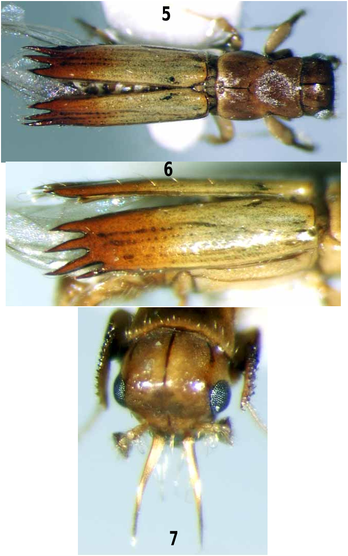
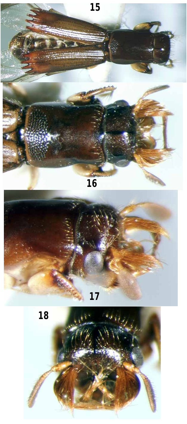
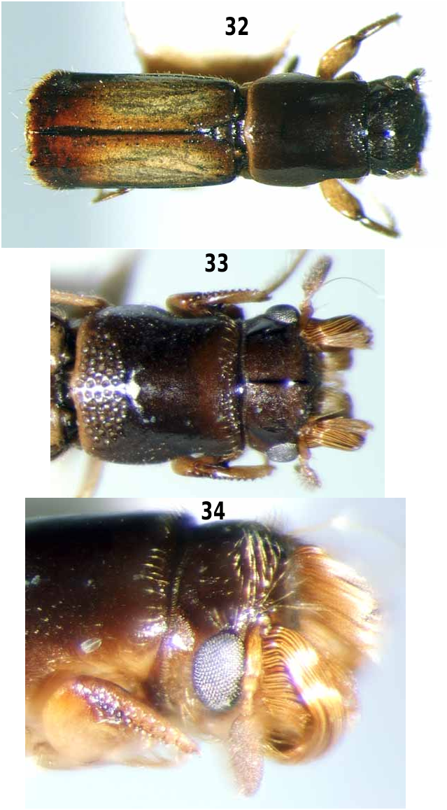
The males of Genyocerus are characterised by the presence of teeth or spines at the apex of the elytral disc; in the females, the elytral apices are subtruncate or transversely rounded. In the males of some species, the apical margin of the fourth ventrite extends as a hyaline membranous structure projecting over the basal part of the fifth ventrite ( Fig. 12 View PLATE 4 ). This probably helps to make the concave fifth ventrite a more effective 'shovel' as the male collects and expels frass from the gallery system. The fifth ventrite of the female is essentially convex, and there is never an extension of the fourth ventrite. The young females bear brushes of hairs on the frons. The arrangement of these is species-specific, and they are probably used in courtship ( Browne 1961a, Roberts 1993) but lost after mating when the female takes over the extension of the gallery system from the male. In the male, the antennae are always inserted below the middle of the frons. In the females, the position of the antennae is associated with the presence or absence of large hair brushes on the upper part of the frons. When these are absent, the antennae are inserted in the same position as in the male ( Fig. 7 View PLATE 2 ); when present, the antennae are inserted on the upper part of the frons at or above the level of the upper margin of the eyes (e.g. Figs 17 View PLATE 5 , 34 View PLATE 10 ). The number of mycangial pores on the pronotum is variable but is almost always greater in the female than in the male (see below).
Arrangement of teeth on male elytral apex
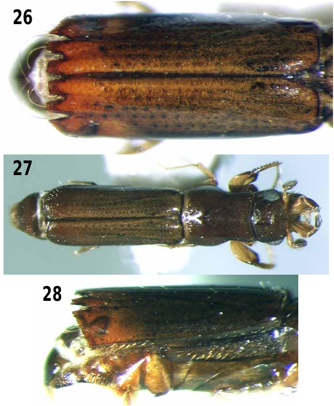
Schedl (e.g. 1966) states that the first and second major teeth at the apex of the male elytra are formed from extensions of interstriae 1 and 3, whereas Browne (1962) writes that they are formed from extensions of interstriae 2 and 4. In fact, the first and second teeth are borne on interstriae 3 and 5. The interstriae in Genyocerus are often impunctate and can only be distinguished by the lines of strial punctures between them. Even the arrangement of the striae on the elytra is often difficult to determine in the smaller species, because the punctures are often small and obsolescent, but it can be seen in larger species such as G. biporus ( Fig. 1 View PLATE 1 ). The first interstriae are normally developed along the suture. The apex may form a small secondary tooth at the inner side of the larger primary tooth on interstriae 3. Striae 1 are very short and usually terminate not more than one-third along the elytra. Thus the second interstriae are very short and taper to a point about onethird from the base of the elytra. Interstriae 3 and 5 are broadened distally and extend over the declivity as teeth. The teeth are usually flattened and blunt or with bifid tips ( Figs 11 View PLATE 4 , 15 View PLATE 5 ), but sometimes are more slen- der, tapering and spine-like ( Figs 5 View PLATE 2 , 30). Interstriae 4 and 6 are narrowed posteriorly and may not reach the end of the elytra. On the posterolateral part of the elytra, striae 6 - 8 end about 1/3 from the elytral apex, and there is a smooth, shining, punctureless area extending across interstriae 7 - 8 and sometimes interstriae 9 ( Fig. 9 View PLATE 3 ). One or both interstriae 7 and 8 and interstriae 9 may bear apical teeth, or interstriae 7 - 9 may be fused apically to form an acute edge bearing several small teeth ( Fig. 27 View PLATE 8 ). In a few species (e.g. G.tenellus , G. exilis ), a tooth is also present on the elytral declivity.
Intraspecific variation
It has become clear during the course of this study that the authors who described the majority of species in the genus Genyocerus underestimated the intraspecific variation in the number of mycangial pores on the pronotum. Both Schedl and Browne in many of their papers (see Wood & Bright 1992 for references) noted that the number of pores differs between the sexes, with the female usually having a greater number (although the reverse is the case in Genyocerus puer (Schedl) , Roberts 1993). In addition, the number of pores can differ between the two sides of the same individual, and there can also be considerable variation between individuals from the same collection site, and from different sites, independently of the differences between males and females. Similarly, in the males of some species, the shape and size of the teeth at the end of the elytral disc can be quite variable intraspecifically. Such variations have sometimes been described as separate species. However, the gradual accumulation of more specimens from a wider range of collection sites has indicated that intermediates exist. The resulting new synonyms are reported below.
Biology
The species of Genyocerus normally breed only in host trees in the family Dipterocarpaceae . The only partial exception to this is G. pendleburyi , which may occasionally attack trees in the family Fagaceae in areas in which dipterocarps are scarce ( Browne 1977). A few other records from non-dipterocarp trees may or may not involve actual breeding. By contrast, species of Diapus , like most platypodines, are generally polyphagous, attacking any trees in which the symbiotic ambrosia fungi will grow. Genyocerus species usually attack dying or felled trees, but Browne (1961a) noted that attacks may occur on stressed trees after drought.
The general features of the life history are given by Beeson (1961) and Browne (1961a). The gallery system is started by the male, but only a short vertical gallery is made. It is probable that the female is attracted by a male-produced pheromone ( Beaver 2000b), although no studies have been made on Genyocerus or Diapus . After mating, the female continues to extend the gallery into the wood. The male remains in the entrance, keeping out potential predators and parasites and removing the frass that accumulates from the boring activities of the female. The gallery system branches mainly in one transverse plane and may extend deeply into the tree. The mycangia on the pronotum transport an ambrosial fungus (or fungi) that is released into the gallery, grows in the wood and provides the only food for both adults and larvae, which feed on the hyphae growing on the gallery walls. The ambrosia fungi associated with Genyocerus and Diapus have not been investigated. Mature larvae construct pupal cells above and below the gallery at the ends of the branches. The adults of the new generation emerge through the parental entrance hole.


No known copyright restrictions apply. See Agosti, D., Egloff, W., 2009. Taxonomic information exchange and copyright: the Plazi approach. BMC Research Notes 2009, 2:53 for further explanation.
|
Kingdom |
|
|
Phylum |
|
|
Class |
|
|
Order |
|
|
Family |
Genyocerus Motschulsky
| Beaver, R. A. & L. - Y 2007 |
Diacavus Schedl, 1939: 363
| Wood, S. L. 1969: 118 |
| Schedl, K. E. 1939: 363 |
Genyocerus
| Motschulsky, V. von 1858: 68 |