
Cherevychnavis, Bochenski & Wertz & Tomek & Gorobets, 2019
|
publication ID |
https://doi.org/ 10.11646/zootaxa.4624.1.3 |
|
publication LSID |
lsid:zoobank.org:pub:84AA439F-5A44-495B-A981-81ED53173690 |
|
persistent identifier |
https://treatment.plazi.org/id/842887D0-EF3E-813F-FF3E-FDFE3B7A17C3 |
|
treatment provided by |
Plazi |
|
scientific name |
Cherevychnavis |
| status |
gen. nov. |
Genus: CHEREVYCHNAVIS gen. nov.
Type species. Cherevychnavis umanskae sp. nov.
Etymology. The genus name is the name of the type locality (Cherevychne) merged with the Latin word for bird (avis). Gender is feminine.
Diagnosis. A member of Charadrii , approximately the size of extant Haematopus ostralegus that differs from all genera by the following combination of characters of the coracoid: robust processus acrocoracoideus with cranio-sternal length about the same as medial projection; the foramen nervi supracoracoidei is very close to the sternal margin of the cotyla scapularis (only in Haematopus is this foramen similarly close, yet is still more widely separated); the medial margin of the facies articularis humeralis is straight almost along its entire length; and sternally, the angulus lateralis is broad with sternal and lateral margins meeting almost at right angles; and on which the dorsal edge of the facies articularis sternalis connects to the angulus lateralis.
Differential diagnosis. The coracoid and humerus differ as follows:
From Haematopus , Recurvirostra and Himantopus in:
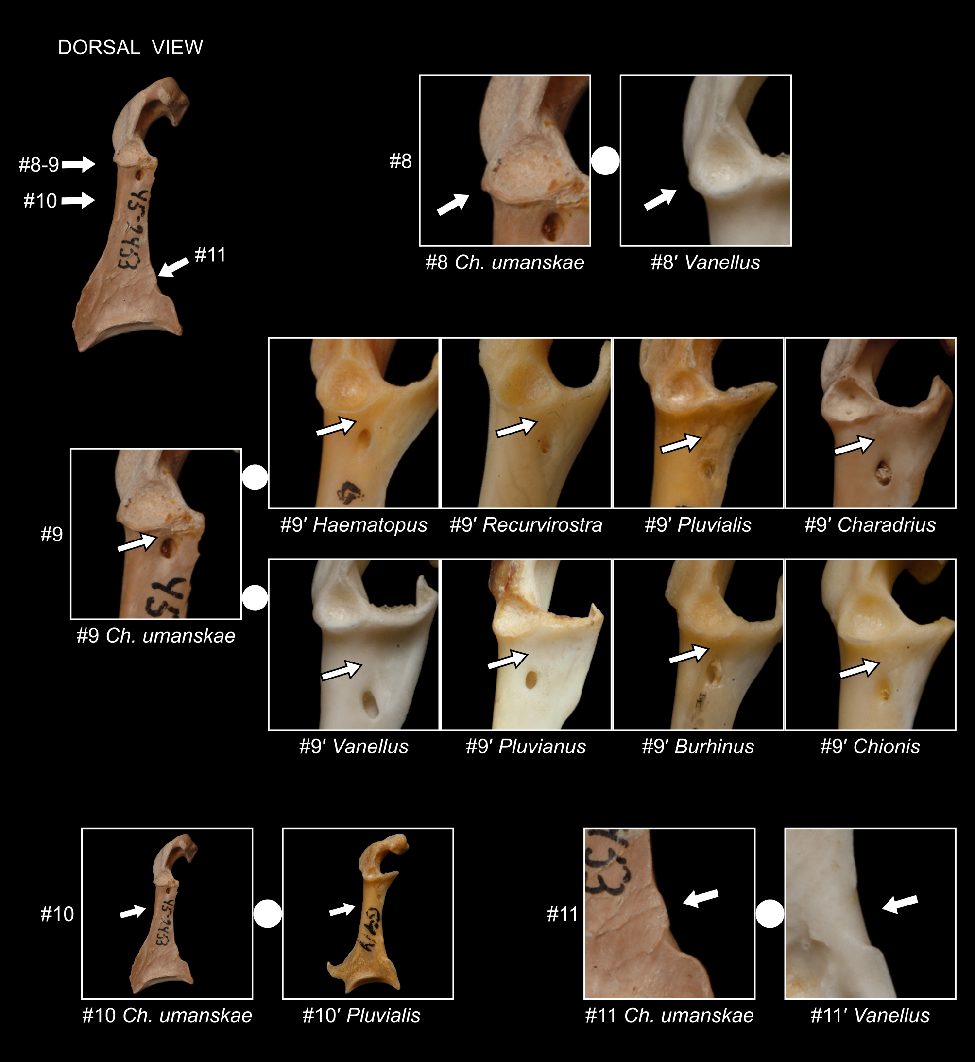
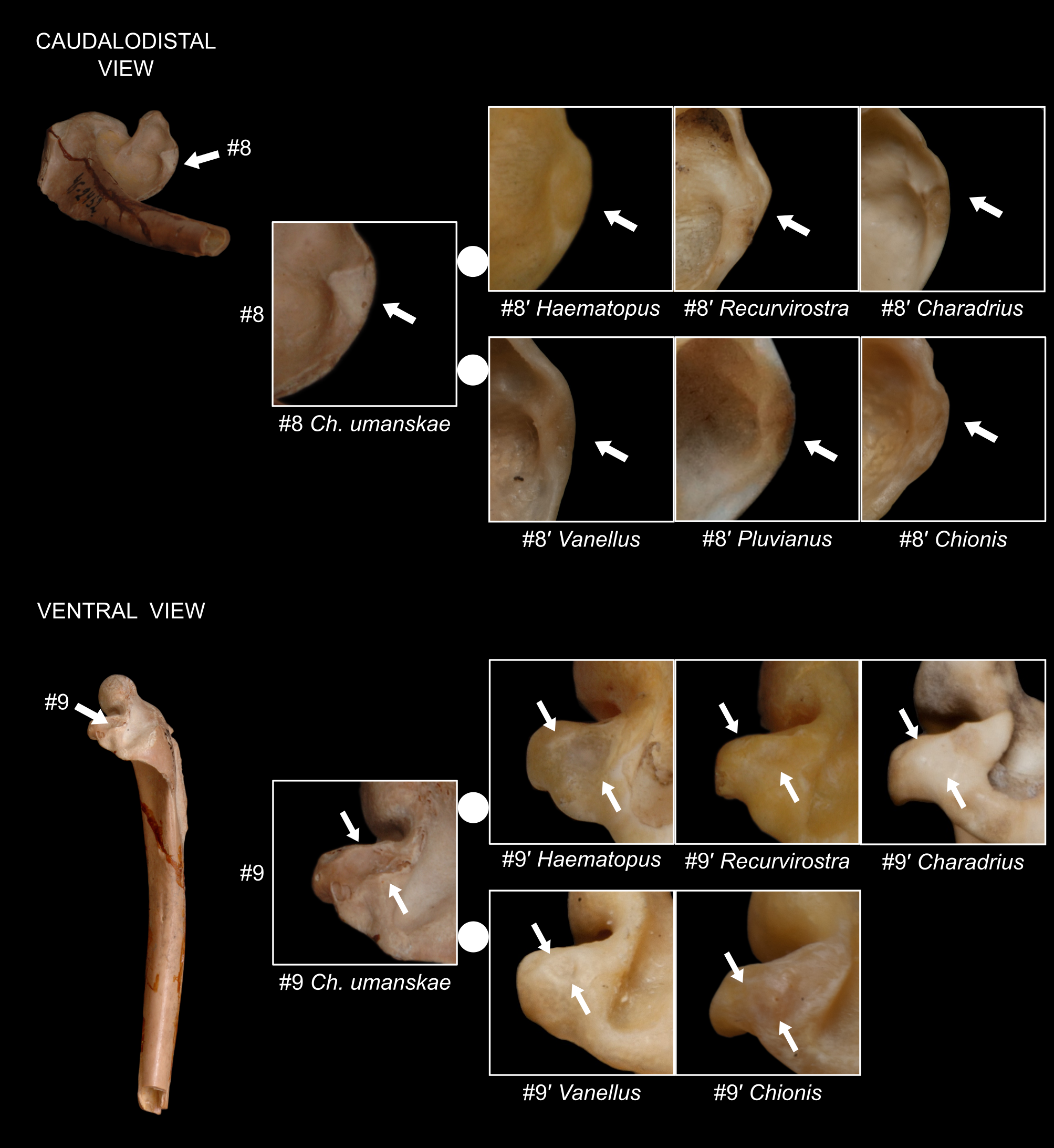
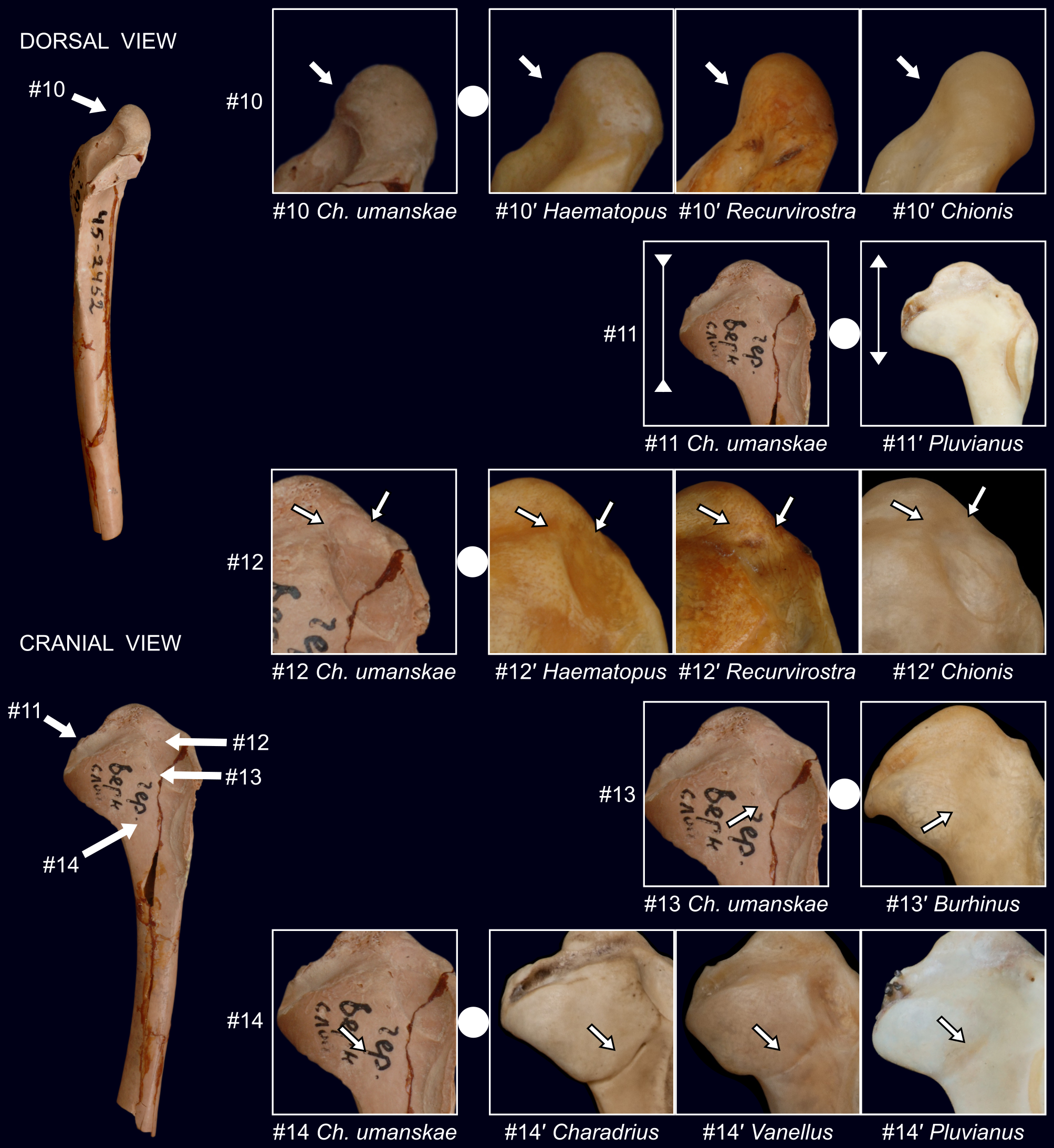
Coracoid ( Figs. 2 View FIGURE 2 –4 View FIGURE 2 View FIGURE 3 View FIGURE 4 ): ● extremitas omalis, dorsal view: The medial margin of the facies articularis humeralis is straight almost along its entire length (#5) (in H. ostralegus , Recurvirostra and Himantopus it arches or curves medially towards the facies articularis clavicularis); ● extremitas omalis, dorsal view: The foramen nervi su- pracoracoidei is very close to the sternal margin of the cotyla scapularis (#9) (in H. ostralegus , Recurvirostra and Himantopus the foramen is somewhat farther sternad from the cotyla); ● extremitas sternalis, dorsal view: The dorsal edge of the facies articularis sternalis connects to the angulus lateralis (#14) (in H. ostralegus , most Recurvirostra and Himantopus the dorsal edge of the facies articularis sternalis disappears well before reaching the angulus lateralis).
Humerus ( Figs. 9–10 View FIGURE 9 View FIGURE 10 ): ● caudalodistal view: The infraspinatus attachment on crista bicipitalis is triangular, very broad dorsoventrally (#8) (in H. ostralegus , it is elongated proximodistally and much narrower; in Recurvirostra it is roundish and in Himantopus it is irregular); ● dorsal view, cranial margin of caput humeri: The caput humeri is more rounded and does not reach far proximally (#10) (in H. ostralegus , Recurvirostra and Himantopus it is more elongated and reaches farther proximally); ● ventral view: There is a deep rounded depression in the crista bicipitalis immediately distal to the tuberculum ventrale (#9) (in H. ostralegus , Recurvirostra and Himantopus the depression is shallower and its edges are less conspicuous); ● cranial view: Proximal part of the impressio musculus coracobrachialis is deeper and its edges are better distinguished (#12) than in extant H. ostralegus , Recurvirostra and Himantopus .
From Pluvialis in:
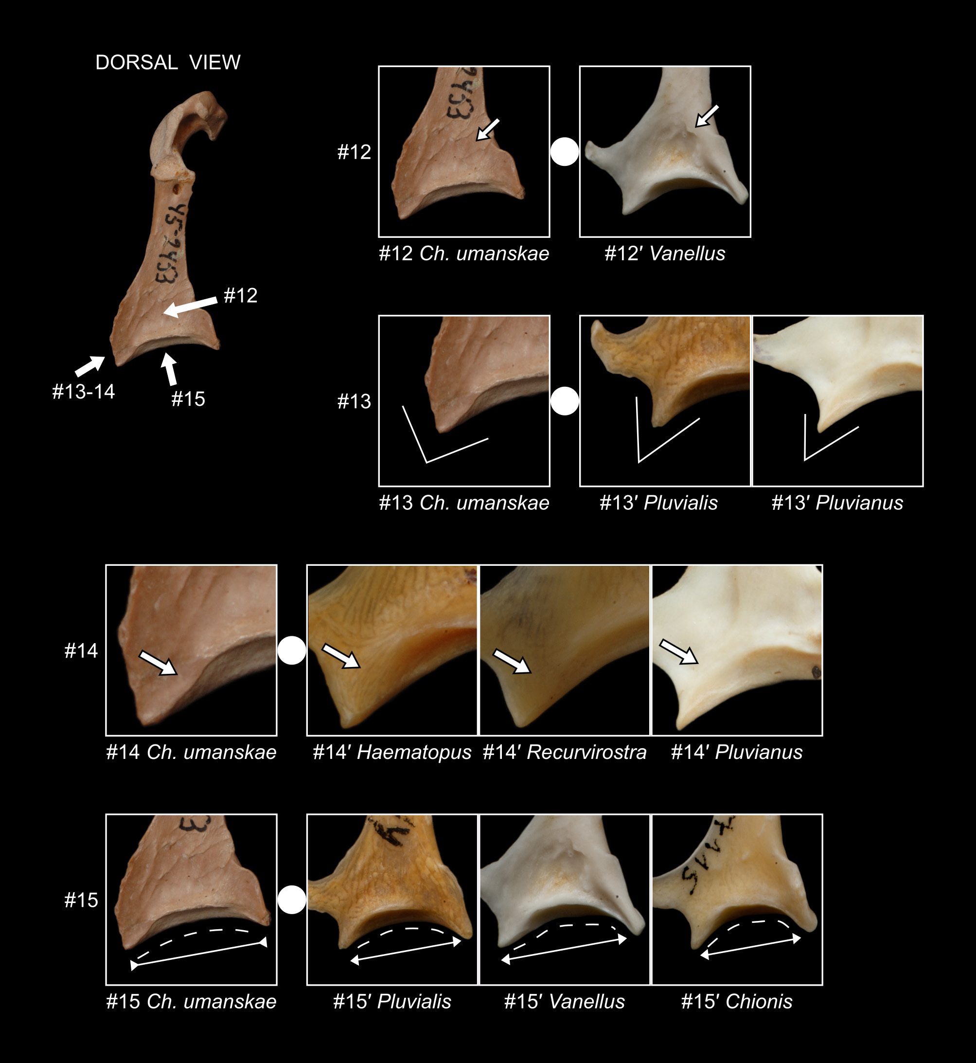
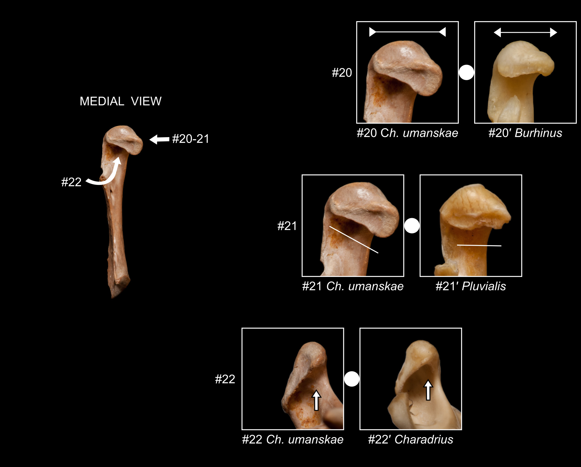
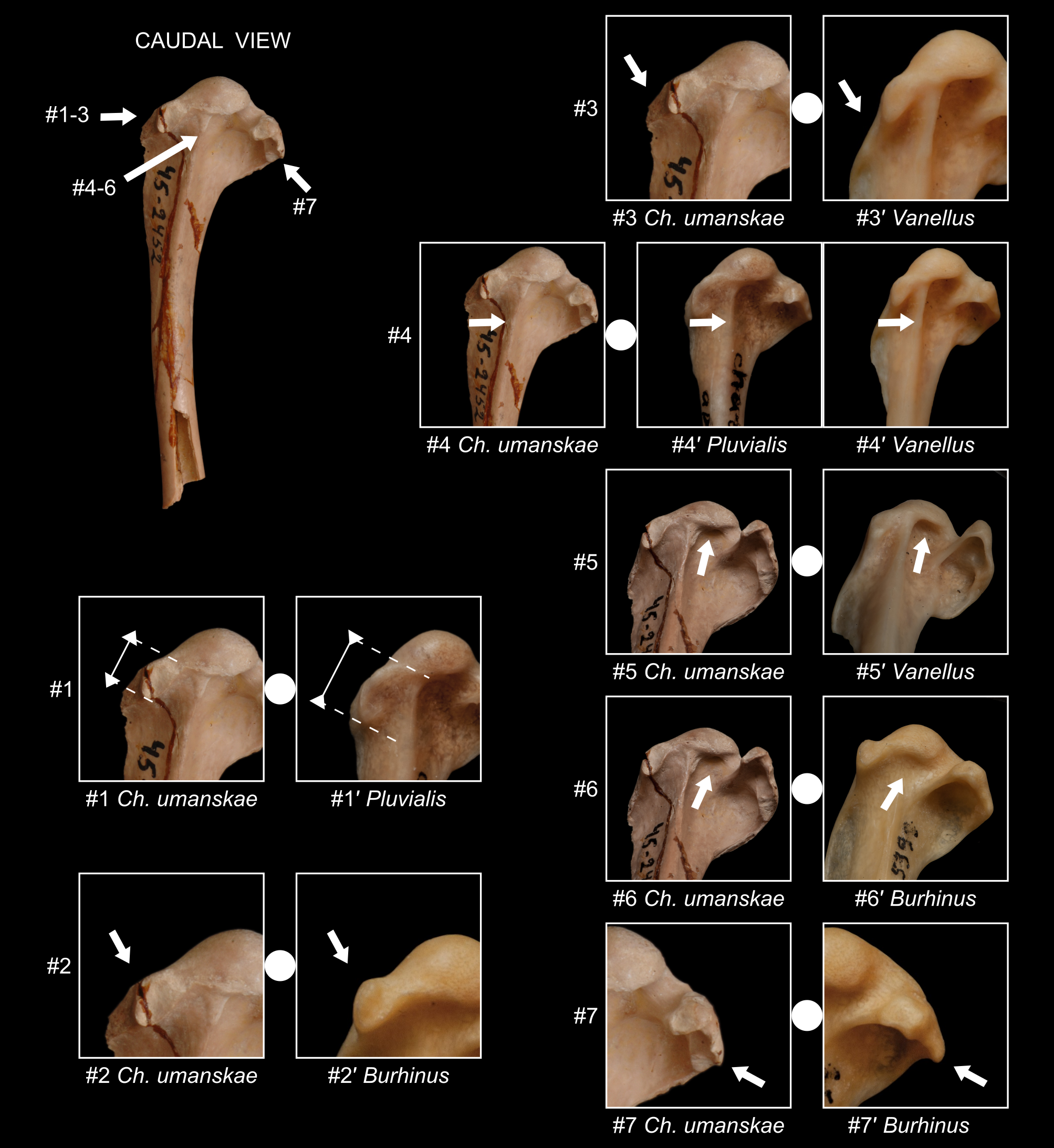
Coracoid ( Figs. 2 View FIGURE 2 –4 View FIGURE 2 View FIGURE 3 View FIGURE 4 and 6 View FIGURE 6 ): ● slender with relatively thinner shaft (#10) (in Pluvialis the bone is stout with thick shaft); ● extremitas omalis, medial view: sternal margin of facies articularis clavicularis is slanting (#21) (in Pluvialis , it is almost perpendicular to the long axis of the bone); ● extremitas omalis: medial area in sulcus supracoracoideus adjacent to the facies articularis humeralis deeply concave (#6) (in Pluvialis , the area is flat or shallowly concave); ● extremitas omalis, dorsal view: the foramen nervi supracoracoidei is very close to the sternal margin of the cotyla scapularis (#9) (in Pluvialis , the foramen is distinctly farther sternad from the cotyla); ● extremitas sternalis, dorsal and/or ventral view: facies articularis sternalis is relatively long (wide) and almost straight lateromedially because angulus medialis and angulus lateralis do not protrude far sternally (#15) (in Pluvialis, facies articularis sternalis is relatively short lateromedially and significantly bent because angulus medialis and angulus lateralis protrude far sternally); ● extremitas sternalis, dorsal and/or ventral view: the angulus lateralis is broad with sternal and lateral margins meeting almost at right angles (#13) (in Pluvialis, it tapers and its edges meet at an acute angle). Humerus ( Fig. 8 View FIGURE 8 ): ● caudal view: proximal part of the capital shaft ridge curves ventrad before reaching the caput humeri (#4) (in Pluvialis, the ridge is straight); ● tuberculum dorsale relatively short proximodistally (#1) (in Pluvialis, it is relatively longer).
From Charadrius in:
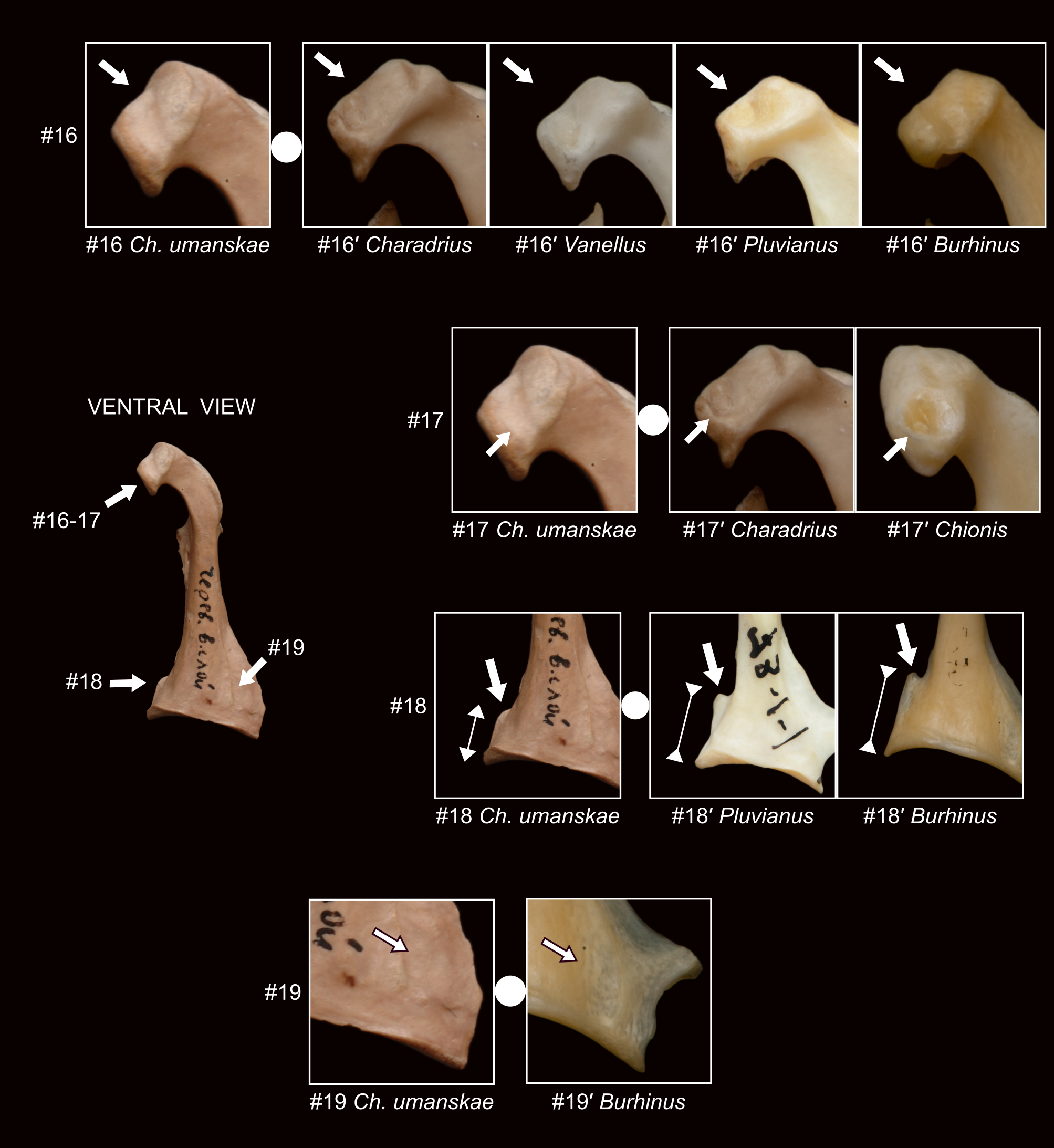
Coracoid ( Figs. 3 View FIGURE 3 and 5–6 View FIGURE 5 View FIGURE 6 ): ● extremitas omalis, ventral view: the processus acrocoracoideus is robust, with cranio-sternal length about the same as medial projection (#16) (in Charadrius , the width of the processus acrocoracoideus is clearly greater than its cranio-sternal length); ● extremitas omalis, ventral view: the ligamental scar on the acrocoracoid (for attachment of ligamentum acrocoracoclaviculare superficiale, Worthy et al. 2009, De Pietri et al. 2018) is shallow and elongated (#17) (in Charadrius, it forms a deep, rounded, conspicuous depression); ● extremitas omalis: the recess below the facies articularis clavicularis, especially in its ventral part, is shallow (#22) (in Charadrius, the recess is very deep, also in its ventral part); ● extremitas omalis, dorsal view: the foramen nervi supracoracoidei is very close to the sternal margin of the cotyla scapularis (#9) (in Charadrius, the foramen is distinctly farther sternad from the cotyla).
Humerus ( Figs. 9–10 View FIGURE 9 View FIGURE 10 ): ● caudalodistal view: the infraspinatus attachment on crista bicipitalis is triangular, very broad dorsoventrally (#8) (in Charadrius , it is elongated proximodistally and much narrower); ● cranial view: the boundary between intumescentia and shaft is difficult to define; the transition is gentle, gradual (#14) (in Charadrius, the transition is clearly marked); ● ventral view: there is a deep rounded depression in the crista bicipitalis immediately distal to the tuberculum ventrale (#9) (in Charadrius, the depression is shallower and its edges are less conspicuous).
From Vanellus in:
Coracoid ( Figs. 3–5 View FIGURE 3 View FIGURE 4 View FIGURE 5 ): ● extremitas omalis, ventral view: the processus acrocoracoideus is robust, with cranio-sternal length about the same as medial projection (#16) (in Vanellus , the width of the processus acrocoracoideus is clearly greater than its cranio-sternal length); ● extremitas omalis, dorsal view: cotyla scapularis very large, its lateral margin protruding laterally beyond the lateral edge of the bone (#8) (in Vanellus , cotyla scapularis relatively small, its lateral margin does not extend laterally beyond the lateral edge of the bone); ● extremitas omalis, dorsal view: the foramen nervi supracoracoidei is very close to the sternal margin of the cotyla scapularis (#9) (in Vanellus, the foramen is distinctly farther sternad from the cotyla); ● extremitas sternalis, dorsal view: the bone surface above the articulation is flat or only slightly concave (#12) (in Vanellus, there is a deep depression there); ● extremitas sternalis: there is a bony blade just above the projection on the margo medialis (#11) (in Vanellus, the blade is missing); ● extremitas sternalis, dorsal and/or ventral view: facies articularis sternalis is long (wide) and almost straight lateromedially because angulus medialis and angulus lateralis do not protrude far sternally (#15) (in Vanellus, facies articularis sternalis is relatively short lateromedially and significantly bent because angulus medialis and angulus lateralis protrude far sternally).
Humerus ( Figs. 8 View FIGURE 8 –10 View FIGURE 8 View FIGURE 9 View FIGURE 10 ): ● caudal view: proximal part of the capital shaft ridge curves ventrad before reaching the caput humeri (#4) (in Vanellus , the ridge is straight); ● caudalodistal view: caput humeri in its ventral part is moderately deeply undercut (#5) (in Vanellus , caput humeri is very deeply undercut); ● caudalodistal view: the infraspinatus attachment on crista bicipitalis is triangular, very broad dorsoventrally (#8) (in Vanellus, it is elongated proximodistally and much narrower); ● cranial view: the boundary between intumescentia and shaft is difficult to define; the transition is gentle, gradual (#14) (in Vanellus, the transition is clearly marked); ● the proximal part of the crista pectoralis slopes dorsally (#3) (in Vanellus, it is almost parallel to the long axis of the bone); ● ventral view: there is a deep rounded depression in the crista bicipitalis immediately distal to the tuberculum ventrale (#9) (in Vanellus, the depression is shallower and its edges are less conspicuous).
From Pluvianus in:
Coracoid ( Figs. 2 View FIGURE 2 –5 View FIGURE 2 View FIGURE 3 View FIGURE 4 View FIGURE 5 ): ● extremitas omalis: the section between processus procoracoideus and processus acrocora- coideus is elongated (#1) (in Pluvianus , it is short in relation to the total length of the coracoid); ● extremitas omalis, ventral view: the processus acrocoracoideus is robust, with cranio-sternal length about the same as medial projection (#16) (in Pluvianus , the width of the processus acrocoracoideus is clearly greater than its cranio-sternal length); ● extremitas omalis, dorsal view: the foramen nervi supracoracoidei is very close to the sternal margin of the cotyla scapularis (#9) (in Pluvianus , the foramen is distinctly farther sternad from the cotyla); ● extremitas sternalis, dorsal view: the dorsal edge of the facies articularis sternalis connects to the angulus lateralis (#14) (in Pluvianus , the dorsal edge of the facies articularis sternalis disappears well before reaching the angulus lateralis); ● extremitas sternalis, dorsal and/or ventral view: the angulus lateralis is broad with sternal and lateral margins meeting almost at right angles (#13) (in Pluvianus , it tapers and its edges meet at an acute angle); ● extremitas sternalis: the projection on the margo medialis is small, dull and ends little above the angulus medialis (#18) (in Pluvianus , it is bigger, sharp and reaches high above angulus medialis).
Humerus ( Figs. 9–10 View FIGURE 9 View FIGURE 10 ): ● caudalodistal view: the infraspinatus attachment on crista bicipitalis is triangular, very broad dorsoventrally (#8) (in Pluvianus , it is elongated proximodistally and much narrower); ● cranial view: the proximal part is approximately as long proximodistally as it is broad dorsoventrally (#11) (in Pluvianus , the proximal part is shorter proximodistally); ● cranial view: the boundary between intumescentia and shaft is difficult to define; the transition is gentle, gradual (#14) (in Pluvianus , the transition is clearly marked).
From Burhinus in:
Coracoid ( Figs. 2 View FIGURE 2 –3 View FIGURE 2 View FIGURE 3 and 5–6 View FIGURE 5 View FIGURE 6 ): ● extremitas omalis: the lateral edge of processus acrocoracoideus is oblique to the long axis of the bone (#4) (in Burhinus , the lateral edge is more vertical); ● extremitas omalis, ventral view: the processus acrocoracoideus is robust, with cranio-sternal length about the same as medial projection (#16) (in Burhinus , the width of the processus acrocoracoideus is clearly greater than its craniosternal length);● extremitas omalis, medial view: sternal margin of facies articularis clavicularis is broad ventrodorsally (#20) (in Burhinus , it is relatively narrow); ● extremitas omalis, dorsal view: the foramen nervi supracoracoidei is very close to the sternal margin of the cotyla scapularis (#9) (in Burhinus , the foramen is distinctly farther sternad from the cotyla); ● extremitas sternalis, ventral view: the bone surface, medial to the linea intermuscularis ventralis, is flat or slightly convex (#19) (in Burhinus , there is a distinct depression there); ● extremitas sternalis: the projection on the margo medialis is small, dull and ends little above the angulus medialis (#18) (in Burhinus , it is bigger, sharp and reaches high above angulus medialis).
Humerus ( Figs. 8 View FIGURE 8 and 10 View FIGURE 10 ): ● caudal view: a well-developed dorsal (second) fossa pneumotricipitalis is present (#6) (in Burhinus , the dorsal fossa is absent); ● caudal view: the proximal edge of tuberculum dorsale slopes gently distally (#2) (in Burhinus , tuberculum dorsale is more pronounced and its proximal edge is often upturned proximally); ● caudal view: crista bicipitalis arches evenly (#7) (in Burhinus , the infraspinatus attachment on crista bicipitalis protrudes clearly more ventrally); ● cranial view: impressio musculus coracobrachialis relatively narrow (#13) (in Burhinus , it is very wide, reaching at least half the width of the proximal humerus).
From Chionis in:
Coracoid ( Figs. 2 View FIGURE 2 –5 View FIGURE 2 View FIGURE 3 View FIGURE 4 View FIGURE 5 ): ● extremitas omalis: the section between processus procoracoideus and processus acrocoracoideus is elongated (#1) (in Chionis , it is short in relation to the total length of the coracoid); ● extremitas omalis, ventral view: the ligamental scar on the acrocoracoid (for attachment of ligamentum acrocoracoclaviculare superficiale, Worthy et al. 2009, De Pietri et al. 2018) is shallow and elongated (#17) (in Chionis, it forms a deep, rounded, conspicuous depression); ● extremitas omalis, ventral view: processus acrocoracoideus protrudes far medially (#7) (in Chionis, the processus protrudes very little medially); ● extremitas omalis, omal view: processus acrocoracoideus is dorsoventrally narrow (#2) (in Chionis, it is very broad); ● extremitas omalis, omal view: ventral edge is slightly bent (#3) (in Chionis, ventral edge is strongly arched because facies articularis humeralis and facets articularis clavicularis protrude far ventrally); ● extremitas omalis, dorsal view: the foramen nervi supracoracoidei is very close to the sternal margin of the cotyla scapularis (#9) (in Chionis, the foramen is distinctly farther sternad from the cotyla); ● extremitas sternalis, dorsal and/or ventral view: facies articularis sternalis is long (wide) and almost straight lateromedially because angulus medialis and angulus lateralis do not protrude far sternally (#15) (in Chionis, facies articularis sternalis is relatively short lateromedially and significantly bent because angulus medialis and angulus lateralis protrude far sternally).
Humerus ( Figs. 9–10 View FIGURE 9 View FIGURE 10 ): ● caudalodistal view: The infraspinatus attachment on crista bicipitalis is triangular, very broad dorsoventrally (#8) (in Chionis , it is elongated proximodistally and much narrower); ● dorsal view, cranial margin of caput humeri: The caput humeri is more rounded and does not reach far proximally (#10) (in Chionis , it is more elongated and reaches farther proximally); ● ventral view: There is a deep rounded depression in the crista bicipitalis immediately distal to the tuberculum ventrale (#9) (in Chionis , the depression is shallower and its edges are less conspicuous); ● cranial view: Proximal part of the impressio musculus coracobrachialis is deeper and its edges are better distinguished (#12) than in most extant Chionis .

No known copyright restrictions apply. See Agosti, D., Egloff, W., 2009. Taxonomic information exchange and copyright: the Plazi approach. BMC Research Notes 2009, 2:53 for further explanation.