
Campodea (Campodea) majorica sicula Condé, 1957
|
publication ID |
https://doi.org/10.11646/zootaxa.4679.2.5 |
|
publication LSID |
lsid:zoobank.org:pub:9173A4B4-1CC2-4857-8628-65E49C3C54BE |
|
DOI |
https://doi.org/10.5281/zenodo.3797729 |
|
persistent identifier |
https://treatment.plazi.org/id/8339879B-BC69-FFB8-EBF0-2C73FCC7F955 |
|
treatment provided by |
Plazi (2020-04-28 07:11:15, last updated 2024-11-28 11:12:42) |
|
scientific name |
Campodea (Campodea) majorica sicula Condé, 1957 |
| status |
|
Campodea (Campodea) majorica sicula Condé, 1957
Figs. 24–48 View FIGURES 24–29 View FIGURES 30–36 View FIGURES 37–42 View FIGURES 43–48 , Table 3
Studied material. 5 females, 2 males and 11 unsexed specimens, Abisso del Gatto , Cefalú, Palermo, Sicily, Italy, 10 June 2017, Giuseppe Nicolosi leg ; 2 ♀♀, 1 juvenile, Abisso della Pietra Selvaggia , Palermo, Sicily, Italy, 17 November 2018. Giuseppe Nicolosi leg.
Additions to original description. Body length 6.4–8.6 mm (females), 5.2–5.3 mm (males); antennae length similar to body length with 44 and 46 antennomeres; medial antennomeres two times longer than wide. Epicuticle of dorsal sclerites of the body and legs covered with microdenticles visible with optical microscopy in addition to a reticulated surface only observed at high magnifications ( Figs. 30–36 View FIGURES 30–36 , 42 View FIGURES 37–42 ); ventral sclerites smooth under optical microscope but surface reticulated at high magnifications ( Figs. 43, 45 View FIGURES 43–48 ); rosette gland formations present along scattered on the body ( Fig. 32 View FIGURES 30–36 ). Dorsal sclerites of body with abundant short, slightly thickened, smooth clothing setae ( Figs. 30–36 View FIGURES 30–36 ); ventral sclerites with longer and thinner clothing setae ( Fig. 43 View FIGURES 43–48 ).
Cupuliform organ occupying 1/8 of the total length of the apical antennomere with about 8 complex olfactory chemoreceptors. Each chemoreceptor is made up of both loosely and tightly packed, multi-perforated folds forming a concentric cupuliform structure with a rim edge. The rim and folds are of varied height and there is a characteristic raised central area, the central pore. All the chemoreceptors are joined by being held within individual cells of a single form ( Figs. 24–26 View FIGURES 24–29 ). Distal and central antennomeres with three whorls of smooth macrosetae smooth or with one or two distal barbs, up to three whorls of smooth setae, and a single distal whorl of 10–12 gouge sensilla 18–22 µm long. Apical antennomere with similar setal arrangement ( Figs. 27–29 View FIGURES 24–29 ). Proximal antennomeres with typical bothriotricha; third antennomere with small subcylindrical ventral sensillum.
Head subtrapezoidal, slightly protuberant on lateral posterior angles. Simple frontal process with three frontal macrosetae and three thicker, longer and well barbed macrosetae along each side of the line of insertion distinguishable.
Thoracic macrosetal distribution ( Figs. 30–36 View FIGURES 30–36 ): 3+3 ma, la, lp macrosetae on pronotum and mesonotum, and 1+1 ma on metanotum; ma and la macrosetae well differentiated from clothing setae, slightly thick and with moderately thick barbs; lp macrosetae long with thin, short barbs along their entire length. Marginal setae well differentiated from clothing setae, thick and with thick barbs. Urotergal macrosetal length, thickness and number variable, 1+1 la and 1+1, 0+1 or 0+0 lp in sixth urotergite ( Figs. 46–48 View FIGURES 43–48 ).
Legs elongate ( Fig. 37 View FIGURES 37–42 ), metathoracic legs reaching the abdominal segments 8 or 9; tibia slightly longer than femur and tarsus, femur and tarsus + pretarsus of similar length ( Table 3). Calcars with barbs of variable length beginning near their base ( Fig. 40 View FIGURES 37–42 ). Tibiae I–III each with one short, smooth ventral macroseta sometimes possessing one minute distal barb ( Fig. 41 View FIGURES 37–42 ). Each tarsus with three smooth, subapical macrosetae, a double ventral row of thicker, weakly barbed ventral setae, and a few setiform sensilla ( Fig. 39 View FIGURES 37–42 ). Subequal slightly curved claws subequal, with setiform lateral processes with a slight apical laminar expansion ( Fig. 38 View FIGURES 37–42 ).
cercal primary articles.
Urosternal macrosetae with a few long distal barbs; 6+6 macrosetae on first urosternite ( Fig. 43 View FIGURES 43–48 ), 4+4 on second to seventh urosternites and 1+1 on eighth urosternite. Stylus setae smooth with two long teeth on apical seta ( Fig. 46 View FIGURES 43–48 ). Cerci 11.5 mm long on 7.2-mm female; cercal base divided in three followed by 14 primary articles ( Table 3). Proximal latero-internal macrosetae with 0–1 distal barbs in the preserved cerci of Abisso del Gatto specimens and with 3–8 barbs in Abisso della Pietra Selvaggia specimens.
Male urosternite I with a posterior field of glandular g 1 setae, and subtrapezoidal appendages with a distal field of glandular a 1 setae. Female subcylindrical appendages with glandular a 1 setae ( Figs. 43, 44 View FIGURES 43–48 ).
Remarks. Campodea (Campodea) majorica sicula belongs to a distinct monophyletic group closely related to the soil-dwelling species C. (C.) grassi Silvestri 1912 living in the Western Mediterranean region from the Iberian to the Italian Peninsulas, southern France, northern Algeria, and a few locations in Corsica, Pontine Islands, Sardinia and Sicily ( Condé 1947a, 1947 b, 1955, 1978; Condé & Mathieu 1957; Sendra 1989; Sendra & Jiménez 1986; Sendra & Moreno 2004; Sendra et al. 2017; Silvestri 1912, 1932). Condé (1955) suggested that this monophyletic group was composed of three others already described species and six subspecies ( C. majorica sicula included), all inhabiting the deep subterranean spaces and found in caves. They occupy the main Western Mediterranean Islands: Corsica ( Campodea (Campodea) cyrnea cyrnea Condé 1946 ; Campodea (Campodea) cyrnea alethae Condé 1948 ; Campodea (Campodea) cyrnea virgolae Condé 1948 and Campodea (Campodea) blandinae blandinae Condé 1948 ), Mallorca ( Campodea (Campodea) majorica majorica Condé 1955 and Campodea (Campodea) majorica interjecta Condé 1955 ), Sardinia ( Campodea (Campodea) blandinae ichnusa Condé 1957 ) and Sicily ( C. (C.) majorica sicula ),; in addition to an inland subspecies from the extreme east of the Pre-Baetic Mountains in the Iberian Peninsula ( Campodea (Campodea) majorica valentina Sendra & Moreno 2004 ) ( Condé 1946 b, 1948, 1955, 1957; Marletta et al. 2015; Sendra & Moreno 2004). The similarities among the taxa of the C. (C.) grassi monophyletic group are based on the pattern of macroseta distribution, their robust dorsal clothing setae, marginal setae and macrosetae, and the micro-denticles on the cuticle surface. The species and subspecies of this group are difficult to separate in spite of their isolated environments and insular distributions. All taxonomic features suggested in the descriptions of the taxa should be reviewed. In fact, the variability observed in the shape of the dorsal thoracic and abdominal macrosetae and in the presence or absence of macrosetae on the sixth urotergite, seen in the studied material of C. (C.) majorica sicula , cast doubt on the validity of the species and subspecies of this monophyletic group.
Scanning electron microscopy observations were used in this study for better examination of taxonomic features, and for future comparation with other taxa. In the future it will be valuable to use molecular data to better understand the current taxa separation and their validity.
Conde, B. (1946 b) Diploures recoltes en Corse par P. Remy (deuxieme note). Collections du Musee de Zoologie de Nancy, 1, 1 - 6.
Conde, B. (1947 a) Diploures recoltes en Corse par P. Remy (3 e note). Bulletin Museum National d'Histoire naturelle, Series 2, 19, 282 - 285.
Conde, B. (1947 b) Campodeides d'Algerie. Bulletin de la Societe entomologique de France, LII (9), 144 - 146.
Conde, B. (1948) Campodeides hypoges de Corse. Bulletin de la Societe Sciences de Nancy, New Series, VII (3), 62 - 68.
Conde, B. (1955) Campodeides cavernicoles des Baleares. Notes biospeologiques, 9, 121 - 132.
Conde, B. & Mathieu, A. (1957) Campodeides Endoges de la Region Pyreneenne. Vie et Milieu, 8 (4), 439 - 472.
Conde, B. (1978) Diplopodes Penicillates, Diploures Campodeides et Palpigrades des iles Ponziane. Revue d'Ecologie et de Biologie du Sol, 15 (2), 273 - 277.
Marletta, A., Nicolosi, G. & Grech, T. (2015) On the presence of Campodea majorica sicula Conde, 1957 (Diplura Campodeidae) in the Abisso della Pietra Selvaggia cave (Mount Pellegrino, Palermo, Italy). Biodiversity Journal, 6 (1), 305 - 308.
Sendra, A. & Jimenez, R. (1986) Contribucion al conocimiento de los Campodeidos endogeos de Espana Peninsular (Insecta, Diplura). Eos, 62, 277 - 284.
Sendra, A. (1989) Datos sobre Campodeidos del Museo Nacional de Ciencias Naturales de Madrid (Insecta, Diplura, Campodeidae). Eos, 65 (1), 115 - 123.
Sendra, A. & Moreno, A. (2004) El subgenero Campodea s. str. en la Peninsula Iberica (Hexapoda: Diplura: Campodeidae). Boletin Sociedad Entomologica Aragonesa, 35, 19 - 38.
Sendra, A., Jimenez-Valverde, A., Gilgado, J. D., Ledesma, E., Baquero, E., Perez-Suarez, G., Cuesta, E., Herrero-Borgonon, J. J., Jordana, R., Tinaut, A., Barranco, P. & Ortuno, V. M. (2017) Diplurans of subsurface terrestrial habitats in the Iberian Peninsula, with a new species description (Diplura: Campodeidae). Zootaxa, 4291 (1), 061 - 080. https: // doi. org / 10.11646 / zootaxa. 4291.1.4
Silvestri, F. (1912) Contribuzione alla conoscenza dei Campodeidae (Thysanura) d'Europa. Bolletino del Laboratorio di Zoologia Generale ed Agraria del R. Istituto Superiore Agrario di Portici, 6, 110 - 147.
Silvestri, F. (1932) Campodeidae (Thysanura) de Espana (primera parte). Eos, 8, 115 - 164.
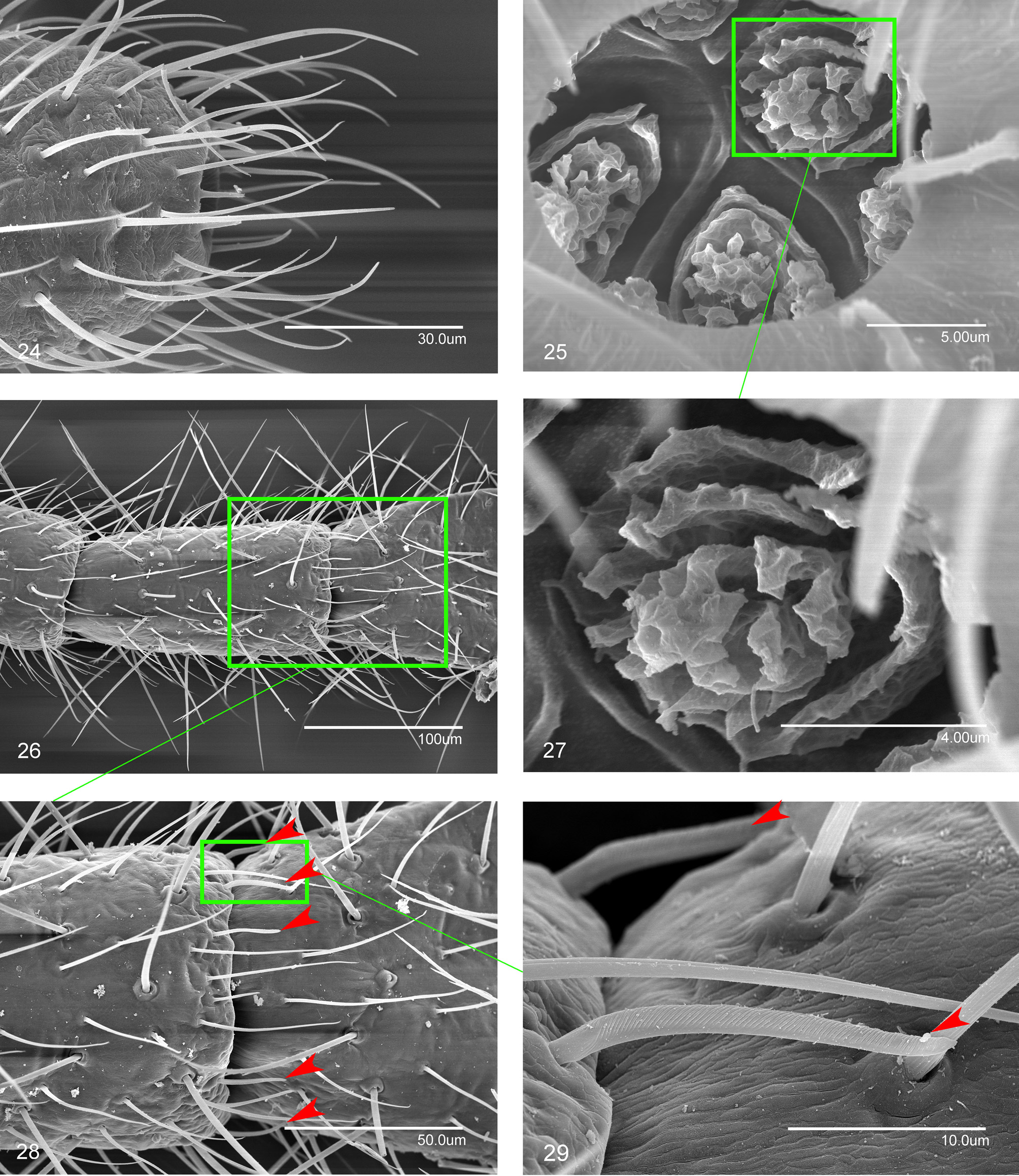
FIGURES 24–29. Campodea (Campodea) majorica sicula Condé, 1957. Specimens from Abisso della Pietra Selvaggia. 24. Distal portion of the last antennomere. 25. Olfactory chemoreceptors within the cupuliform organ. 26. Medial antennomeres. 27. Detail of an olfactory chemoreceptor. 28. Distal portion of a medial antennomere. 29. Gouge sensillum from the distal whorl of a medial antennomere (red arrows indicate gouge sensilla on antennae in Figs. 28–29; f: fold; ca: central pore).
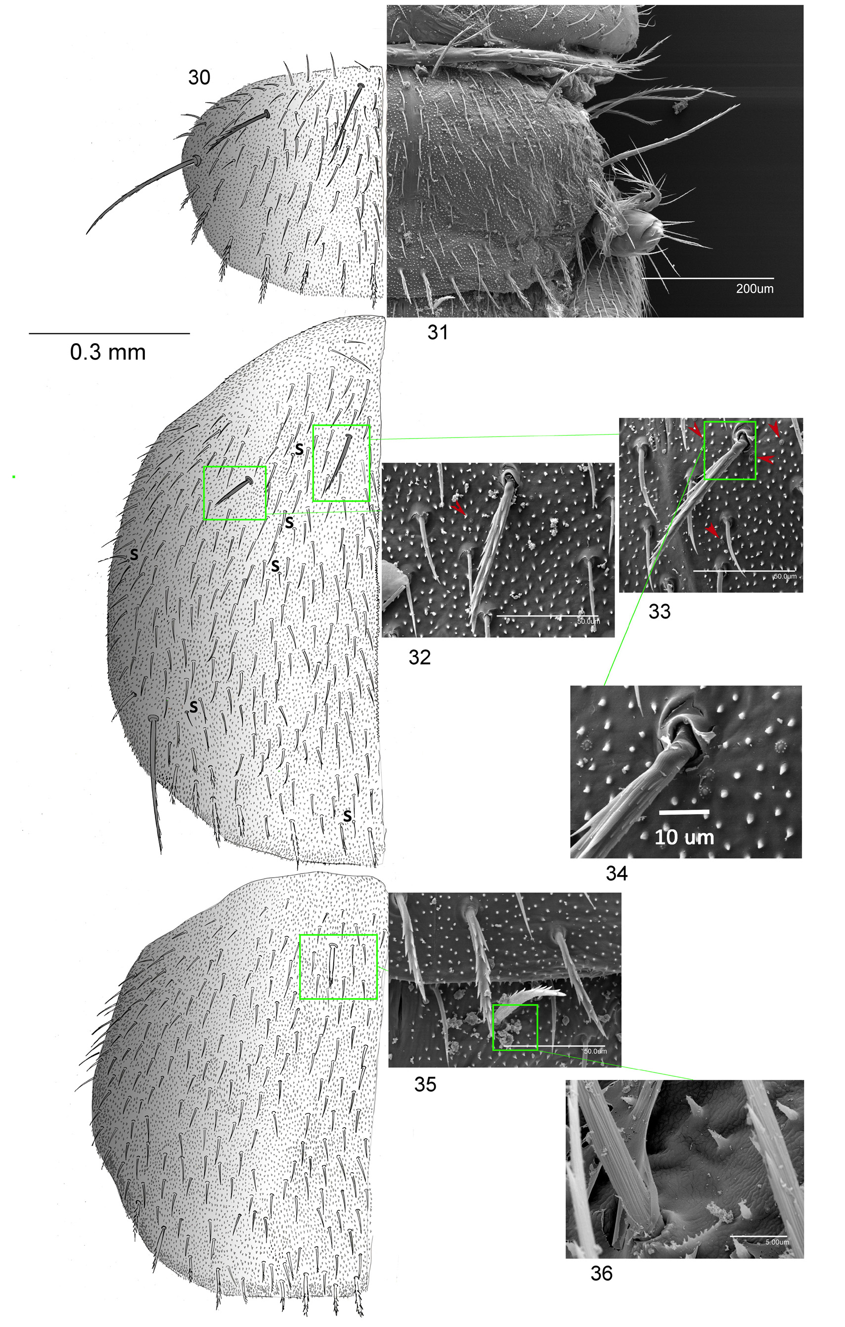
FIGURES 30–36. Campodea (Campodea) majorica sicula Condé, 1957. Specimens from Abisso della Pietra Selvaggia. 30. Pronotum, mesonotum and metanotum, left side; scale bar = 0.3 mm. 31. Pronotum, right side. 32. Lateral anterior macroseta on mesonotum. 33. Medial anterior macroseta on mesonotum. 34. Detail of rosette gland formations. 35. Medial anterior macroseta on metanotum. 36. Surface of metanotum (red arrows indicate rosette gland formations on mesonotum in Figs. 32 and 33).
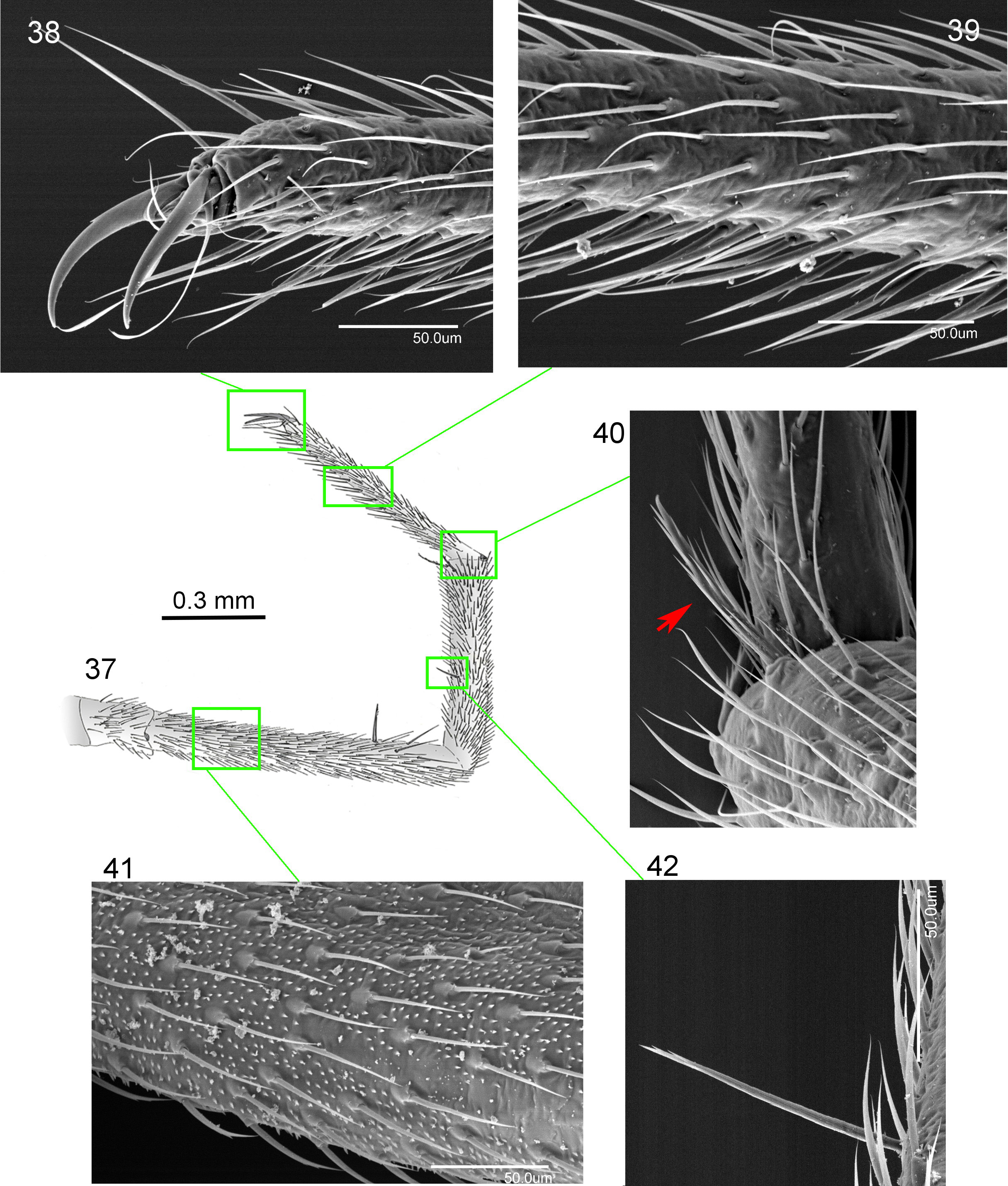
FIGURES 37–42. Campodea (Campodea) majorica sicula Condé, 1957. Specimens from Abisso della Pietra Selvaggia. 37. Metathoracic leg; scale bar = 0.5 mm. 38. Pretarsus and apical portion of tarsus. 39. Medial portion of tarsus. 40. Tibio-tarsal joint with calcar, indicated by red arrow. 41. Tibial macroseta. 42. Femur surface.
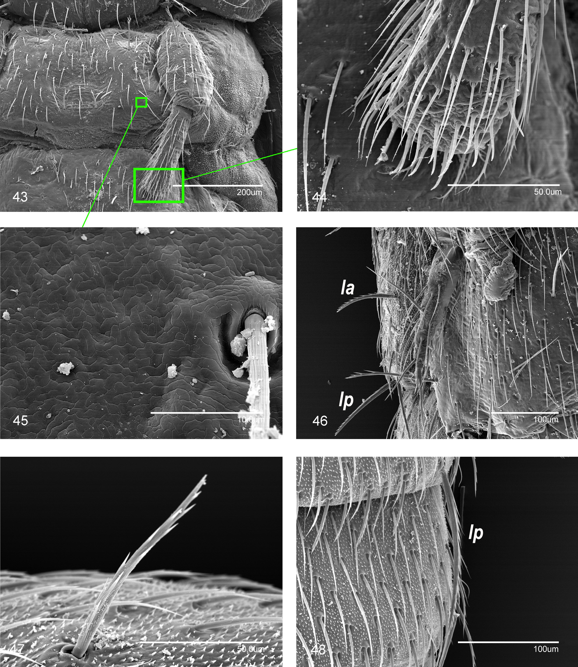
FIGURES 43–48. Campodea (Campodea) majorica sicula Condé, 1957. Specimens from Abisso della Pietra Selvaggia. 43. First urosternite of female, left side. 44. Distal portion of the left appendage from the first urosternite of female. 45. Reticulated surface of first urosternite. 46. Latero-ventral view of the seventh abdominal segment; lateral anterior (la) and lateral posterior (lp) urotergal macrosetae. 47. Lateral anterior macroseta on sixth urotergite. 48. Lateral posterior (lp) macroseta on the sixth urotergite.
No known copyright restrictions apply. See Agosti, D., Egloff, W., 2009. Taxonomic information exchange and copyright: the Plazi approach. BMC Research Notes 2009, 2:53 for further explanation.
|
Kingdom |
|
|
Phylum |
|
|
Class |
|
|
Order |
|
|
Family |
|
|
Genus |
1 (by plazi, 2020-04-28 07:11:15)
2 (by ExternalLinkService, 2020-04-28 07:21:21)
3 (by ExternalLinkService, 2020-04-28 07:38:37)
4 (by ExternalLinkService, 2020-05-06 11:13:57)
5 (by ExternalLinkService, 2022-01-29 13:13:11)
6 (by plazi, 2023-10-31 11:09:01)