
Sphaerodoropsis megatuberculata, Capa, Maria & Bakken, Torkild, 2015
|
publication ID |
https://doi.org/ 10.11646/zootaxa.4000.2.3 |
|
publication LSID |
lsid:zoobank.org:pub:7EDEDAEE-642C-4F9D-A04D-141815D73343 |
|
DOI |
https://doi.org/10.5281/zenodo.5667471 |
|
persistent identifier |
https://treatment.plazi.org/id/81007D79-8D50-2544-FF0F-F833FDED1D89 |
|
treatment provided by |
Plazi |
|
scientific name |
Sphaerodoropsis megatuberculata |
| status |
sp. nov. |
Sphaerodoropsis megatuberculata View in CoL n. sp.
Fig. 4 View FIGURE 4 Q–R, 5K, 13
Sphaerodoropsis sp. MoV632 Wilson & Bakken 2003 (in part).
Material examined. Holotype: NMV F.69689, 15.5 km SW of Pt Ricardo, Eastern Bass Strait, 37° 53' 08'' S, 148° 28' 56'' E, 45 m, Feb 1991, medium sand. Paratypes: NMV F.69687 (1 spec. for SEM), 15.5 km SW of Pt Ricardo, Eastern Bass Strait, 37° 53' 08'' S, 148° 28' 56'' E, 45 m, Feb 1991, medium sand; NMV F.69688 (1 spec.), 15.5 km SW of Pt Ricardo, Eastern Bass Strait, 37° 53' 08'' S, 148° 28' 56'' E, 45 m, 4 Jun 1991, medium sand; NMV F.69686 (1 spec. for SEM), 15.5 km SW of Pt Ricardo, Eastern Bass Strait, 37° 53' 08'' S, 148° 28' 56'' E, 45 m, 4 Jun 1991, medium sand; NMV F.63844 (1 spec.), 15.5 km SW of Pt Ricardo, Eastern Bass Strait, 37° 53' 10'' S, 148° 28' 57'' E, 45 m, 26 Sep 1990, sand-shell; NMV F.132659 (1 spec.), 14 km SW of Marlo, Eastern Bass Strait, 37° 54' 00'' S, 148° 25' 07'' E, 26 m, 12 Aug 1989, sand-shell; NMV F.63845 (1 spec.), 3.2 km S of Cape Conran, Eastern Bass Strait, 37° 50' 37'' S, 148° 43' 28'' E, 49 m, 28 Sep 1990, sand-shell; NMV F.132902 (1 spec.), 43 km SE of Port Albert, Bass Strait, 38° 53' 42'' S, 147° 06' 30'' E, 58 m, 18 Nov 1981.
Diagnosis. Ellipsoid body with 17 longitudinal rows of large ellipsoid to cylindrical macrotubercles in two transversal rows per segment, with eight macrotubercles between parapodia and nine in posterior transversal row. Dorsum with additional spherical papillae in 2–3 transversal rows per segment; ventrum with spherical papillae in four longitudinal rows. Two parapodial papillae, one on each of anterior and posterior surfaces. Parapodia with 5–6 chaetae per fascicle, blades 2–3 times as long as wide. Females with an oval tubercle between chaetigers 5–6, males with modified ventral cirrus in chaetigers 4–7.
Description. Measurements and general morphology. Holotype female with ellipsoid body measuring 2.5 mm long and 0.5 mm wide, for 20 chaetigers, convex dorsum and flattened ventrum ( Fig. 13 View FIGURE 13 A). Segmentation unremarkable, tegument with transverse wrinkles ( Fig. 13 View FIGURE 13 C–E). Preserved specimen lacking pigmentation.
Head. Anterior end bluntly rounded. Prostomium with short appendages, including a pair of digitiform blunt lateral antennae, a pair of palps similar in shape and length to lateral antennae, median antenna half the length than lateral antennae ( Fig. 13 View FIGURE 13 B). Tentacular cirri similar to median antenna. Antenniform papillae not observed. A few scattered spherical papillae on the head, in different sizes ( Fig. 13 View FIGURE 13 B).
Tubercles. First chaetiger with two dorsal macrotubercles, sessile large, ellipsoid to cylindrical. Number increases towards chaetiger 5. Following chaetigers with 17 longitudinal rows of macrotubercles, arranged in double transverse rows on each segment in a zig-zag pattern ( Figs 4 View FIGURE 4 Q, 13A–B, E), with eight macrotubercles in rows between parapodia and nine in posterior transverse rows. Macrotubercles ellipsoidal to cylindrical of different sizes ( Fig. 13 View FIGURE 13 B, D–F), with pores mainly over the distal surface ( Fig. 13 View FIGURE 13 H). Dorsum with additional small spherical papillae arranged in 2–3 transverse rows per segment, with 6–8 papillae per row ( Figs 4 View FIGURE 4 Q, 13D–E). Ventral surface with spherical papillae in 4–6 (zig-zag) longitudinal rows, two rows close to parapodia and 2–4 rows in mid-body, arranged in more or less two segmental and one intersegmental transverse row, where the mid-body longitudinal rows are skewed in position compared to outer lines giving a zig-zag pattern ( Figs 4 View FIGURE 4 R, 13C). Body epithelium with microscopic rounded granules ( Fig. 13 View FIGURE 13 G).
Parapodia. Parapodia sub-conical, about 1–2 times longer than wide. Digitiform acicular lobe projecting anterior to chaetae, projecting beyond ventral cirri, ventral cirri slender digitiform. Two parapodial papillae, one close to base on anterior surface and one on posterior surface ( Figs 5 View FIGURE 5 K, 13C–F).
Chaetae. Compound chaetae present in all chaetigers, arranged in a curved transverse row posterior to acicular lobe, numbering 5–7 per fascicle ( Figs 5 View FIGURE 5 K, 13D–F). Shaft with slender distal end and strong spinulation on edge ( Fig. 13 View FIGURE 13 I–K). Blades mostly similar in length within each fascicle and along chaetigers (2–3 times longer than maximum width), with recurved distal end and conspicuous spinulation along the proximal 3/4th length of blade ( Fig. 13 View FIGURE 13 I–K). Some blades observed with fine distal spines on the edge opposite to serration ( Fig. 13 View FIGURE 13 J).
Pygidium . Pygidium sub-terminal, with a mid-ventral digitiform anal cirrus ( Fig. 13 View FIGURE 13 C).
Internal features. Eyes present in holotype, as dark brown to black spots positioned deep in integument in chaetigers 1–2. Muscular pharynx not observed.
Reproductive features. Holotype and one paratype female with ‘copulatory organs’ as oval tubercle between chaetigers 5 and 6. Males with modified ventral cirri swollen at the base, with a distal knob and surface with pores in chaetigers 4–7 ( Fig. 13 View FIGURE 13 F). Eggs not observed.
Variation. Specimens measuring 1.5–4.5 mm long and 0.3–0.6 mm wide, for 18–23 chaetigers. Prostomial appendages barely visible in contracted specimens, in specimens relaxed appendages clearly visible and longer than shown in SEM ( Fig. 13 View FIGURE 13 B), shape and relative length of appendages as described for holotype. Macrotubercles similar in shape in all specimens examined but in some individuals they are relatively larger than those described for holotype and observed under SEM ( Fig. 13 View FIGURE 13 D–F). Number of macrotubercles in examined specimens agree with holotype; being six on second chaetiger, remaining chaetigers with eight between parapodia and nine in rows posterior to "parapodial row", per segment; last chaetiger with four macrotubercles. Parapodial papilla on anterior surface of parapodia difficult to observe in stereo microscope at its position at the base of parapodia, it gives an appearance of this papilla sit on body surface in contracted specimens.
Remarks. A distinct and unique feature of Sphaerodoropsis megatuberculata n. sp. is the large ellipsoid to cylindrical macrotubercles, not described in any other Sphaerodoropsis species. This species belongs in Sphaerodoropsis Group 3 (sensu Borowski 1994), and resembles S. solis , but the latter has ventral ‘macrotubercles’ which are absent in S. megatuberculata n. sp., and has double transverse rows of six and seven macrotubercles per segment, while S. megatuberculata n. sp. has eight and nine. The new species also resembles S. bisphaeroserialis , S. garciaalvarezi and S. arctowskyensis in arrangement of macrotubercles and dorsal and ventral papillae ( Moreira et al. 2004). With its 8+9 macrotubercles in double transverse rows per segment S. megatuberculata n. sp. has higher number of macrotubercles than the group of three closely related species which all have 6+7. Further, there are differences in number and distribution of dorsal and ventral papillae between the species ( Moreira et al. 2004). Moreira et al. (2004) use "microtubercles" for dorsal and "papillae" for ventral papillae, for what is termed "papillae" here. Sphaerodoropsis megatuberculata n. sp., S. bisphaeroserialis , S. garciaalvarezi and S. arctowskyensis , seem to be a group of species with close affinities ( Moreira et al. 2004). The group also shares an austral distribution where respective species have been found in southern Australia, Sub-Antarctic and Antarctic environments.
Sexual dimorphism is observed in this species. A structure believed to be genital opening in female specimens has been observed as a disc-like tubercle immediately ventral to parapodia between chaetigers 5 and 6, being similar to what has been observed in other Sphaerodoropsis species ( Moreira et al. 2004; Reuscher & Fiege 2011). In male specimens ‘copulatory organs’ appear as modified ventral cirri in chaetigers 4–7.
Etymology. The species is named after its characteristic enlarged and uniquely looking macrotubercles.
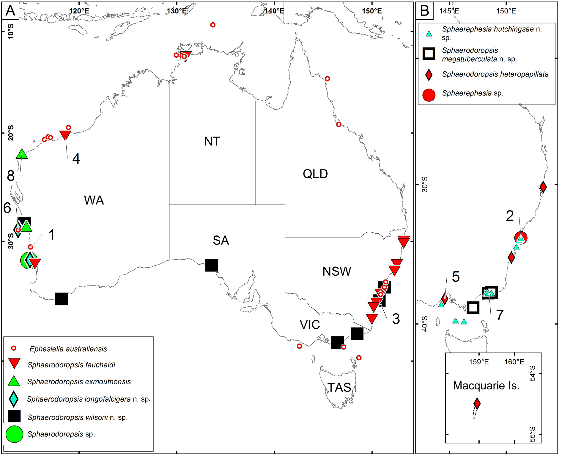
Type locality. 15.5 km SW of Pt Ricardo, Eastern Bass Strait ( Fig. 15 View FIGURE 15 ).
Distribution Bass Strait ( Fig. 15 View FIGURE 15 ).
Habitat. Sand, 26–58 m depth.


| NMV |
Museum Victoria |
No known copyright restrictions apply. See Agosti, D., Egloff, W., 2009. Taxonomic information exchange and copyright: the Plazi approach. BMC Research Notes 2009, 2:53 for further explanation.
|
Kingdom |
|
|
Phylum |
|
|
Class |
|
|
Order |
|
|
Family |
|
|
Genus |