
Stilbops
|
publication ID |
https://doi.org/ 10.5281/zenodo.211080 |
|
DOI |
https://doi.org/10.5281/zenodo.6178670 |
|
persistent identifier |
https://treatment.plazi.org/id/8079878D-FFB2-FF88-4DAD-6F68FA1B701B |
|
treatment provided by |
Plazi |
|
scientific name |
Stilbops |
| status |
|
Key to Japanese species of the genus Stilbops View in CoL View at ENA
(Males of auster , japonicus and michinokuensis are unknown)
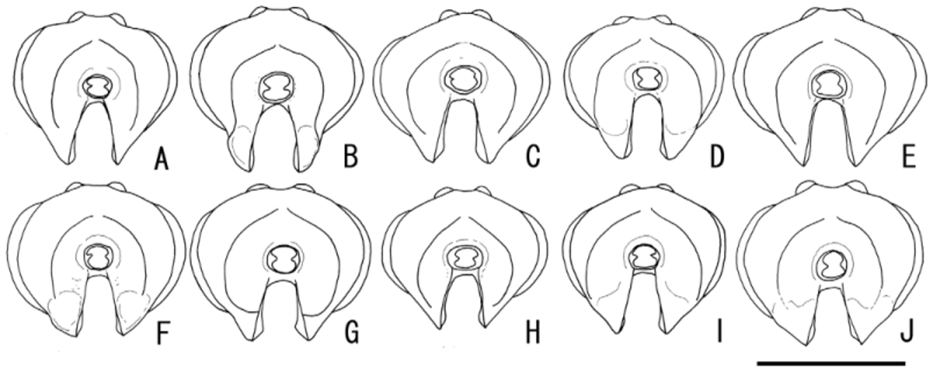
1. Anterior tentorial pit large, its maximum diameter more than 1/3 length of OD. Apical margin of clypeus sometimes notched medially in anterior view ( Fig. 2 View FIGURE 2 C, G). Basal portion of mandible convex ( Fig. 3 View FIGURE 3 K, M). Face covered with dense and long setae................................................................................................ 2
-. Anterior tentorial pit normal size, its maximum diameter less than 1/3 length of OD. Apical margin of clypeus convex or subtruncate in anterior view ( Fig. 2 View FIGURE 2 A, B, D-F, H-J). Basal portion of mandible flat ( Fig. 3 View FIGURE 3 L). Setae on lower part of face not remarkably denser and longer than upper area............................................................... 3
2. Apical margin of clypeus with median notch ( Fig. 2 View FIGURE 2 C). Lower end of occipital carina effaced ( Fig.4 View FIGURE 4 C). Hind femur black (or sometimes blackish-brown) ( Fig. 12 View FIGURE 12 D, E)................................................ S. coeloclypeus sp. nov.
-. Apical margin of clypeus without median notch ( Fig. 2 View FIGURE 2 G). Lower end of occipital carina complete ( Fig. 4 View FIGURE 4 G). Hind femur brown, not black ( Fig. 12 View FIGURE 12 K)................................................... S. mandibularis Kasparyan, 1999 View in CoL
3. Lower part of gena with convex profile in anterior view ( Fig. 2 View FIGURE 2 B); in lateral view, strongly concave ( Fig. 3 View FIGURE 3 B). Lower end of occipital carina reaching base of mandible ( Fig. 4 View FIGURE 4 B). Female with tergites red except for base of TI ( Fig. 9 View FIGURE 9 B). Hind leg largely yellow including hind coxa ( Fig. 12 View FIGURE 12 B, C)............................................ S. cavigena Kasparyan, 1984 View in CoL
-. Lower part of gena with flat profile in anterior view ( Fig. 2 View FIGURE 2 A, D-F, H-J); in lateral view, varying from weakly concave to flat ( Fig. 3 View FIGURE 3 A, D-F, H-J). Lower end of occipital carina effaced, not reaching mandibular base ( Fig. 4 View FIGURE 4 A, D-F, H-J). Hind coxa always largely black, remainder of hind leg black or yellow ( Fig. 12 View FIGURE 12 A, F-J, L-O)................................... 4
4. Hind femur and tibia largely black or blackish-brown ( Fig. 12 View FIGURE 12 A, F, G, M, N)....................................... 5
-. Hind femur and tibia largely yellow or brown ( Fig. 12 View FIGURE 12 C, H-J, O)................................................ 8
5. Tegula yellow ( Fig. 6 View FIGURE 6 A). Lower part of gena not concave ( Fig. 3 View FIGURE 3 A)................................ S. auster sp. nov.
-. Tegula black ( Fig. 6 View FIGURE 6 D, I). Lower part of gena concave ( Figs. 3 View FIGURE 3 D, I)............................................. 6
6. Antenna with 16 flagellomeres. OOD 1.4 times as long as OD. Vein Cu-a of fore wing distant from Rs&M by less than 0.5 times length of Cu-a............................................................. S. femoralis Kasparyan, 1999 View in CoL
-. Antenna with 17 or 18 flagellomeres. OOD 1.4–1.8 times as long as OD. Vein Cu-a of fore wing distant from Rs&M by more than 0.6 times length of Cu-a............................................................................ 7
7. Hind tibia 8.7–8.9 times as long as deep in lateral view. TI 1.2–1.3 (in female) or 1.7 (in male) times as long as wide. Metasomal tergite usually almost black......................................................... S. ezoensis sp. nov.
-. Hind tibia 7.8–8.5 times as long as deep in lateral view. TI 1.3–1.4 (in female) or 1.4–1.6 (in male) times as long as wide. Metasomal tergite usually broadly tinged with red............................................ S. montanus sp. nov.
8. Body densely punctate. Punctures on lateral lobes of mesoscutum separated by 0.3 to 0.9 times their diameter. Lower parts of gena not concave in lateral view ( Fig. 2 View FIGURE 2 E).................................................. S. japonicus sp. nov.
-. Body moderately punctate. Punctures on lateral lobes of mesoscutum separated by 1.0 to 1.2 times their diameter. Lower parts of gena more or less concave in lateral view ( Fig. 2 View FIGURE 2 F, H, J).................................................... 9
9. Body length about 5.0 mm. Area dentipara of propodeum with lateromedian longitudinal carina complete ( Fig. 8 View FIGURE 8 H). Tergites completely black ( Fig. 9 View FIGURE 9 I). Clypeus partly yellow. Tegula yellow ( Figs. 6 View FIGURE 6 H, 7 H).............. S. michinokuensis sp. nov.
-. Body length about 5.5–6.5 mm. Area dentipara of propodeum with lateromedian longitudinal carina sometimes absent ( Fig. 8 View FIGURE 8 F). TII and following tergites red (some specimens of kunashiricus View in CoL black) ( Fig. 9 View FIGURE 9 F, G, K). Clypeus completely black. Tegula yellow or black...................................................................................... 10
10. Pronotum nearly entirely punctate ( Fig. 5 View FIGURE 5 F). Area superomedia of propodeum incomplete ( Fig. 8 View FIGURE 8 F). Propodeum largely covered with punctures ( Fig. 8 View FIGURE 8 F). Tegula yellow ( Figs. 6 View FIGURE 6 F, 7 F). Tergites sometimes black in female ( Fig. 9 View FIGURE 9 G).......................................................................................... S. kunashiricus Kasparyan, 1999 View in CoL
-. Pronotum with large smooth area on lower half ( Fig. 5 View FIGURE 5 J). Area superomedia of propodeum complete ( Fig. 8 View FIGURE 8 J). Propodeum covered with fine, small punctures ( Fig. 8 View FIGURE 8 J). Tegula black ( Figs. 6 View FIGURE 6 J, 7 J). Tergites always red in females ( Fig. 9 View FIGURE 9 J)......................................................................................... S. orientalis Kasparyan, 1984 View in CoL


No known copyright restrictions apply. See Agosti, D., Egloff, W., 2009. Taxonomic information exchange and copyright: the Plazi approach. BMC Research Notes 2009, 2:53 for further explanation.
|
Kingdom |
|
|
Phylum |
|
|
Class |
|
|
Order |
|
|
Family |
|
Kingdom |
|
|
Phylum |
|
|
Class |
|
|
Order |
|
|
Family |
|
|
Genus |