
Pleurobranchus iouspi Ev. Marcus, 1984
|
publication ID |
https://doi.org/ 10.5281/zenodo.7225407 |
|
persistent identifier |
https://treatment.plazi.org/id/7B5BA125-7B4D-0274-4DC1-7AE8FC6D28B0 |
|
treatment provided by |
Valdenar |
|
scientific name |
Pleurobranchus iouspi Ev. Marcus, 1984 |
| status |
|
Pleurobranchus iouspi Ev. Marcus, 1984 View in CoL
( Figs. 1 View Fig E-F; 2; 11-17)
Pleurobranchus iouspi Ev. Marcus (1984: 68 View in CoL , figs. 57-61); Rios (2009: 417).
Oscanius View in CoL (?) testudinarius View in CoL auct. non Cantraine, 1835: Ev. Marcus (1970: 939, fig. 30).
Pleurobranchus testudinarius View in CoL auct. non Cantraine, 1835: Rios (1994: 206); Cunha et al. (2014: 47); Goodheart et al. (2015: 340, figs. 7H-I, in part).
Pleuroranchus evelinae auct. non Abbott, 1949: Ev. Marcus (1984: 63, in part).
Pleurobranchus (Susania) emys Ev. Marcus (1984: 70 View in CoL , figs. 57- 61). syn. nov.
Pleurobranchus emys View in CoL : Rios (2009: 417).
Pleurobranchus atlanticus View in CoL auct. non Abbott, 1949: Rios (2009: 417).
Type material: Holotype, as five microscopic slides: MZSP 119936. Each microscopic slide presents one of the following features: radula ( Fig. 11A View Fig ); jaw ( Fig. 11B View Fig ); portions of mantle ( Fig. 11C View Fig ); unrecognizable parts of reproductive system ( Fig. 11D View Fig ); and penis ( Figs. 11 View Fig E-H).
Type locality: 24°47'S, 45°15'W; São Paulo State, Brazil.
Material examined: Holotype; Syntypes of P. emys : Colombia: Santa Marta: MZSP 119934 [one microscope slide with radula and jaw platelets]; MZSP 119935 [one histological microscope slide with sections of the mantle]. Brazil: Maranhão state: 2°05'S, 42°44'W: CMPHRM-A 693, 46 m; Rio de Janeiro state: Arraial do Cabo: Oratório: MNRJ 33242, 20/iii/2014, A. Kassuga coll. [2; 1 dissected]; Rio de Janeiro: Cagarras Archipelago: Redonda Island: MNRJ 10708, P. S. Young & C. S. Serejo coll. [1]; MNRJ 10707, P. S. Young & C. S. Serejo coll. [1]; SAE ML 95, 04/iv/2012, 15 m, F. Moraes coll. [1; 1 dissected]; Rasa Island: MNRJ 33069, 07/iii/2014, 7 m, F. Moraes coll. [1; 1 dissected]; Maricá: Maricás Islands: MNRJ 33070, 20/ii/2014, Carlos Rangel and Jéssica Pinho coll. [1; 1 dissected].
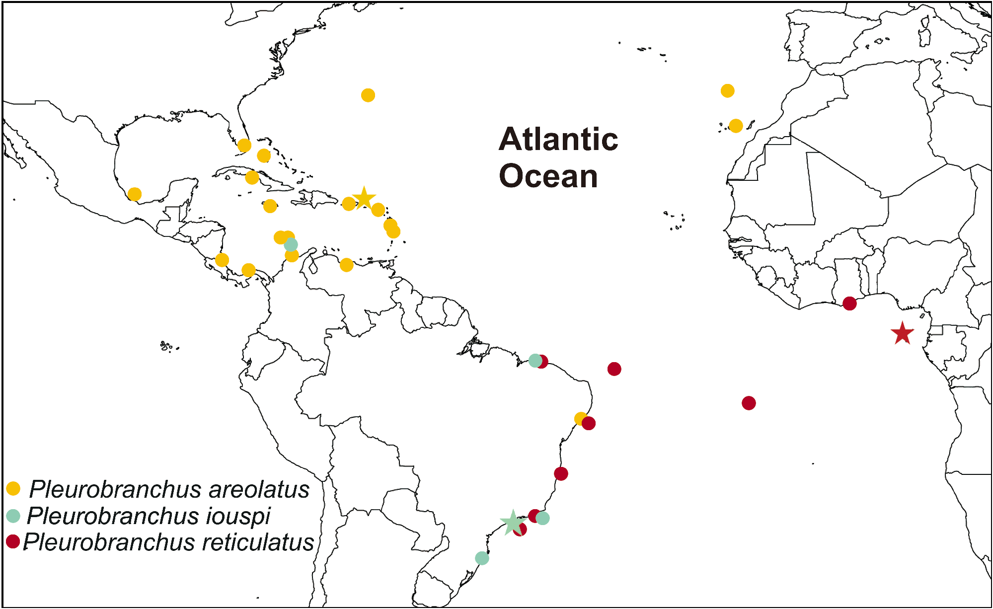
Specimen records ( Fig. 2 View Fig ): Colombia: Santa Marta (Ev. Marcus 1984); Brazil: Maranhão state (Ev. Marcus 1970); Rio de Janeiro state ( Goodheart et al. 2015; present study); São Paulo state (Ev. Marcus 1984); Santa Catarina state ( Cunha et al. 2014).
Redescription: External morphology ( Figs. 1 View Fig E-F; 12A-C): Coloration of living specimens with highly variable, with two main types. First type completely pale orange or bright yellow ( Fig. 1E View Fig ), sometimes with white marks; some specimens with a bright pink ring surrounding pointed (high) tubercles; mantle edge completely orange or white; rhinophores, tentacles, gill and foot same color pattern of mantle. Second color type is reddish (red, orange, dark red) ( Fig. 1F View Fig ); space between tubercles white; pointed (high) tubercles usually with darker pigment (dark red), sometimes a bright pink ring surrounding it; mantle edge same pattern of rest of body or white; rhinophores, tentacles, gill and foot same color pattern of mantle. Length of preserved specimens 51-99 mm; width 46-73 mm; length of foot 34-74 mm; width of foot 16-58 mm. Body subquadrate, slightly depressed, narrows posteriorly. Mantle covered foot partially, sometimes projecting beyond notum. Mantle covered by medium to large tubercles (1-7 mm) irregularly arranged all around dorsum; tubercles shape can be rounded or pointed (high); usually at mantle edge is located tubercles with lower diameter; pointed tubercles usually located in middle of dorsum ( Fig. 1F View Fig ); tubercles can be contracted when specimen is disturbed. Oral veil sturdy and broad that connects with head region; laterally, oral tentacles with a deep notch over almost its length. Rhinophores rolled and joined at their bases, up to one quarter of its length. Gill exposed laterally ( Fig. 12A View Fig ); 1/2 length of body; main rachis with rounded tubercles at point of origin of pinnae, forming zig-zag line; small rounded tubercles at base of pinnules; tubercles composed by spicules; tripinnate pinnae; 18- 24 pinnae; 7-12 pinnae free from the body wall, attached by branchial membrane. Anal opening lying over end of gill membrane. Pre-branchial pore opening approximately beside main rachis. Nephropore lying under/near first pinnae or between first and second pinnae. Elongated penis with 21.6 mm in length (in a preserved specimen with 51 mm in length) and 27 mm in length (in a preserved specimen with 75 mm in length); penis and female aperture surrounded by a very wide sheath (0.9 mm high) composed of three flaps ( Figs. 12 View Fig A-C). Posteriorly rounded foot with elongated metapodial gland, corresponding 1/7 to 1/4 of body length; anteriorly bilabiated, upper lip notched and smaller than the other one.
Mantle ( Figs. 13 View Fig A-D): Mantle, tubercles, rhinophores and dorsal portion of oral veil densely covered by spicules, which makes it seems coriaceous. Two types of spicules: linear, rod-like (length: 314.0-427.2 μm; thickness: 13.0-16.0 μm) ( Fig. 13A View Fig ); stellate spicules with three similar rays (ray length: 13.8-18.9 μm) ( Fig. 13B View Fig ) and with five rays, four of which are arranged in same plane (ray length: 45.4-102.2 μm; ray thickness: 22.7- 34.0 μm) ( Fig. 13C View Fig ). Mantle densely covered by spicules with five rays ( Fig. 13D View Fig ); perpendicular ray usually smaller than others rays (ray length: 45.4- 56.8 μm). Rod-like and stellate spicules with three rays calcareous; spicules with five rays partially calcareous, but not entirely formed by calcium carbonate. As sodium hypochlorite dissolve it, also composed of an unidentified organic matrix.
Shell ( Fig. 14A View Fig ): White to brownish with light golden tones in some parts of shell, depending on incidence of light; fragile; brownish periostracum; not observed in some specimens; subquadrangular in outline. Shell approximately 1.5 times longer than wide; length 2.2 mm, width 1.6 mm (in a preserved specimen with 51 mm in length); and, length: 6.6 mm, width 3.7 mm (in a preserved specimen with 75 mm in length). Spire has 1.5 whorls. Protoconch smooth. Growth lines distinct; sculptured with longitudinal lines transverse to growth lines. Shell in anterior/left portion of body, immediately anterior to heart, partially above pericardium and blood gland.
Circulatory system: Circulatory system of P. iouspi identical to the P. reticulatus as described above.
Digestive system ( Figs. 14 View Fig B-F; 15-16): Digestive system of P. iouspi very similar to the P. reticulatus as described above, with the following exceptions: Each jaw plate showing alternate rows formed by elongated elements with slight cruciform lateral expansion (preserved specimen measuring 51 mm in length: 30 elements transversally, 50 elements longitudinally) ( Figs. 14 View Fig B-C) (Holotype: 43 elements transversally, 60 elements longitudinally); elements consist on a main cusp with 1-5 denticles in each side ( Figs. 14 View Fig B-C) (Holotype with 2-5 denticles in each side), which could be of different sizes and asymmetrical in relation to main cusp ( Figs. 14 View Fig B-C). Pair of m4, main dorsal tensor muscle of radula, short, originating in lateral region of cartilages, surrounding them ventrally, inserting into subradular membrane ( Figs. 15 View Fig A-C). Pair of m5, secondary dorsal tensor muscle of radula large and broad, covering median portions of cartilage, extending up to dorsal region; originating in posterior surface of cartilages; inserting laterally in mj ( Figs. 15 View Fig A-C). Pair of strong retractor muscles that originates in most posterior portion of m5 ( Figs. 15A, C View Fig ) separated in almost its total length. Radula cream; formula 107 × 255.0.255 (holotype, specimen with 60 mm preserved length), 76 × 260.0.260 (from preserved specimen with 75 mm length) and 64 × 148.0.148 (from preserved specimen with 51 mm long). Duct of acid gland thin and short (same width as salivary duct) ( Figs. 15 View Fig AB); not convoluted. Esophagus internally with thin longitudinal folds ( Fig. 16A View Fig ). Stomach voluminous and rounded filled with food ( Fig. 16A View Fig ); internally, anteriorly with thin and smooth wall, except by the ventral furrow; posteriorly with large flaps and a central-ventral furrow that leads to intestine ( Fig. 16A View Fig ); stomach passes ventrally into digestive gland. Intestine long and thin walled; internally, smooth, with thin longitudinal folds near anus.
(A)
Identifiable stomach contents: Small bivalves (in most part of the cases juveniles; e.g. Arcidae ); small crustaceans (e.g., Tanaidacea : Paratanais coelhoi Araújo-Silva and Larsen, 2012 ; Ostracoda); arborescent bryozoans; algae; grains of sand.
R e p r o d u c t i v e s y s t e m ( Figs. 12 View Fig B-F): Triaulic. Ampulla elongated, same width as vas deferent and vagina; curved, but not convoluted. Spermoviduct branching into two ducts, shorter oviduct and other duct leading to prostate. Prostate tubular and convoluted ( Figs. 12 View Fig D-E); four times wider than deferent duct ( Fig. 12F View Fig ). Deferent duct thin, narrowing into a long and non-contractile penis; vas deferens forming loops inside penis ( Fig. 12F View Fig ). Penis (in a preserved specimen with 51 mm length: length 14 mm; width 1.5 mm) (in a preserved specimen with 75 mm length: length 20.8 mm; width 2.3 mm) ( Figs. 12 View Fig AC; E). Basal penial lobe asymmetrical ( Fig. 12B View Fig ). Penis with some sparse rod-like spicules. Vaginal duct convoluted with two allosperm vesicles arranged semi-serially. Proximally, vagina divides into seminal receptacle and bursa copulatrix as a short duct, both allosperm receptacles lie close to each other, their stalks are crossed ( Fig. 12D View Fig ). Seminal receptacle elongated; not convoluted ( Fig. 12D View Fig ). Bursa copulatrix rounded. Vaginal opening immediately ventral to penis ( Fig. 12A View Fig ). Vagina about same diameter of prostate. Genital aperture surrounded by three well-distinct large penial flaps; two of these flaps are bifurcated after their base ( Figs. 12 View Fig A-B).
Nervous system ( Fig. 12G View Fig ): Nervous system of P. iouspi very similar to P. reticulatus as described above, except for: the absence of cp6, cp7 and cp8; the connective between buccal and cerebro-pleural ganglia leaving the most anterior portion of cerebro-pleural ganglia in ventral area and inserts in buccal ganglia; and, np3 innervates laterally the body until enters into foot.
Remarks on the type material of P l e u r o b r a n c h u s i o u s p i ( Figs. 11 View Fig A-H): The histological slides of the type of Pleurobranchus iouspi mentioned in the original description were rediscovered in the Malacological Collection of the MZSP, as well as most of species dealt in Ev. Marcus (1984), they were all on the same slide collection. Although the slides are not appropriately labeled, all have the same handwritten symbol (“ IO ”, probably referring to the name iouspi ) ( Figs. 11 View Fig A-E). Furthermore, the original figure of the penis (Ev. Marcus 1984: 69, fig. 61) closely resembles the slide-mounted material ( Figs. 11 View Fig FH), which has a peculiar anterior basal bulb, making it rather asymmetrical ( Fig. 11G View Fig ); a thin deferent duct, narrowing into a long and non-contractile penis ( Fig. 11H View Fig ); and a vas deferens forming loops inside the penis ( Fig. 11H View Fig ). Ev. Marcus (1984) described P. iouspi based on a single specimen, therefore, since most of species dealt in Ev. Marcus (1984) were on the same slide collection found and the fact that the original figure of the penis closely resembles the slide-mounted material lead us to believe that the remaining slides should also correspond to the holotype. The radula formula was found to be 107 × 255.0.255, although Ev. Marcus (1984) described it as 100 × 250.0.250. The probable difference in the number of rows is due to the slightly broken condition of the posterior-most portion of the radula.
Remarks on the type material of P l e u r o b r a n c h u s e m y s ( Figs. 17 View Fig A-D): The original material studied by Ev. Marcus (1976b, 1984) was searched for in the recently located slides belonging to the collection of Er. Marcus and Ev. Marcus, housed in the MZSP. It was find some histological slides of the syntypes of Pleurobranchus emys Ev. Marcus, 1984 : MZSP 119934, syntype, Santa Marta, Colombia [one microscope slide with radula and jaw platelets]; MZSP 119935, probable syntype, Santa Marta, Colombia [one histological microscope slide with sections of the mantle]. Pleurobranchus emys Ev. Marcus, 1984 was described based on the material studied by Ev. Marcus (1976b) from Colombia (then named Oscanius testudinarius ). Ev. Marcus (1984) clearly stated that the type of P. emys corresponds a microscope slide with radula and jaw platelets, and two color slides of living specimens. Part of the above-mentioned type series was rediscovered in the MZSP. One slide with radula and jaw ( Fig. 17A View Fig ) is clearly the microscope slide mentioned by Ev. Marcus (1984), and is herein considered as a syntype lot ( MZSP 119934); one histological slide with sections of the mantle ( Fig. 17B View Fig ), not mentioned in original description, is herein considered a probable syntype ( MZSP 119935). Evidence that these histological slides correspond to the material originally used to describe P. emys is: the slide with sections of mantle shows the dense layer of radiating spicules as described by Ev. Marcus (1984) ( Fig. 17D View Fig ), and is labeled as “ Susania emys 1983”, one year before publication, and as “ Oscanius testudidarius ”, the name originally used by Ev. Marcus in 1976 ( Fig. 17B View Fig ); the slide with radula and jaw is labeled “ Oscanius testudinarius ”, with the name “ testudinarius ” scratched over ( Fig. 17A View Fig ), indicating that this was the previous determination by Ev. Marcus (1976b), subsequently changed to P. emys ; this slide also matches the description and drawings of Ev. Marcus (1984).


| MZSP |
Sao Paulo, Museu de Zoologia da Universidade de Sao Paulo |
| MNRJ |
Museu Nacional/Universidade Federal de Rio de Janeiro |
| ML |
Musee de Lectoure |
| R |
Departamento de Geologia, Universidad de Chile |
| AC |
Amherst College, Beneski Museum of Natural History |
| IO |
Instituto de Oceanografia da Universidade de Lisboa |
| FH |
Fort Hays |
No known copyright restrictions apply. See Agosti, D., Egloff, W., 2009. Taxonomic information exchange and copyright: the Plazi approach. BMC Research Notes 2009, 2:53 for further explanation.
|
Kingdom |
|
|
Phylum |
|
|
Class |
|
|
Order |
|
|
Family |
|
|
Genus |
Pleurobranchus iouspi Ev. Marcus, 1984
| Grzelak, Katarzyna & Sørensen, Martin V. 2022 |
Pleurobranchus emys
| Rios E. 2009: 417 |
Pleurobranchus atlanticus
| Rios E. 2009: 417 |
Pleurobranchus testudinarius
| Goodheart J & Camacho-Garcia Y & Padula V & Schrodl M & Cervera JL & Gosliner TM & Valdes A. 2015: 340 |
| Cunha CM & Padula V & Magalhaes ARM & Ferreira FM & Ferreira JF 2014: 47 |
| Rios E. 1994: 206 |
Pleurobranchus iouspi Ev. Marcus (1984: 68
| Rios E. 2009: 417 |
| Marcus Ev. 1984: 68 |
Pleuroranchus evelinae
| Marcus Ev. 1984: 63 |
Pleurobranchus (Susania) emys Ev. Marcus (1984: 70
| Marcus Ev. 1984: 70 |
Oscanius
| Marcus Er & Marcus Ev. 1970: 939 |