
Torula phytolaccae Y.X. Li, C.F. Liao & Doilom, 2023
|
publication ID |
https://doi.org/ 10.11646/phytotaxa.584.1.1 |
|
DOI |
https://doi.org/10.5281/zenodo.7624047 |
|
persistent identifier |
https://treatment.plazi.org/id/7B4A472C-FFAB-B828-FF5D-FF4C8932FAF7 |
|
treatment provided by |
Plazi |
|
scientific name |
Torula phytolaccae Y.X. Li, C.F. Liao & Doilom |
| status |
sp. nov. |
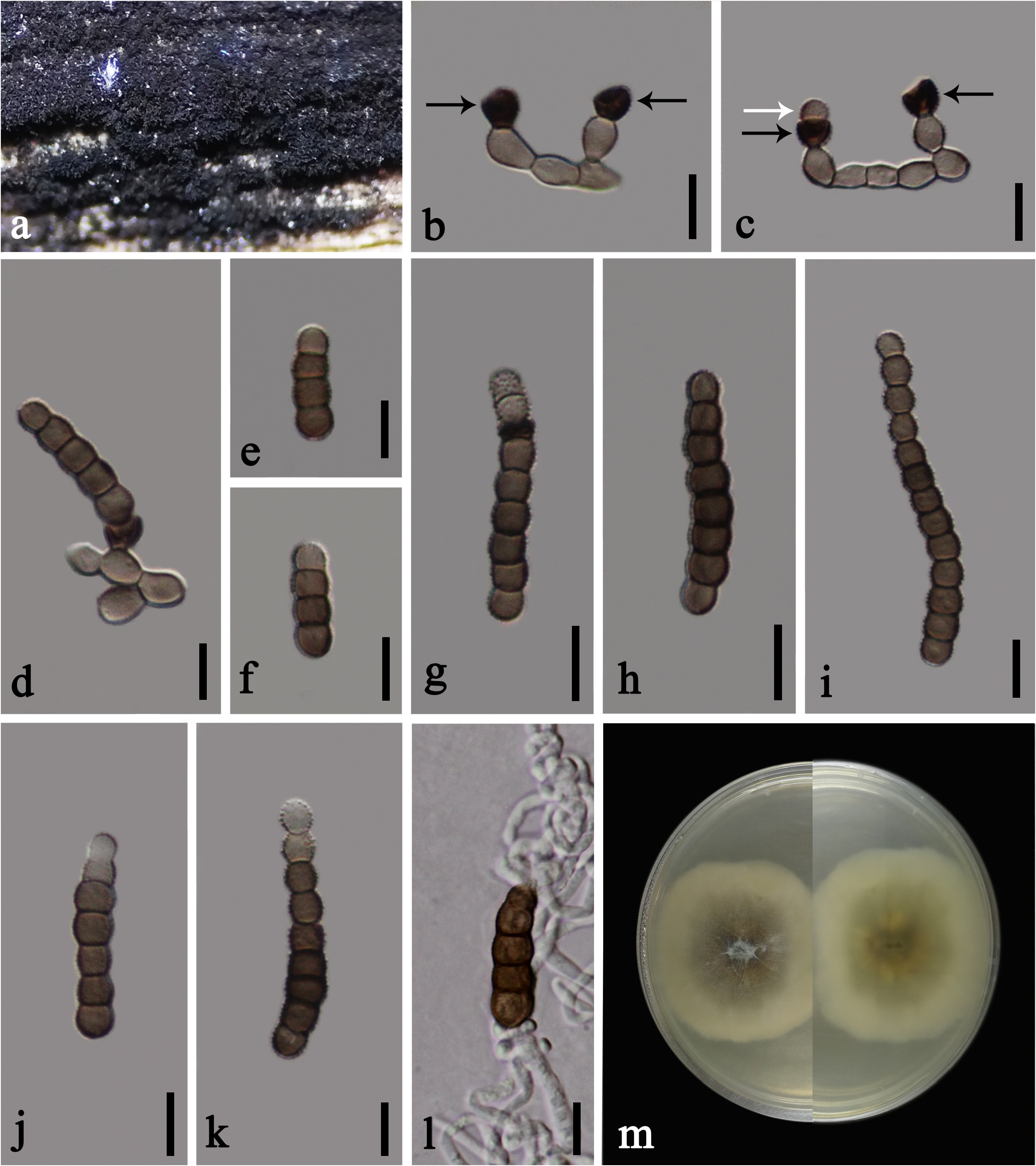
Torula phytolaccae Y.X. Li, C.F. Liao & Doilom , sp. nov. ( FIGURE 2 View FIGURE 2 )
Index Fungorum number: IF559683; Facesoffungi number: FoF 11438
Etymology:—Name refers to the host genus Phytolacca on which the fungus was collected.
Holotype:— ZHKU 22-0056
Saprobic on dead stems of Phytolacca acinosa . Sexual morph: undetermined. Asexual morph: Colonies effuse, dense, velvety, black on the host. Mycelium 2–3 µm thick, immersed to superficial on the substrate, septate, branched, smooth to minutely verruculose, brown. Conidiophores 10–24 µm long × 3–7 µm wide (x = 18.5 × 5 µm, n = 15), semi-macronematous, mononematous, flexuous, unbranched, verruculose, thick-walled, doliiform to subcylindrical, consisting of 1–3 cells or reduced to conidiogenous cells, pale brown, arising from lateral and terminal on a hypha. Conidiogenous cells 4–6.7 µm long × 5–7.7 µm diam. (x = 5.3 × 6.5 µm, n = 20), monoblastic, lateral to terminal, dark brown to black, verruculose, thick-walled, doliiform to cupulate. Conidia (9–)20–34(−63) × 5–9 (x = 27 × 6 µm, n = 40), phragmosporous, solitary to catenate, acrogenous, straight or slightly curved, dark brown to black, verrucose, 2–12-septate, predominantly 3–5-septate, rounded at both ends, mostly subcylindrical, composed of subglobose cells, constricted at the septa, subhyaline to pale brown at the apex, thick-walled.
Culture characteristics: —Colonies on PDA reaching 28–30 mm diam. after 14 days at 25 ˚C, medium dense, mycelium partly immersed to superficial, flat or effuse, edge entire, cottony, hairy at the centre, initially floccose at the centre with white and brown aerial mycelia, greyish green circle at the edge with pale creamy outward, becoming a purple-red circle, whitish at the margin from above; greenish-grey the centre with pale creamy outward from below, without pigment produced in PDA. Asexual morph did not produce on PDA at 25 ˚C within 2 months.
Material examined:— CHINA. Yunnan Province: Kunming City, 25.1339867°N 102.7418331°E, on dead stems of Phytolacca acinosa Roxb. (Phytolaccaceae) , 28 June 2019, C.F. Liao (ZHKU 22-0056, holotype); ex-type living culture ZHKUCC 22-0107, ibid., living culture ZHKUCC 22-0108.
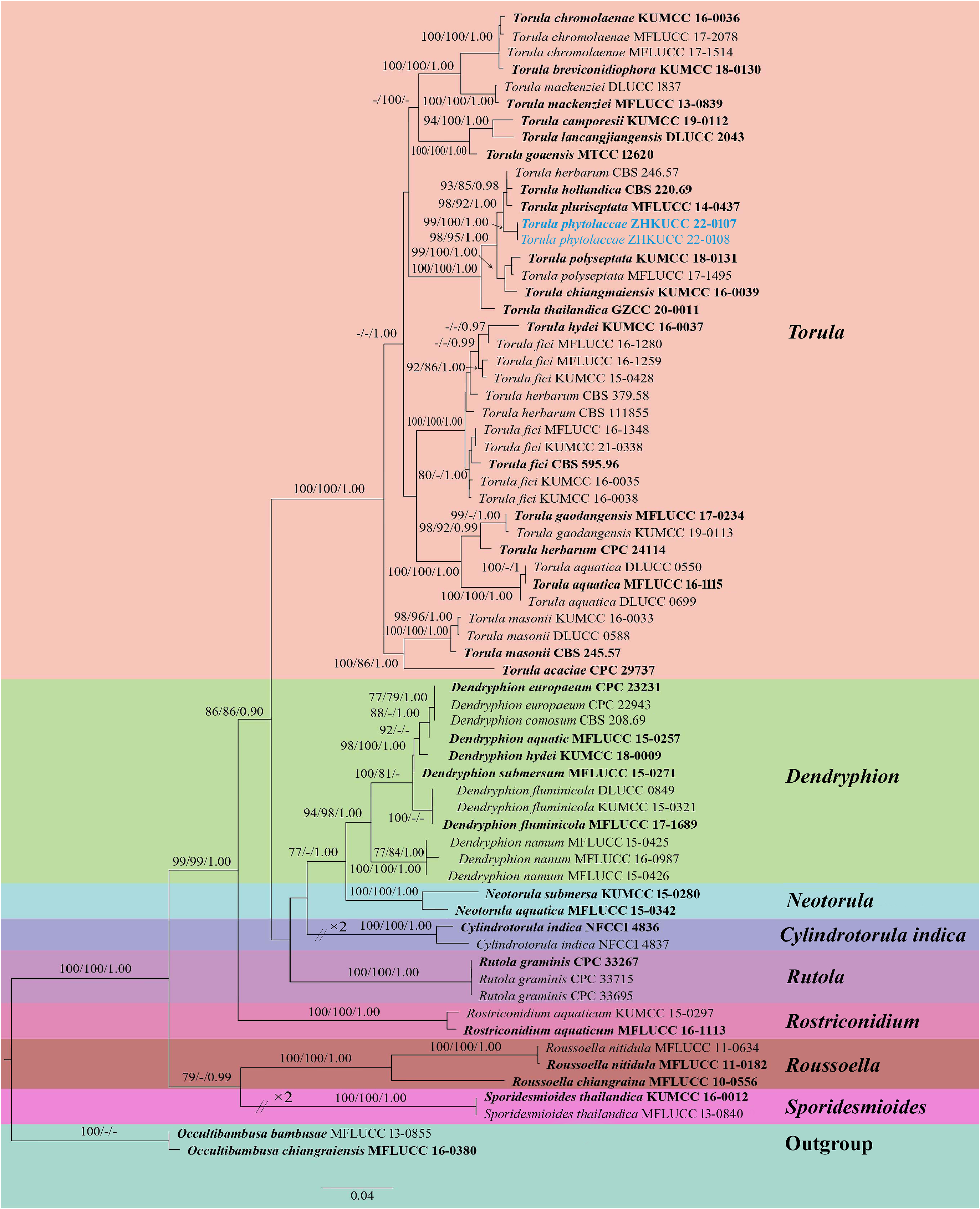
Notes:—In our multi-locus phylogeny, Torula phytolaccae clusters with T. herbarum (CBS 246.57), T. hollandica (CBS 220.69) and T. pluriseptata (MFLUCC 14-0437), but forms a distinct branch with 98% ML, 92% MP and 1.00 PP values ( FIGURE 1 View FIGURE 1 ). The description of T. herbarum CBS 246.57 is unavailable for morphological comparison, but the type strain CPC 24114 resided in a phylogenetically unrelated clade. Torula phytolaccae differs from T. hollandica in having more septa of conidia (2–12 vs. 2–4) and different conidial pigmentation (dark brown to black vs. pale to reddish brown) ( Crous et al. 2015). Torula phytolaccae differs from T. pluriseptata in having longer conidiophores (10–24 µm vs. 2.8–4.3 µm) and larger conidiogenous cells (4–6.7 µm long × 5–7.7 µm diam. vs. 3–3.5 µm long × 3.8– 4.6 µm diam.) (Li et al. 2018). In addition, T. phytolaccae has subhyaline to pale brown, verrucose cells at the apex of conidia, which were absent in T. hollandica and T. pluriseptata ( Crous et al. 2015, Li et al. 2017). Torula phytolaccae resembles T. monilis in having doliiform conidiogenous cells and verrucose conidia comprising subglobose cells, but differs in having more septa of conidia (2–12 vs. 3), different conidial pigmentation (dark brown to black vs. brown), longer conidiophores (10–24 µm vs. 14 µm) ( Crous et al. 2015). A morphological comparison of T. phytolaccae and similar species is also provided in TABLE 2 View TABLE 2 . Based on morphology and phylogenetic evidence, here we introduce Torula phytolaccae as a novel species from Phytolacca acinose .
No known copyright restrictions apply. See Agosti, D., Egloff, W., 2009. Taxonomic information exchange and copyright: the Plazi approach. BMC Research Notes 2009, 2:53 for further explanation.
|
Kingdom |
|
|
Phylum |
|
|
Class |
|
|
Order |
|
|
Family |
|
|
Genus |