
Gamispatulus ferrilongus, Narciso & Silva, 2020
|
publication ID |
https://doi.org/ 10.11646/zootaxa.4803.3.3 |
|
publication LSID |
lsid:zoobank.org:pub:45FB73BA-DF09-4603-AC61-0C1BDA825833 |
|
persistent identifier |
https://treatment.plazi.org/id/7A7D879C-FFCA-FF9C-1BE7-6915FC7EF9E9 |
|
treatment provided by |
Plazi |
|
scientific name |
Gamispatulus ferrilongus |
| status |
sp. nov. |
Gamispatulus ferrilongus n. sp.
(ZooBank registration: urn:lsid:zoobank.org:act:CCB1F932-89B1-4494-B429-CC7D8)
( Figs 5–8 View FIGURE 5 View FIGURE 6 View FIGURE 7 View FIGURE 8 )
Type host. Schizodon intermedius Garavello & Britski, 1990 (Anostomidae)
Type Locality. Veados River , Jurumirim Reservoir , Upper Paranapanema River (23° 16′2.49″ S, 48° 38′15.72″ W), municipality of Itatinga, São Paulo State, Brazil GoogleMaps .
Additional locality. Paranapenema River , Jurumirim Reservoir, Upper Paranapanema River (23° 29′16.54″ S, 48° 37′12.88″ W), municipality of Angatuba, São Paulo State, Brazil GoogleMaps .
Site in host. Nostrils.
Specimens deposited. Holotype INPA 2521 View Materials (adult female) and Paratypes INPA 2522 View Materials to INPA 2525 View Materials (7 adult females) deposited in the Invertebrate Collection of the Instituto Nacional de Pesquisas da Amazônia ( INPA), municipality of Manaus, Amazonas State, Brazil .
Prevalence and mean intensity in nostrils. Seven infected hosts in 28 analyzed fish (or 25%) and 2 ± 0,4 copepods per infected fish.
Prevalence and mean intensity on the gill filaments. N one of the 28 analyzed fish.
Etymology. The specific name results from the combination of two Latin words: ferri (= any iron tool or weapon, including a sword) and longus (= long), in reference to the shape and size of the rostral spine, resembling an ancient Roman sword.
Differential diagnosis. Rostrum well-developed, armed with long rostral spine; rostral spine with acute tip; tip extending up to half of cephalothorax. Retrostylets simple, without adjacent spatulate processes. Dorsal surface of the genital double-somite with 2 rounded processes (= anterior and posterior process) on both lateral margins.
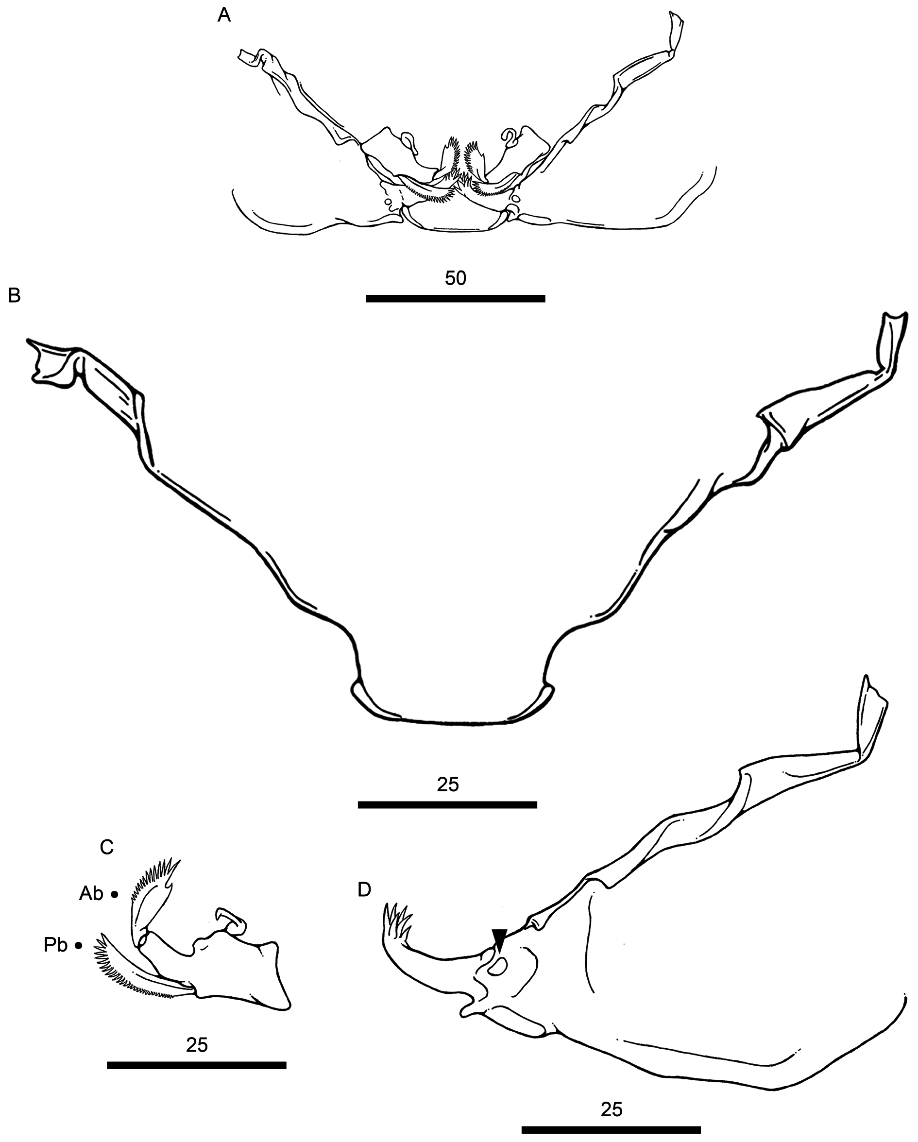
Description of adult female. Based on 11 female specimens, no males observed. Body cyclopiform ( Fig. 5A View FIGURE 5 ), comprising prosome, urosome, and caudal rami; prosome consisting of cephalosome and PS-1; PS-1 fused to cephalosome; and 3 free pedigerous somites (PS-2 to PS-4). Cephalothorax triangular ( Fig. 5B View FIGURE 5 ), decreasing in width anteriorly, maximum width at level of retrostylets tip ( Table 2), dorsal eyespot, rostrum well-developed and protruded anteriorly, dorsal surface with several pores; pores distributed in anterior half of cephalothorax; and armed with paired dorsolateral stylets (= retrostylets). Rostrum armed with ventral rostral spine; rostral spine long, extending up to half of cephalothorax, with sharp tip ( Fig. 5D View FIGURE 5 ). Retrostylets simple ( Fig. 5B View FIGURE 5 ), without adjacent spatulate processes; stylet process broad, with acute tip. Free pedigerous somites tapering posteriorly ( Fig. 5A View FIGURE 5 ); PS- 2 narrower than PS-1, with paired integumental windows laterally on tergite ( Fig. 5C View FIGURE 5 ); PS-3 and PS-4, both lacking such integumental windows ( Fig. 5A View FIGURE 5 ).
Urosome consisting of PS-5, genital double-somite, and 3 free abdominal somites (AS-1 to AS-3) ( Fig. 6A View FIGURE 6 ); PS-5 ( Figs. 6A, C View FIGURE 6 ) reduced, smaller and thinner than other prosome somites, unornamented; genital double-somite ( Figs. 6A, C View FIGURE 6 ), 1.5 times wider than long, bearing paired slit-like genital apertures dorsally, ornamented with transverse row of spinules on ventral surface, dorsal surface with 2 rounded processes (= anterior and posterior process) on both lateral margins ( Fig. 6C View FIGURE 6 ); abdominal somites decreasing in width from anterior to posterior, each somite ornamented with posterior spinule row along ventral margin ( Fig. 6A View FIGURE 6 ); AS-3 (= anal somite) deeply incised posteriorly (= anus).
Caudal rami ( Fig. 6A View FIGURE 6 ), about 1.5 times longer than wide; each ramus ornamented with paired spinule rows on ventral surface and armed with 4 setae, all naked: seta 1 and 3 shortest, both setae inserted on ventral surface; seta 2 and 4, both setae inserted on posterior margin; seta 4 longest.
Antennule 5-segmented ( Fig. 5E View FIGURE 5 ), setal formula: 10, 4, 4, 2, 5 + 2 ae (total 27). Antenna ( Fig. 6D View FIGURE 6 ) 4-segmented comprising coxobasis, and 3-segmented enp; coxobasis (= first segment) broad, unornamented; enp-1 (= second segment) ornamented with spinule row along outer margin and large spine near middle of inner margin; enp-2 (= third segment) slightly curved, with single pore on concave margin (arrowed in Fig. 26D); enp-3 (= fourth segment) reduced, unornamented; and 2 terminal claws (= inner and middle claw); middle claw curved, with fossa on concave margin (arrowed in Fig. 6D View FIGURE 6 ); inner claw without fossa.
Buccal apparatus ( Fig. 7A View FIGURE 7 ) comprising labrum, mandible, and maxilla; labrum broad, truncated posteriorly, partially covering other buccal components ( Fig. 7B View FIGURE 7 ); mandible armed with 2 blades (= anterior and posterior blade); anterior blade ornamented with spinules along posterior margin and armed with apical tooth; posterior blade longer and thinner than previous blade, ornamented with spinules along posterior margin; maxilla 2-segmented, comprising syncoxa (= first segment) and basis (= second segment); syncoxa broad, with large subdistal pore (arrowed in Fig. 7D View FIGURE 7 ); basis with multiples spinules.
P1 to P4 biramous ( Figs. 8 View FIGURE 8 A-D), each leg comprising coxa, basis, endopod (inner ramus) and exopod (outer ramus). P1 ( Fig. 8A View FIGURE 8 ); coxa unornamented; basis with bare outer seta; enp 2-segmented, both segments with spinules along outer margin and lacking any ornament on inner margin; enp-1 (= proximal segment) armed with 1 plumose seta on inner margin; enp-2 (= distal segment) about 2 times longer than previous segment, armed with 2 serrated spines and 5 plumose setae; exp 3-segmented; exp-1 and -2, both with spinules along outer margin; all segments lacking any ornament on inner margin; exp-1 (= proximal segment) about 1.5 times longer than following segments, armed with single distal spine on outer margin; exp-2 (= middle segment) protrude laterally, armed with 1 plumose seta on inner margin; exp-3 (= distal segment) ornamented with few spinules (3-4 spinules) located immediately next to first seta (arrowed in Fig. 8A View FIGURE 8 ), armed with 2 simple spines (not serrated) and 5 plumose setae.
P2 ( Fig. 8B View FIGURE 8 ); coxa ornamented with spinules (4 spinules); basis with bare outer seta; enp 3-segmented, all segments with spinules along outer margin and lacking any ornament on inner margin; enp-1 (= proximal segment) armed with 1 plumose seta on inner margin; enp-2 (= middle segment) armed with 2 plumose setae on inner margin; enp-3 (= distal segment) armed with simple spine (not serrated) and 4 plumose setae; exp 3-segmented; exp-1 and -2, both with spinules on outer margin; all segments lacking any ornament on inner margin; exp-1 (= proximal segment) about 1.5 longer than following segments, armed with single distal spine on outer margin; exp-2 (= middle segment) armed with 1 plumose seta on inner margin; exp-3 (= distal segment) armed with 2 minute spines; spines smaller than those present in P1 exp-3; and 6 plumose setae. P3 ( Fig. 8c View FIGURE 8 ) with same ornamentation and armament described for P2.
P4 ( Fig. 8D View FIGURE 8 ); coxa ornamented with spinules (3 spinules); basis with bare outer seta; enp 2-segmented, both segments lacking any ornament on outer and inner margin; enp-1 (= proximal segment) armed with 1 plumose seta on inner margin; enp-2 (= distal segment) armed with 4 plumose setae distally, lacking any spines; exp 1-segment- ed; exopodal segment lacking any ornament on outer and inner margin, armed with 2 minute spines; spines smaller than those present in P1 exp-3; and 4 plumose setae.
P5 reduced and represented by 2 naked setae ( Figs. 6A, C View FIGURE 6 ). Spine and setal formula of biramous swimming legs is presented in Table 4.
Intercoxal sclerites slender, unornamented, with both ends directed posteriorly ( Fig. 6B View FIGURE 6 ). Intercoxal plates from P1 to P3 with paired lateral pores; intercoxal plate of P4, absent ( Fig. 6B View FIGURE 6 ). Egg sac paired ( Fig. 8E View FIGURE 8 ), uniseriate.
Remarks. The new copepod was identified as member of the Ergasilidae based on the absence of maxillipeds and the presence of uniramous antennae comprising coxobasis (= first segment) and 3-segmented enp with fourth segment (= enp-3) armed with 1 or more terminal claws, mandible bearing 2 spinulate blades, 2-segmented maxilla with the distal segment (= basis) ornamented with multiple spinules, and P4 exp 2-segmented in adult females ( Boxshall & Halsey 2004). Among ergasilids, the new copepod resembles species from the vaigamid subgroup ( Gamidactylus , Gamispatulus , Gamispinus , Pseudovaigamus , and Vaigamus ) in having the combination of a 2-segmented enp for P1 and cephalothorax armed with a pair of dorsolateral stylets (or retrostylets).
The new copepod was identified as member of Gamispatulus for possessing the following combination of diagnostic features: (1) rostrum armed with rostral spine (lacking in Gamidactylus and Gamispinus ); (2) 5-segmented antennule (6-segmented in species of Gamidactylus , Pseudovaigamus , and Vaigamus ); (3) antenna with 2 terminal claws (a single claw is present in Pseudovaigamus and Vaigamus ); (4) third antennary segment unornamented (ornamented with long spinules in Gamispinus ), and (5) P4 with 2-segmented enp and 1-segmented exp. In addition to these generic features, the new copepod also resembles its congener, G. schizodontis (type species), in possessing smooth intercoxal plates in P1-P3, second antennary segment (or enp-2) ornamented with spinule row along outer margin and large spine near middle of inner margin, and egg sacs uniseriate.
The new copepod, Gamispatulus ferrilongus n. sp., can be readily separated from its congener in having simple retrostylets, thus diverging from restrostylets with medial spatulate process of G. schizodontis . Furthermore, the size of rostral spine is also different in these two species: in Gamispatulus ferrilongus n. sp. it is about three times longer than that in G. schizodontis : 180 in Gamispatulus ferrilongus n. sp. vs. ≅ 60 in G. schizodontis [see figure 5 in Thatcher & Boeger (1984b)]. The morphology of the genital double-somite in G. ferrilongus n. sp. also differs from that of G. schizodontis : in G. ferrilongus n. sp. the dorsal surface of this somite carries 2 rounded processes (= anterior and posterior process) with similar size on both lateral margins ( Fig. 6C View FIGURE 6 ), whereas in G. schizodontis this somite, even though it carries similar processes (see Fig. 1A), they are relatively smaller and different in size from each other. Another distinct difference between both species is the armature of the caudal rami: in G. ferrilongus n. sp. each ramus bears 4 setae being 2 smaller (= seta 1 and 3) and 2 longer (= seta 2 and 4), whereas in G. schizodontis each ramus bears only 2 long setae (due to their position these setae are putative equivalent to seta 2 and 4 from G. ferrilongus n. sp.), lacking any short setae. Finally, the ornamentation of the legs of G. ferrilongus n. sp. differs in several aspects from those present in G. schizodontis , as follow: (1) coxa from P2 to P4 carries minute spinules (3-4 spinules) laterally in G. ferrilongus n. sp. vs. coxa with long spinules (2-5 spinules) in G. schizodontis ; (2) P1 exp-1 armed with single distal spine on outer margin in G. ferrilongus n. sp. vs. P1 exp-1 armed with 2 unequal spines (i.e. anterior spine short and triangular, and posterior spine long and thinner) in G. schizodontis ; and (3) exp-1 of P2 and P3 ornamented with a row of minute spinules along outer margin in G. ferrilongus n. sp. vs. exp-1 of P2 and P3 ornamented with three prominent spines located in the distal half of the segment as in G. schizodontis .
Based on the morphological differences listed above, the present specimens were considered as a new species, Gamispatulus ferrilongus n. sp., of the ergasilid genus Gamispatulus .


| INPA |
Instituto Nacional de Pesquisas da Amazonia |
No known copyright restrictions apply. See Agosti, D., Egloff, W., 2009. Taxonomic information exchange and copyright: the Plazi approach. BMC Research Notes 2009, 2:53 for further explanation.
|
Kingdom |
|
|
Phylum |
|
|
Class |
|
|
Order |
|
|
Family |
|
|
Genus |