
Gymnothorax mucifer Snyder, 1904
|
publication ID |
https://doi.org/ 10.11646/zootaxa.4559.1.6 |
|
publication LSID |
lsid:zoobank.org:pub:929DF43D-DA2B-409B-82EF-6200E8DF2086 |
|
DOI |
https://doi.org/10.5281/zenodo.5932782 |
|
persistent identifier |
https://treatment.plazi.org/id/663987F8-D611-FFDD-FF15-FE97FC4CF85A |
|
treatment provided by |
Plazi |
|
scientific name |
Gymnothorax mucifer Snyder, 1904 |
| status |
|
Gymnothorax mucifer Snyder, 1904 View in CoL
( Figs. 1–5 View FIGURE 1 View FIGURE 2 View FIGURE 3 View FIGURE 4 View FIGURE 5 , Table 2)
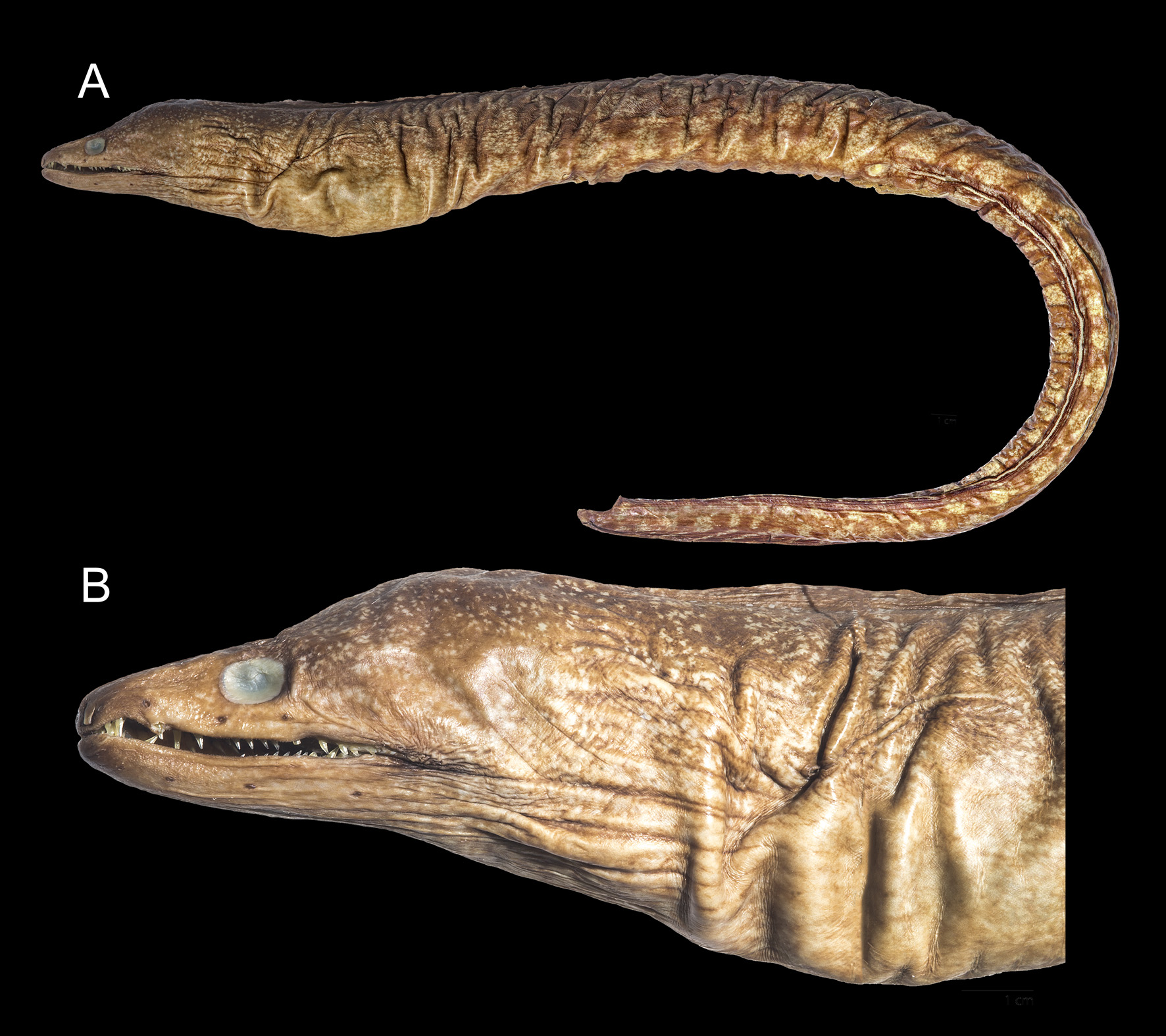
Diagnosis. A moderately sized moray (to at least 715 mm TL) with yellowish white spots on dark brown body and fins. Spots small and scattered evenly on top of head, grading into snowflake-like blotches on trunk and anterior part of tail, centralizing as separate rounded blotches on posterior part of tail. White margin on anal fin, replaced by serial pale blotches on posterior part of tail. Peripheral intermaxillary and median intermaxillary teeth uniserial. Maxillary teeth uniserial in larger males, biserial in immatures and females. Vomerine teeth small and stout in a staggered row, sometimes biserial centrally. Dentary teeth in one row, an additional peripheral row with small teeth present in immatures and females. Predorsal vertebrae 4–6, preanal vertebrae 51–55 and total vertebrae 130–141.
Description. Measurements from Taiwanese and Hawaiian specimens. A moderately stout moray, depth at gill opening 13.2–27.0 and at anus 16.9–28.6 in TL. Anus anterior to mid-point of body, preanal length 2.1–2.4 in TL. Dorsal fin moderately high, the origin anterior to gill opening, predorsal length 9.5–11.9 in TL and 1.1–1.5 in HL. Anal fin shallow, originating just behind anus. Gill opening slightly below mid-body. Head length 7.3–8.7 in TL. Eyes above middle of jaw with diameter 8.2–12.2 in HL. Jaws subequal, 1.9–2.7 in HL, teeth not visible when mouth closed. Snout moderately elongate, 4.4–5.9 in HL. Anterior nostril at snout tip, moderately elongate and tubular, shorter than eye diameter in length; posterior nostril above the anterior margin of eye as a pore with raised rim. Head pores typical and with dark rim; three supraorbital pores, first on tip of snout, second just above anterior nostril, and third above and between first and second infraorbital pores; four infraorbital pores, first behind anterior nostril, second midway between anterior nostril and third infraorbital pore, third below anterior margin of eye, and fourth pore below posterior margin of eye; six mandibular pores along lower jaw, all before rictus; two branchial pores on postero-dorsal head before gill opening. Predorsal vertebrae 4–6 (5), preanal vertebrae 51–55 (53), total vertebrae 130–141 (137).
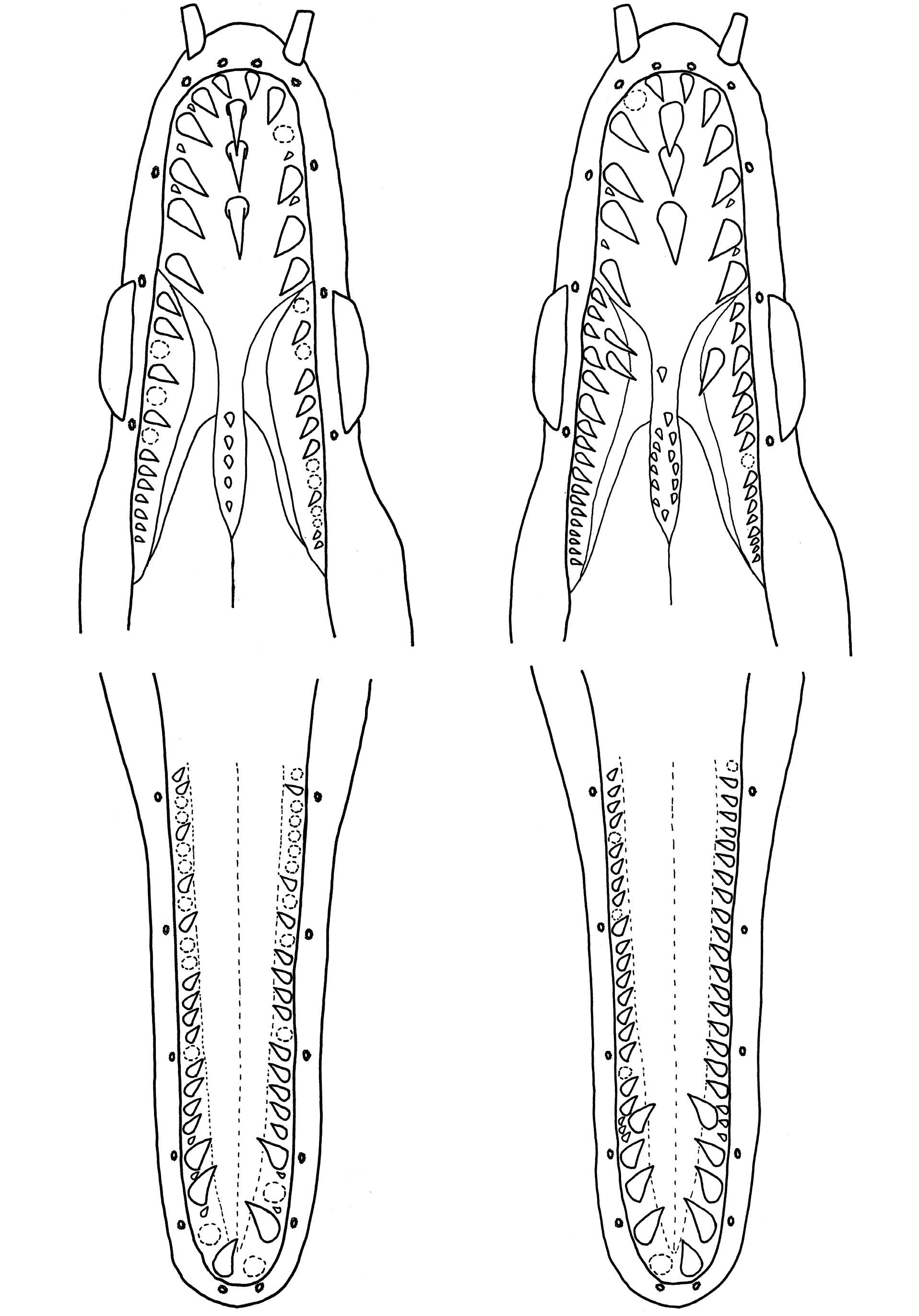
Dentition ( Fig. 4 View FIGURE 4 ): teeth smooth, pointed and slightly retrorse. Peripheral intermaxillary teeth uniserial, with 5– 7 canines on each side. Median intermaxillary teeth very long, slender and uniserial, 2–3 in number, larger posteriorly. Maxillary teeth uniserial, 10–18 on each side, shorter than peripheral intermaxillary teeth, triangular, smaller on anterior and posterior sides, largest centrally; an additional inner row with 1–4 very long and pointed teeth on each side in females and immatures. Vomerine teeth small and stout, 5–20 in a staggered row, sometimes biserial centrally. Dentary teeth 17–27 on each side, anterior 4–5 pairs large, strong and retrorse, the remaining teeth smaller and subequal in size; an additional peripheral row with 3–6 pairs of small teeth present at 3 rd –5 th anterior dentary teeth in females and immatures. Several small teeth in spaces between larger teeth on peripheral intermaxillary and anterior dentary.
Color of fresh specimens (Figs. 1,3,5): dark brown in background and covered with moderate-sized yellowish white spots on top of head, body and fins. Spot arrangements variable among individuals. In general, spots aggregate to form numerous snowflake blotches on trunk and anterior part of tail, but centralize to form separate rounded blotches on posterior part of tail. Spots small and scattered evenly on top of head, scarcely perceptible on snout and lower jaw. Lower jaw, throat and ventral body lighter in color. Anal fin with prominent white margin and a dark brown submarginal band. White margin ends before tip of tail and replaced by several continuously pale blotches. Gular folds, gill opening and corner of mouth slightly dark. Iris of eye yellowish in fresh specimens.
Phylogenetic analysis and comparisons. The topology of ML and Bayesian phylogenetic analyses based on the 2,438 bp of concatenated genetic markers revealed that material from Taiwan are three well recognized species. Gymnothorax mucifer is sister to G. niphostigmus while G. kidako is a sister taxon of the other two species ( Fig. 6 View FIGURE 6 ). The inter- and intra-species K2P genetic distances based on 647 bp COI fragment are 5.7–7.8% and 0.2–0.8%, respectively ( Table 3). The high inter-species genetic distances which far exceed the intraspecific variation of moray eels (1.0–1.6% in average and maximum to 3.7%; Reece et al., 2011; Huang et al., 2018b) also support the recognition of three valid species. In each clade, color patterns of lower jaw and posterior tail are consistent and therefore used as diagnostic characters.
The holotype (USNM 50868) of G. mucifer is well preserved and its color pattern is in good condition as originally described ( Fig. 2 View FIGURE 2 ). It has dark brown background and pale spots scattered evenly on head, grading into snowflake-like patches on trunk, and more rounded and farther apart on tail. The white margin on the anal fin is interrupted by serial pale blotches on posterior part of tail although the damaged tail may bring uncertainty (Dr. D.G. Smith, pers. comm.). Pale spots are scarcely perceptible on lower jaw and snout. Dentition and vertebral formula were not available in the original description, but recently provided by Böhlke & Smith (2002). Teeth uniserial, six in each side of peripheral intermaxillary, three in median intermaxillary, 15–16 in maxillary, 10 in vomer and 23–24 in dentary. The vertebral formula is 5–53–123+. Although there is no sequence available from Hawaii, the type locality of G. mucifer , all specimens from Hawaii identified as G. “ kidako ” and those from Taiwan fit the diagnosis based on the holotype of G. mucifer and can be well distinguished from G. kidako .
Gymnothorax mucifer differs from G. kidako by having a plain lower jaw (vs. mottled), white margin of the anal fin with serially pale blotches on the posterior part of tail (vs. continuous to the tip of tail), more dentary teeth (17–27 vs. 16–20), and fewer total vertebrae (130–141 vs. 140–143) ( Table 2, Fig. 5 View FIGURE 5 ). Color patterns on lower jaw and posterior part of tail are reliable characters to separate G. mucifer from G. kidako even though blotches on body are quite variable in both species ( Figs. 1 View FIGURE 1 , 5 View FIGURE 5 ). The total vertebrae of G. mucifer and G. kidako are discrete in Taiwanese specimens (130–138 vs. 140–143) but slightly overlapped after including the Hawaiian specimens ( Table 2). The Hawaiian G. mucifer tend to have slightly more vertebrae than the Taiwanese material. Böhlke & Randall (2000) noted that some Hawaiian muraenids had more vertebrae than usual depending on habitat difference or genetic isolation. Similar to the vertebral formula, the number of dentary teeth is discrete and can be used to separate the Taiwanese specimens into two species (21–27 in G. mucifer vs. 16–20 in G. kidako ) ( Table 2).
Gymnothorax niphostigmus View in CoL has similar color pattern of dark brown background with pale snowflake blotches on body, plain lower jaw and white margin on the anal fin. However, G. mucifer View in CoL can be easily separated from G. niphostigmus View in CoL by more numerous and larger blotches on tail (vs. blotches small, fewer and farther apart), white margin of anal fin discontinuous (vs. continuous and ends before tail tip), and fewer total vertebrae (130–141 vs. 140–146) ( Table 2, Fig. 5 View FIGURE 5 ). Gymnothorax nasuta De Buen, 1961 View in CoL also has similar pale spots, white anal fin margin and overlapping vertebral formula, but is characterized by exceptionally long anterior nostrils (6–7% HL), yellowish posterior nostrils when alive, small pale spots on lower jaw and continuously white anal fin margin ( De Buen, 1961; Randall and McCosker, 1975). Many Indo-Pacific congeners with similar pale spots and overlapping vertebral formula can be easily separated by lacking of the white anal fin margin, for instance, G. baranesi Smith, Brokovich & Einbinder, 2008 View in CoL , G. johnsoni ( Smith, 1962) View in CoL , G. prionodon Ogilby, 1895 View in CoL and G. punctatus Bloch & Schneider, 1801 View in CoL . Based on evidence of color pattern, morphometrics and molecular analysis, G. mucifer View in CoL is clearly a valid species well separated from G. kidako View in CoL and other congeners.
Among our examined material, a specimen identified as G. “ kidako ” from Society Island (BPBM 31625, 900 mm TL) was not able to be identified to either species of Gymnothorax . This specimen is similar to G. kidako in having pale spots on body and continuously white margin of anal fin extended to tip of tail, but its dentition and plain lower jaw and snout are similar to G. mucifer . Furthermore, this specimen differs from both species by longer snout (4.2 in HL), the beginning of dorsal fin slightly anterior to gill opening, and significantly more vertebrae (VF 6–58–149) ( Table 2). In addition to morphological characters, this specimen was collected from 350 m depth, a deepest record of G. “ kidako ”. However, G. kidako is a common species abundant in shallow water according to local Japanese records and its reproduction behavior has also been reported at a shallow depth of 12 m in Sagami Bay, Japan ( Moyer & Zaiser, 1982). It is not common for a muraenid to have such a wide vertical distribution. Further study is needed to confirm the identity of this specimen.
Distribution and capture depth. The phylogenetic tree based on 647 bp COI gene shows that all G. kidako sequences from Taiwan are grouped with a G. kidako sequence ( AP002976 View Materials ) from Japan, the type locality of G. kidako , and three sequences recorded as G. kidako and G. prionodon from New Caledonia (RESIC677-11) and Western Australia (FOAI571-09 and FOAO1360-18) are grouped with G. mucifer from Taiwan ( Fig. 7 View FIGURE 7 ). Furthermore, six Australian specimens are apparently G. mucifer in morphological characters and vertebral formulae, with VF 4–6, 51–55, 136–140 (QM I 5075, QM I 5141 and QM I 20041 from Queensland; CSIRO H 6465-01 and CSIRO H 8275-01, voucher specimens of FOAI571-09 and FOAO1360-18, respectively, from Western Australia; and AMS I 19103-064 with unknown VF from New South Wales). Depending on molecular and morphological characters, the distribution ranges of G. kidako , G. niphostigmus and G. mucifer can be summarized as: (1) G. kidako is restricted to the area from Japan to Taiwan; (2) G. niphostigmus is currently known from Taiwan and Vietnam; and (3) G. mucifer is distributed over a wider range, including Hawaii, New Caledonia, Australia, and Taiwan ( Fig. 8 View FIGURE 8 ). The capture depths of G. mucifer specimens are 20–150 m in Taiwan, 12–100 m in Australia and 20–192 m in Hawaii. In general, G. mucifer inhabits a wide range of depth from 12 to 192 m.
Sexual dimorphism in dentition. Sexual dimorphism in the dentition has been reported in several muraenids ( Hatooka, 1986; Smith et al., 2008). Females and juveniles have more maxillary and dentary teeth than adult males and long inner maxillary teeth are lost in adult males. In our examined G. mucifer , all specimens less than 431 mm TL have inner maxillary teeth. But in specimens longer than or equal to 431 mm TL, only females have inner maxillary teeth except that one male (576 mm TL) has one on left side. There are two mid-sized male specimens (332 and 402 mm TL) with embedded inner maxillary teeth, implying a transformational stage of dentition. In addition, only immatures shorter than 339 mm and females have an additional peripheral row of small teeth outside the 3 rd –5 th anterior dentary teeth ( Fig. 4 View FIGURE 4 ). The dentition of G. mucifer is roughly consistent with previous studies on other sexually dimorphic morays. Adult male does lose some rows of teeth, but numbers of teeth of the remaining rows in adult males are not lower than those of females and immatures.
Material examined. Gymnothorax mucifer Snyder, 1904 . Twenty- nine specimens, 210–666 mm TL. TAIWAN: DOS 0 6261 (552+ mm), DOS 0 6264 (592+ mm*), DOS 0 6265 (662+ mm), DOS 0 6266 (587+ mm*), DOS 0 6267 (666 mm), DOS 0 6268 (558+ mm), Hoping Island, Keelung; DOS 0 6262 (519+ mm), Aodi, New Taipei City; DOS 0 6263 (576 mm), Daxi, Yilan; TOU-AE 1949 (314 mm), TOU-AE 1950 (270 mm), TOU-AE 2350 (246 mm), TOU-AE 3471 (290 mm), TOU-AE 3665 (331 mm), TOU-AE 3692 (332 mm), TOU-AE 3776
(339 mm), TOU-AE 3782 (431 mm), TOU-AE 3783 (402 mm), TOU-AE 4163 (272+ mm), Changbin, Taitung. AUSTRALIA: AMS I 19103-064 (274 mm), New South Wales; QM I 5075 (576+ mm), QM I 5141 (626 mm), QM I 20041 (432 mm), Queensland; CSIRO H 6465-01 (210 mm), CSIRO H 8275-01 (220 mm), Western Australia. HAWAIIAN ISLANDS: BPBM 29284 (314 mm), Hawaii; BPBM 28625 (221 mm), Molokai; BPBM 8511 (504 mm), BPBM 37046 (485 mm), BPBM 37047 (318 mm), Oahu. *Tail tip is damaged but hypural remains.
Gymnothorax kidako ( Temminck & Schlegel, 1846) View in CoL . Eight specimens, 572–676 mm TL. TAIWAN: DOS 0 3540 (658 mm), Aodi, New Taipei City; DOS 0 6258 (631 mm), DOS 0 6259 (572 mm), DOS 0 6360 (676 mm), DOS 0 6361 (626 mm), Hoping Island , Keelung ; TOU-AE 4828 (633 mm), TOU-AE 4829 (595 mm), TOU-AE 4830 (666 mm), Daxi, Yilan.
Gymnothorax niphostigmus Chen, Shao & Chen, 1996 View in CoL . Eleven specimens, 635–950 mm TL. TAIWAN: DOS 0 3536 (950 mm), Magong, Penghu; DOS 0 3537 (842 mm), TOU-AE 5582 (820 mm), TOU-AE 5606 (745+ mm), ASIZP 0 56940 (713 mm, holotype), Hoping Island , Keelung ; TOU-AE 0 238 (882 mm), TOU-AE 4981 (743 mm), TOU-AE 4982 (754 mm), TFRI-TT 0 71 (635 mm, paratype), Changbin , Taitung ; ASIZP 0 56941 (757+ mm, paratype), Aodi , New Taipei City ; TFRI-TT 0 63 (737+ mm, paratype), Chenggong , Taitung .
Gymnothorax View in CoL sp. 900 mm TL. Society Islands: BPBM 31625, Moorea.


| ASIZP |
Academia Sinica Institute of Zoology, Ichthyology Collection |
No known copyright restrictions apply. See Agosti, D., Egloff, W., 2009. Taxonomic information exchange and copyright: the Plazi approach. BMC Research Notes 2009, 2:53 for further explanation.
|
Kingdom |
|
|
Phylum |
|
|
Class |
|
|
Order |
|
|
Family |
|
|
Genus |
Gymnothorax mucifer Snyder, 1904
| Huang, Wen-Chien, Chen, Hong-Ming & Liao, Te-Yu 2019 |
G. baranesi
| Smith, Brokovich & Einbinder 2008 |
Gymnothorax niphostigmus
| Chen, Shao & Chen 1996 |
G. niphostigmus
| Chen, Shao & Chen 1996 |
Gymnothorax niphostigmus
| Chen, Shao & Chen 1996 |
G. johnsoni (
| Smith 1962 |
Gymnothorax nasuta
| De Buen 1961 |
G. mucifer
| Snyder 1904 |
G. mucifer
| Snyder 1904 |
G. prionodon
| Ogilby 1895 |
Gymnothorax kidako (
| Temminck & Schlegel 1846 |
G. punctatus
| Bloch & Schneider 1801 |